. 2025 May 16;31(1):192.
doi: 10.1186/s10020-025-01245-y.

Endogenous Galectin-8 protects against Th17 infiltration and fibrosis following acute kidney injury

Affiliations

Endogenous Galectin-8 protects against Th17 infiltration and fibrosis following acute kidney injury

Elisa Perez-Moreno et al. Mol Med. .

Abstract

Background: Acute kidney injury (AKI) is a serious clinical condition characterized by a rapid decline in renal function, often progressing to chronic kidney disease (CKD) and fibrosis. The endogenous mechanisms influencing kidney injury resolution or maladaptive repair remain poorly understood. Galectin-8 (Gal-8), a tandem-repeat β-galactoside-binding lectin, plays a role in epithelial cell proliferation, epithelial-mesenchymal transition, and immune regulation, all of which are critical in AKI outcomes. While exogenous Gal-8 administration has shown renoprotective effects, its endogenous role in kidney injury progression and resolution remains unclear.

Methods: To investigate the endogenous role of Gal-8 in AKI, we compared the responses of Gal-8 knockout (Gal-8-KO; Lgals8-/- bearing a β-gal cassette under the Lgals8 gene promoter) and wild-type (Lgals8+/+) mice in a nephrotoxic folic acid (FA)-induced AKI model. Renal Gal-8 expression was assessed by β-galactosidase staining, lectin-marker colocalization, and RT-qPCR. Renal function, structure, and immune responses were evaluated at the acute (day 2) and fibrotic (day 14) phases of injury. Plasma creatinine levels were measured to assess renal function, while histological analyses evaluated tubular damage, renal inflammation, and extracellular matrix deposition. Flow cytometry was performed to characterize the immune response, focusing on pro-inflammatory T cells.

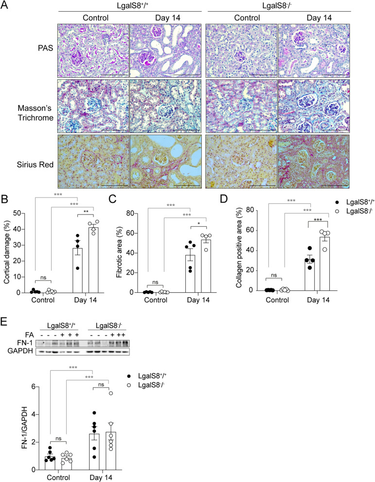
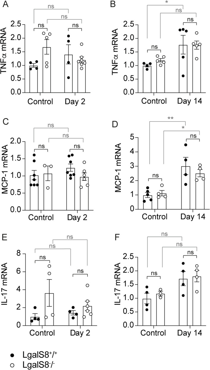
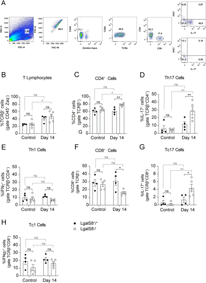
Results: Galectin-8 was predominantly expressed in the renal cortex, localizing to tubules, glomeruli, and blood vessels, with its levels decreasing by half following AKI. Both Lgals8+/+ and Lgals8-/- mice exhibited similar renal function and structure impairments during the acute phase, though Lgals8+/+ mice showed slightly worse damage. By the fibrotic phase, Lgals8-/- mice exhibited more pronounced cortical damage and fibrosis, characterized by increased type I and III collagen deposition and enhanced Th17 cell infiltration, while myofibroblast activation remained comparable to that of Lgals8+/+ mice.

Conclusions: Endogenous Gal-8 does not significantly protect the kidney during the acute phase and is dispensable for cell proliferation and death in response to AKI. However, it is crucial in preventing maladaptive repair by regulating extracellular matrix homeostasis and mitigating fibrosis. Additionally, Gal-8 contributes to inflammation resolution by limiting persistent immune cell infiltration, particularly IL-17-secreting cells.

Keywords: AKI; Acute kidney injury; CDK; Chronic kidney disease; Fibrosis; Galectin-8; Inflammation and Th17.


Conflict of interest statement

Declarations. Ethics approval and consent to participate: This study has been approved by the Ethical Committee of Universidad San Sebastián (Protocol number 01–2021-10). Consent for publication: Not applicable. Competing interests: The authors declare no competing interests.

Figures

Fig. 1
Fig. 1
Gal-8 is expressed in the kidney and its levels decrease following AKI. A β-galactosidase staining in kidneys from Lgals8−/− mice, reflecting endogenous Gal-8 expression. The inset shows a magnified view of the renal cortex. g = glomeruli, t = tubule, v = blood vessel. Scale bar: 100 μm. B Gal-8 transcript levels in the renal medulla and cortex of Lgals8+/+ mice, measured by RT-qPCR and normalized to β-actin. C Lectin staining of kidney structures in Lgals8−/− mice: collecting ducts (DBA, red), proximal tubules (LTA, green), and whole kidney (WGA, magenta). β-galactosidase staining is shown in grayscale. Arrows indicate colocalization of lectin and β-galactosidase staining. Scale bar: 50 μm. D β-galactosidase staining in kidneys from Lgals8−/− mice at 2 and 14 days after AKI induction. Scale bar: 50 μm. E Gal-8 transcript levels in kidneys from Lgals8+/+ mice at 2 and 14 days after AKI induction, assessed by RT-qPCR and normalized to β-actin. F Gal-8 transcript levels in the renal medulla and cortex of Lgals8+/+ mice at 2 days post-AKI induction, measured by RT-qPCR and normalized to β-actin. Data is presented as mean ± SEM of n = 3–9. *p < 0.05, **p < 0.01
Fig. 2
Fig. 2
Renal impairment during the acute phase after AKI in Lgals8−/− and Lgals8+/+ mice. A Plasma creatinine levels measured 2 days after AKI induction in Lgals8+/+ and Lgals8−/− mice. B H&E and PAS staining in renal cortex from Lgals8+/+ and Lgals8−/− mice. Representative images captured at 40X magnification. Scale bar: 100 μm. C Tubular dilation quantified in histological sections from Lgals8+/+ and Lgals8−/− mice. D Transcript levels of the tubular injury marker KIM-1, determined by RT-qPCR and normalized to β-actin. E Immunoblot quantification of NGAL protein levels in Lgals8+/+ and Lgals8−/− mice, normalized to GAPDH. Mean ± SEM of n = 4–7. *p < 0.05, **p < 0.01, ***p < 0.001
Fig. 3
Fig. 3
Similar levels of cell proliferation and death during the acute phase of AKI in Lgals8−/− and Lgals8+/+ mice. A Immunofluorescence (IF) and immunohistochemistry (IHQ) for the proliferation marker Ki67 assessed 2 days after AKI induction in Lgals8+/+ and Lgals8−/− mice. Scale bar: 50 μm. B Quantification of total Ki67-positive nuclei. C Quantification of Ki67-positive cells in tubular epithelial and (D) interstitial cells of Lgals8+/+ and Lgals8−/− mice. E Cell death assessment by TUNEL assay in kidney sections from Lgals8+/+ and Lgals8−/− mice, 2 days after AKI induction. Scale bar 50 μm. F Quantification of TUNEL-positive cells. Data is presented as mean ± SEM of n = 4–6. **p < 0.01, ***p < 0.005
Fig. 4
Fig. 4
Cortical damage and fibrosis are increased during the fibrotic phase in Lgals8−/− mice. A PAS, Masson´s Trichrome and Sirius Red staining in representative images of kidney sections from Lgals8+/+ and Lgals8−/− mice 14 days after damage induction. Scale bar: 100 μm. B Renal cortical damage measurements show the extent of the injured area in Lgals8+/+ and Lgals8−/− mice. C Fibrotic area quantified by collagen stained blue in kidney sections from Lgals8+/+ and Lgals8−/− mice. D Collagen-positive area was quantified by collagen stained red in kidney sections. E Immunoblot quantification of fibronectin-1 in the kidney of Lgals8+/+ and Lgals8−/− mice normalized to GAPDH. Data represent mean ± SEM of n = 4–6, *p < 0.05, **p < 0.01, ***p < 0.001
Fig. 5
Fig. 5
Similar fibroblast activation in the fibrotic phase in Lgal8+/+ and Lgals8−/− mice. (A) Vimentin and α-SMA protein levels compared by immunoblot 14 days after AKI induction in Lgals8+/+ and Lgals8−/ mice, normalized to GAPDH. (B) and (C) graphs quantifying the relative levels of vimentin and α-SMA, respectively. D Immunofluorescence (IF) and immunohistochemistry (IHQ) representative images of α-SMA localization in the kidney of Lgals8+/+ and Lgals8−/ mice. Scale bar: 50 μm. E Quantification of fluorescence intensity in α-SMA IF staining. F Cell proliferation activity shown by Ki67 IF and IHQ staining. G Quantification of total Ki67-positive nuclei. H Ki67-positive cells in tubular epithelial and (I) interstitial cells of Lgals8+/+ and Lgals8−/ mice. Data is presented as mean ± SEM of n = 4–6. *p < 0.05, **p < 0.01, ***p < 0.001
Fig.6
Fig.6
Expression of pro-inflammatory markers in Lgal8+/+ and Lgals8−/− mice. mRNA levels of TNF-α (A-B), MCP-1 (C-D), and IL-17 (EF) were measured at 2 and 14 days after injury induction in Lgals8+/+ and Lgals8−/ mice by RT-qPCR and normalized to β-actin. Data is presented as mean ± SEM of n = 4–7. *p < 0.05, **p < 0.01
Fig.7
Fig.7
Enhanced Th17 kidney infiltration in Lgals8−/− mice. A A gating strategy was used to analyze cytokine production on stimulated CD4 and CD8 T cells from kidneys. Lymphocytes were gated based on size and granularity, and then doublet exclusion was performed. Events from these gates were plotted against Zombie Aqua viability dye and CD45 marker. Cells were further selected for CD45 and TCRβ positivity before CD4 and CD8 gating was applied. Finally, IFNγ and IL-17 production were evaluated in CD4+ and CD8+ populations. Fourteen days after damage induction, mononuclear cells from kidneys were isolated and analyzed by flow cytometry to detect the infiltration of (A) total T cells, (B) CD4+ T cells, (C) Th1 cells, (D) Th17 cells, (E) CD8+ T cells, (F) Tc1 cells and (G) Tc17 cells in Lgals8+/+ and Lgals8−/− mice. Mean ± SEM of n = 4–5. *p < 0.05, **p < 0.005

References

    1. Abu El-Asrar AM, Missotten L, Geboes K. Expression of myofibroblast activation molecules in proliferative vitreoretinopathy epiretinal membranes. Acta Ophthalmol. 2011;89:e115-121. - PubMed
    1. Anginot A, Espeli M, Chasson L, Mancini SJ, Schiff C. Galectin 1 modulates plasma cell homeostasis and regulates the humoral immune response. J Immunol. 2013;190:5526–33. - PMC - PubMed
    1. Basile DP. Rarefaction of peritubular capillaries following ischemic acute renal failure: a potential factor predisposing to progressive nephropathy. Curr Opin Nephrol Hypertens. 2004;13:1–7. - PubMed
    1. Basile DP, Anderson MD, Sutton TA. Pathophysiology of acute kidney injury. Compr Physiol. 2012;2:1303–53. - PMC - PubMed
    1. Basile DP, Donohoe D, Roethe K, Osborn JL. Renal ischemic injury results in permanent damage to peritubular capillaries and influences long-term function. Am J Physiol Renal Physiol. 2001;281:F887-899. - PubMed

LinkOut - more resources