. 2025 May 15;31(1):191.
doi: 10.1186/s10020-025-01255-w.

Inhibition of diacylglycerol O-acyltransferase 1 provides neuroprotection by inhibiting ferroptosis in ischemic stroke

Affiliations

Inhibition of diacylglycerol O-acyltransferase 1 provides neuroprotection by inhibiting ferroptosis in ischemic stroke

Youjie Zeng et al. Mol Med. .

Abstract

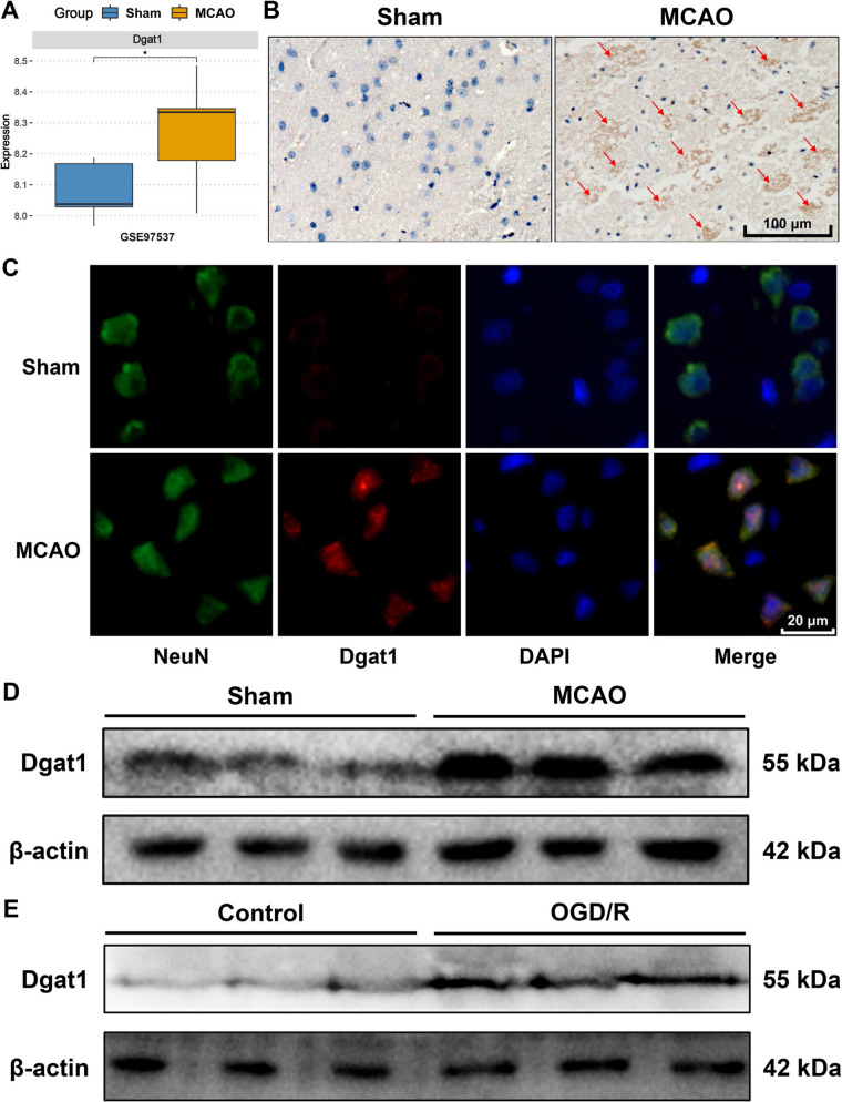
Background: Diacylglycerol O-acyltransferase 1 (DGAT1) is crucial for triglyceride synthesis, yet its role in ischemic stroke remains unclear. This study investigated DGAT1 in ischemic stroke using middle cerebral artery occlusion (MCAO) rat models and highly differentiated PC12 cells subjected to oxygen-glucose deprivation/reoxygenation (OGD/R).

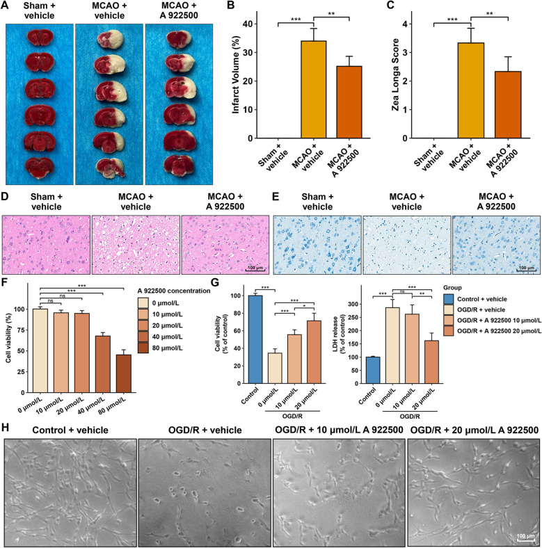
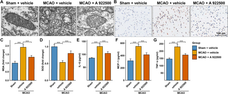
Methods: The therapeutic effects of DGAT1 inhibition in MCAO rats were assessed using the Zea-Longa score and 2,3,5-Triphenyltetrazolium chloride (TTC) staining. The effects on highly differentiated PC12 cells subjected to OGD/R were evaluated using the Cell Counting Kit-8 (CCK-8) and lactate dehydrogenase (LDH) assays. Ferroptosis-related mitochondrial damage was evaluated using transmission electron microscope. Additionally, the mechanisms by which DGAT1 inhibition regulates ferroptosis were further explored via immunohistochemistry, immunofluorescence, Western blotting, qPCR, JC-1 assay, and reactive oxygen species (ROS) detection.

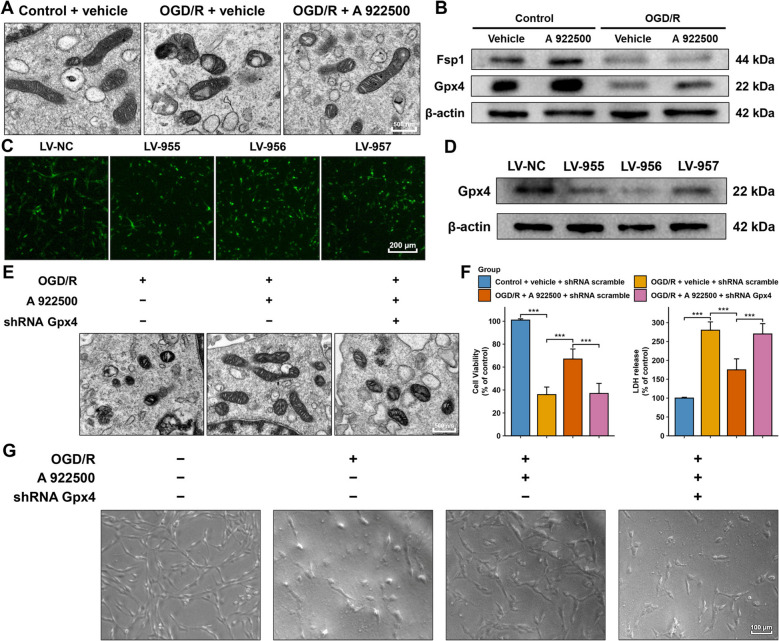
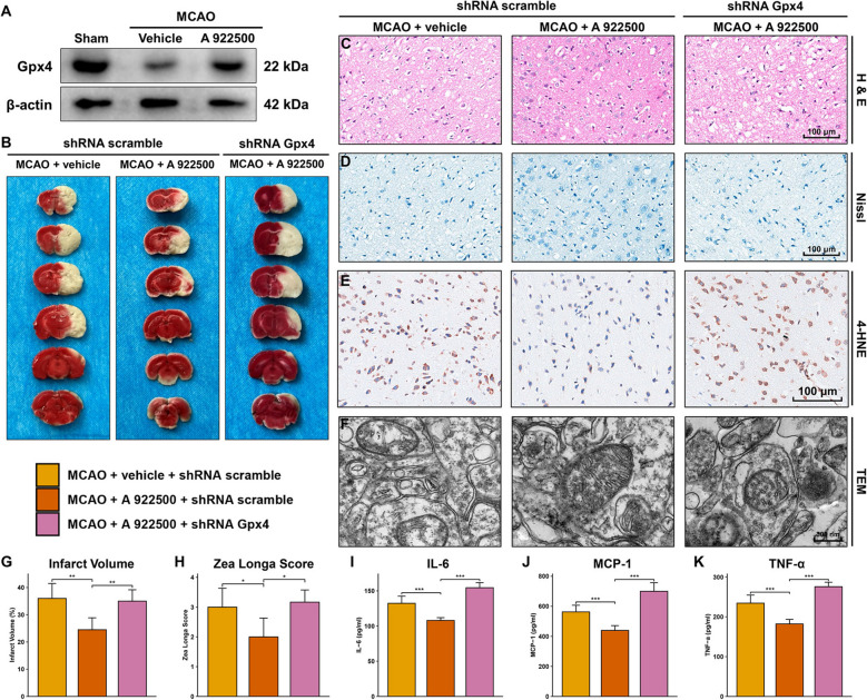
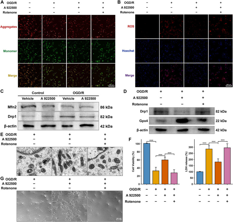
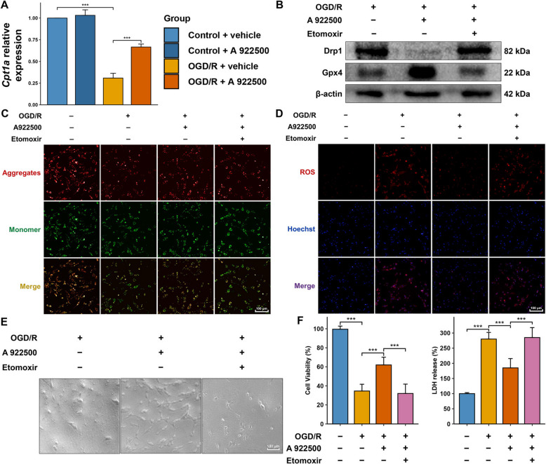
Results: DGAT1 expression was elevated in both MCAO and OGD/R models. The DGAT1 inhibitor A 922500 improved neurological deficits, reduced infarct volume, and minimized neuronal loss in MCAO rats, while also enhancing cell viability and reducing LDH levels in OGD/R-treated PC12 cells. DGAT1 inhibition significantly alleviated ferroptosis in MCAO rats, as indicated by (i) reduced mitochondrial shortening and cristae disruption, (ii) decreased 4-HNE levels, (iii) reduced MDA and increased SOD, and (iv) lowered levels of inflammatory factors (IL-6, MCP-1, and TNF-α). Moreover, both in vivo and in vitro experiments showed that DGAT1 inhibition significantly increased Gpx4 levels, whereas lentiviral delivery of Gpx4 shRNA markedly reversed its beneficial effects. In MCAO rats, Gpx4 shRNA significantly elevated 4-HNE levels and exacerbated ferroptosis-related mitochondrial damage. In vitro, DGAT1 inhibition increased mitochondrial membrane potential and reduced ROS, whereas rotenone, a mitochondrial function inhibitor, decreased Gpx4 and impaired cell viability. Furthermore, DGAT1 inhibition significantly upregulated the key β-oxidation gene Cpt1a, whereas etomoxir, a β-oxidation inhibitor, reduced cell viability and mitochondrial membrane potential, increased ROS, and downregulated Gpx4.

Conclusions: Our study suggests that DGAT1 inhibition may enhance β-oxidation and mitochondrial function, thereby increasing Gpx4 levels, suppressing ferroptosis, and ultimately exerting neuroprotective effects in ischemic stroke.

Keywords: DGAT1; Ferroptosis; Ischemic stroke; Lipid metabolism; Mitochondrial dysfunction.


Conflict of interest statement

Declarations. Ethics approval and consent to participate: The animal experiments in this study were approved by the Ethics Committee of the Department of Laboratory Animals of Central South University. Consent for publication: Not applicable. Competing interests: The authors declare no competing interests.

Figures

Fig. 1
Fig. 1
DGAT1 is upregulated in MCAO and OGD/R models. A In the GSE97537 dataset, DGAT1 mRNA levels were significantly upregulated in MCAO rat models compared to the sham group. B IHC analysis showing significantly increased DGAT1 expression in the brain tissue of MCAO rat compared to the sham group. C Fluorescence co-localization analysis demonstrating significant co-expression of DGAT1 with the neuronal marker NeuN, indicating predominant DGAT1 expression in neurons in the MCAO model. D Western blot analysis showing significant upregulation of DGAT1 expression in MCAO group compared to the sham controls. E Western blot analysis showing significant upregulation of DGAT1 expression in highly differentiated PC12 cells subjected to OGD/R compared to the control group
Fig. 2
Fig. 2
DGAT1 inhibition exerts neuroprotective effects in both in vivo and in vitro models of ischemic stroke. A TTC staining showing the effect of DGAT1 inhibition on MCAO-induced infarct volume in rat brains. B Quantification of infarct volume in rat brain tissues from different groups based on TTC staining results. C Bar plot demonstrating the effect of DGAT1 inhibition on Zea Longa scores in MCAO rats. D H&E staining illustrating the improvement in brain pathological damage caused by MCAO following DGAT1 inhibition. E Nissl staining showing that DGAT1 inhibition alleviates neuronal damage induced by MCAO. F CCK-8 cytotoxicity assay determining the candidate concentrations of the DGAT1 inhibitor A 922500 for in vitro experiments. G CCK-8 and LDH assays assessing the protective effects of DGAT1 inhibition on OGD/R-induced injury in highly differentiated PC12 cells. H Optical microscopy images showing the effects of DGAT1 inhibition on cell morphology and survival in OGD/R-treated highly differentiated PC12 cells
Fig. 3
Fig. 3
DGAT1 inhibition attenuates ferroptosis in the MCAO model. A TEM showing that DGAT1 inhibition improved ferroptosis-associated mitochondrial abnormalities. B IHC showing that DGAT1 inhibition alleviated the MCAO-induced upregulation of 4-HNE. C Bar plot showing that DGAT1 inhibition reduced the MCAO-induced MDA elevation. D Bar plot showing that DGAT1 inhibition reversed the MCAO-induced reduction in SOD levels. E Bar plot showing that DGAT1 inhibition reduced the MCAO-induced elevation of IL-6. F Bar plot showing that DGAT1 inhibition reduced the MCAO-induced elevation of MCP-1. G Bar plot showing that DGAT1 inhibition reduced the MCAO-induced elevation of TNF-α
Fig. 4
Fig. 4
Inhibition of DGAT1 protected highly differentiated PC12 cells in the OGD/R model by attenuating ferroptosis through upregulating Gpx4. A TEM images showing that the DGAT1 inhibitor A 922500 alleviated ferroptosis-related mitochondrial damage in highly differentiated PC12 cells following OGD/R treatment. B Western blot analysis showing the effect of DGAT1 inhibition on two ferroptosis resistant proteins (Fsp1 and Gpx4). C Lentiviral infection carrying Gpx4 shRNA was successfully achieved in highly differentiated PC12 cells. D The most effective Gpx4 shRNA sequence was identified by western blot analysis. E TEM showing that Gpx4 shRNA significantly reversed the improvements in ferroptosis-related mitochondrial morphology induced by DGAT1 inhibition in OGD/R-treated highly differentiated PC12 cells. F CCK-8 and LDH assays showing that Gpx4 shRNA reversed the protective effects of DGAT1 inhibition in OGD/R-treated highly differentiated PC12 cells. G Optical microscopy showing that Gpx4 shRNA reversed the improvements in cell morphology and number induced by DGAT1 inhibition in OGD/R-treated highly differentiated PC12 cells
Fig. 5
Fig. 5
DGAT1 inhibition mitigates neurological impairment in MCAO rats by attenuating ferroptosis through upregulating Gpx4. A Western blot analysis showing that DGAT1 inhibition upregulated Gpx4 expression, which was decreased in MCAO rats. B TTC staining analysis showing that the reduction in infarct volume caused by DGAT1 inhibition was reversed by Gpx4 shRNA. C H&E staining showing that Gpx4 shRNA reversed the protective effects of DGAT1 inhibition on cerebral tissue damage in MCAO rats. D Nissl staining showed that Gpx4 shRNA reversed the protective effects of DGAT1 inhibition on neuronal injury in MCAO rats. E Gpx4 shRNA increased 4-HNE levels, which had been reduced by DGAT1 inhibition in the MCAO model. F TEM analysis showing that Gpx4 shRNA reversed the improvements in ferroptosis-related mitochondrial damage induced by DGAT1 inhibition. G Bar plot of TTC staining results showing that Gpx4 shRNA reversed the DGAT1 inhibition-induced reduction of infarct volume in MCAO rats. H Gpx4 shRNA reversed the improvement in Zea Longa neurological scores induced by DGAT1 inhibition. I Gpx4 shRNA increased IL-6 levels, which had been reduced by DGAT1 inhibition in the MCAO model. J Gpx4 shRNA increased MCP-1 levels, which had been reduced by DGAT1 inhibition in the MCAO model. K Gpx4 shRNA increased TNF-α levels, which had been reduced by DGAT1 inhibition in the MCAO model
Fig. 6
Fig. 6
DGAT1 inhibition protects highly differentiated PC12 cells in the OGD/R model by resisting ferroptosis through improving mitochondrial function. A JC-1 assay showing the effect of DGAT1 inhibition on mitochondrial membrane potential in the OGD/R model. B ROS assay showing the effect of DGAT1 inhibition on reducing ROS levels in the OGD/R model. C Western blot analysis showing the effect of DGAT1 inhibition on mitochondrial fission (Drp1) and fusion (Mfn2) proteins in the OGD/R model. D Western blot analysis showing the influence of rotenone treatment (mitochondrial dysfunction) on Gpx4 expression in the OGD/R model. E TEM showing that rotenone (inhibitor of mitochondrial function) significantly reversed the protective effects of DGAT1 inhibition against ferroptosis-related mitochondrial damage in the OGD/R model. F CCK-8 and LDH assays showing that rotenone reduced cell viability and increased LDH release, reversing the protective effects of DGAT1 inhibition in the OGD/R model. G Optical microscopy images showing that DGAT1 inhibition improved cell morphology and number, and that these effects were reversed by rotenone in OGD/R-treated highly differentiated PC12 cells
Fig. 7
Fig. 7
DGAT1 inhibition improves mitochondrial function by upregulating β-oxidation in the OGD/R model. A qPCR showing that DGAT1 inhibition increased Cpt1a mRNA expression compared to the OGD/R group. B Western blot analysis showing that the β-oxidation inhibition by etomoxir reversed the DGAT1 inhibition-induced decrease in Drp1 levels and the increase in Gpx4 levels in the OGD/R model. C JC-1 assay showing that β-oxidation inhibition decreased mitochondrial membrane potential, which had been improved by DGAT1 inhibition in the OGD/R model. D ROS assay showing that β-oxidation inhibition increased ROS levels, which had been reduced by DGAT1 inhibition in the OGD/R model. E Optical microscopy showing that the improvements in PC12 cell morphology and number caused by DGAT1 inhibition were reversed by β-oxidation inhibition in the OGD/R model. F CCK-8 and LDH assays showing that β-oxidation inhibition decreased cell viability and increased LDH levels, reversing the protective effects of DGAT1 inhibition in the OGD/R model

References

    1. Adibhatla RM, Hatcher JF. Tissue plasminogen activator (tPA) and matrix metalloproteinases in the pathogenesis of stroke: therapeutic strategies. CNS & Neurolog Disorders-Drug Targets (Formerly Current Drug Targets-CNS & Neurological Disorders). 2008;7:243–53. - PMC - PubMed
    1. Alim I, et al. Selenium drives a transcriptional adaptive program to block ferroptosis and treat stroke. Cell. 2019;177:1262-1279.e1225. - PubMed
    1. Andrabi SS, Parvez S, Tabassum H. Ischemic stroke and mitochondria: mechanisms and targets. Protoplasma. 2020;257:335–43. - PubMed
    1. Bae YS, Oh H, Rhee SG, Yoo YD. Regulation of reactive oxygen species generation in cell signaling. Mol Cells. 2011;32:491–509. - PMC - PubMed
    1. Bakthavachalam P, Shanmugam PST. Mitochondrial dysfunction - Silent killer in cerebral ischemia. J Neurol Sci. 2017;375:417–23. - PubMed

MeSH terms

Substances

LinkOut - more resources