

Diagnostic journey and genetic analysis of a novel homozygous CYP2U1 mutation causing autosomal recessive spastic paraplegia type 56 (SPG56) in a consanguineous family
- PMID: 40375209
- PMCID: PMC12083119
- DOI: 10.1186/s12883-025-04211-7
Diagnostic journey and genetic analysis of a novel homozygous CYP2U1 mutation causing autosomal recessive spastic paraplegia type 56 (SPG56) in a consanguineous family
Abstract
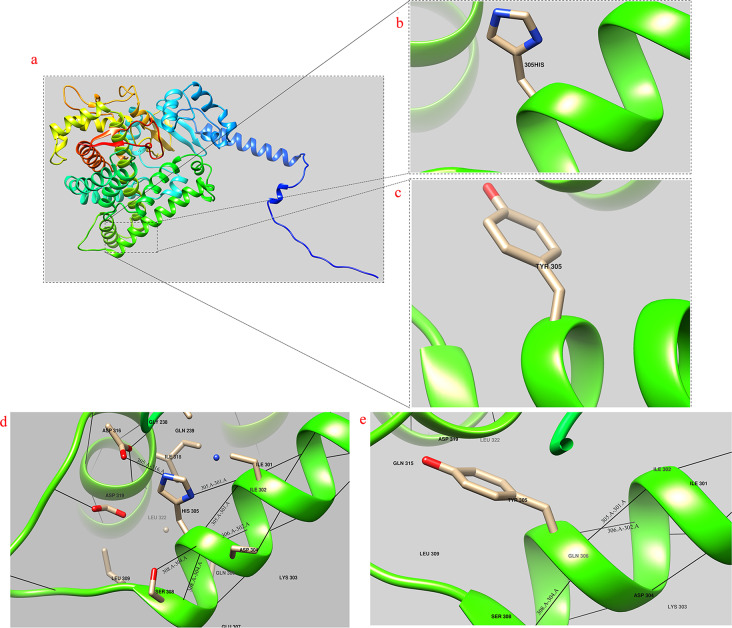
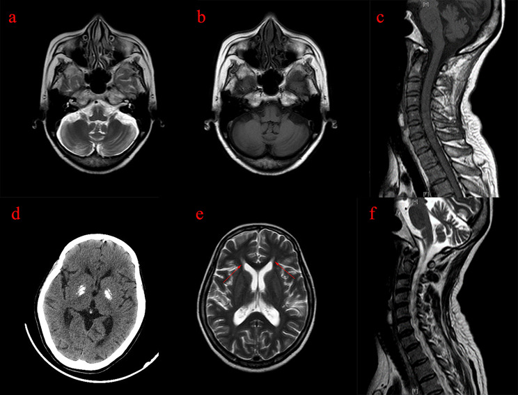
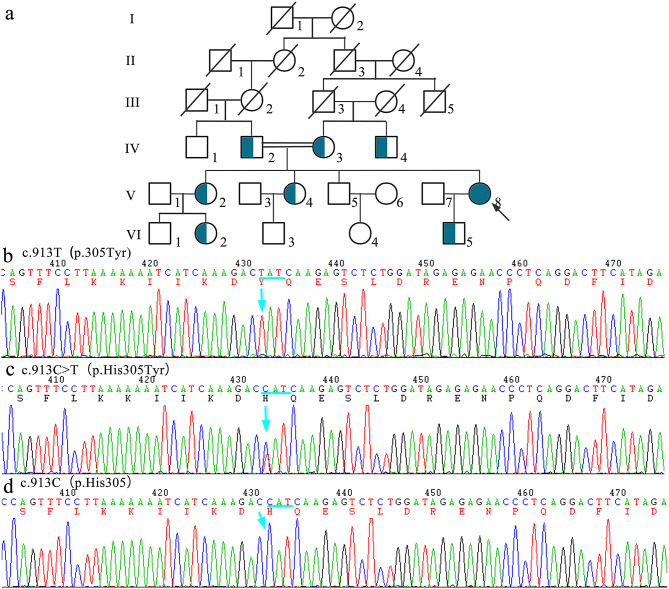
Hereditary spastic paraplegia (HSP) is a neurodegenerative disorder, with spastic paraplegia type 56 (SPG56) being an exceptionally rare, autosomal recessive subtype caused by mutations in the CYP2U1 gene. This study reports a complex case of an adult female from a consanguineous family who presented with cognitive developmental delays, short stature, and progressive neurological symptoms. At age 39, she developed unilateral tremors, which progressed to generalized tremors and leg weakness with a tiptoe gait. The clinical findings included hypertonia in the upper limbs, exaggerated reflexes in the lower limbs, vague speech, and emotional disturbances. Brain MRI revealed corpus callosum thinning, "ears of the Lynx" sign, bilateral globus pallidus calcifications, and mild brain atrophy. Comprehensive genomic analysis, including whole exome sequencing (WES), copy number variation (CNV) assessment, mitochondrial DNA sequencing, variant filtering, and Sanger sequencing, identified a homozygous c.913 C > T (p.His305Tyr) mutation in CYP2U1 (NM_183075). The heterozygous carriers presented no symptoms. This case contributes to the phenotypic spectrum of SPG56, offering new insights into its diagnosis and genetic underpinnings.
Keywords: CYP2U1; Complex HSP; Consanguinity; Ears of the Lynx; Hereditary spastic paraplegia; SPG56.
© 2025. The Author(s).
Conflict of interest statement
Declarations. Ethics approval and consent to participate: This study was approved by the Bioethics Committee of Fujian Provincial Hospital, Fuzhou, China (Approval Number: No. K2023-12-018). All participants provided written informed consent prior to their inclusion in the study, in accordance with the Declaration of Helsinki. Consent for publication: Written informed consent was obtained from the patient for the publication of any potentially identifiable images or data included in this article. Competing interests: The authors declare no competing interests.
Figures
Similar articles
-
The clinical and molecular spectrum of ZFYVE26-associated hereditary spastic paraplegia: SPG15.Brain. 2023 May 2;146(5):2003-2015. doi: 10.1093/brain/awac391. Brain. 2023. PMID: 36315648 Free PMC article.
-
Charting the genetic landscape of autosomal recessive hereditary spastic paraplegia: A deep dive into 10 exceptionally rare cases.Neurogenetics. 2025 Aug 9;26(1):58. doi: 10.1007/s10048-025-00841-8. Neurogenetics. 2025. PMID: 40782215
-
Rare novel CYP2U1 and ZFYVE26 variants identified in two Pakistani families with spastic paraplegia.J Neurol Sci. 2020 Apr 15;411:116669. doi: 10.1016/j.jns.2020.116669. Epub 2020 Jan 11. J Neurol Sci. 2020. PMID: 32006740
-
[FA2H gene-associated spastic paraplegia (SPG35) - familial case with late onset].Zh Nevrol Psikhiatr Im S S Korsakova. 2025;125(5):137-144. doi: 10.17116/jnevro2025125051137. Zh Nevrol Psikhiatr Im S S Korsakova. 2025. PMID: 40457680 Review. Russian.
-
A novel homozygous variant in RNF170 causes hereditary spastic paraplegia: a case report and review of the literature.Neurogenetics. 2022 Apr;23(2):85-90. doi: 10.1007/s10048-022-00685-6. Epub 2022 Jan 18. Neurogenetics. 2022. PMID: 35041108 Review.
References
-
- Mereaux JL, Banneau G, Papin M, Coarelli G, Valter R, Raymond L, et al. Clinical and genetic spectra of 1550 index patients with hereditary spastic paraplegia. Brain. 2022;145(3):1029–37. 10.1093/brain/awab386. - PubMed
-
- Minase G, Miyatake S, Nabatame S, Arai H, Koshimizu E, Mizuguchi T, et al. An atypical case of SPG56/CYP2U1-related spastic paraplegia presenting with delayed myelination. J Hum Genet. 2017;62(11):997–1000. 10.1038/jhg.2017.77. - PubMed
MeSH terms
Substances
Grants and funding
- 2023Y9284/Joint Funds for the innovation of science and Technology in Fujian province
- 2022J01409, 2022J01996, 2021J02053, 2023Y9320/Fujian Province Natural Science Fund Project
- 2021QNB001/Fujian Provincial Youth Scientific Program on health
- 2022CXB002, 2021CXB001, 2022CXA001/Fujian Province Medical Innovation Foundation
- 2020-822, 2021-848, 2022-840/the Special Research Foundation of Fujian Provincial Department of Finance, China
LinkOut - more resources
Full Text Sources
