Abdominal fat distribution in endometrial cancer: from diagnosis to follow-up
- PMID: 40375215
- PMCID: PMC12079972
- DOI: 10.1186/s12885-025-14155-3
Abdominal fat distribution in endometrial cancer: from diagnosis to follow-up
Abstract
Background: The objective of this study is to quantify abdominal obesity markers from computed tomography (CT) scans at primary diagnosis and follow-up in a large endometrial cancer cohort, and to assess temporal change in obesity markers in relation to surgicopathological patient characteristics and outcome.
Methods: Total- (TAV), subcutaneous- (SAV), visceral (VAV) abdominal fat volumes, and visceral-to-total fat percentage (VAV%) were derived from CT scans acquired in an endometrial cancer patient cohort at primary diagnosis (nprimary=293). Temporal (delta, δ) changes in CT obesity markers from primary diagnosis to follow-up were assessed for all patients with a follow-up CT 13 (7, 19) [median (interquartile range)] months after diagnosis (nfollow-up=152/293 patients). The CT obesity markers were assessed in relation to clinicopathological features and progression-free survival (PFS) using Mann-Whitney U-test, and Cox hazard ratios (HRs), respectively.
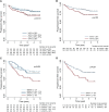
Results: At primary diagnosis, VAV% was the only marker significantly associated with high-risk histology (median of 33% for endometrioid endometrial carcinoma (EEC) grade 1-2, 36% for EEC grade 3 and 36% for non-endometrioid EC, p = 0.003), myometrial invasion (MI) (median of 34% for MI < 50% vs. 35% for MI ≥ 50%, p = 0.03) and lymphovascular space invasion (LVSI) (median of 34% for no LVSI vs. 36% for LVSI, p = 0.009). High VAV% (≥ 35%) also predicted poor PFS both in univariable analysis (HR = 1.8, p = 0.02), and when stratified for surgicopathological FIGO stage (HR = 3.1, p = 0.03). At follow-up, median TAV, VAV, SAV, and VAV% were significantly lower than at primary diagnosis (p < 0.001 for all). Furthermore, patients with progression had larger reductions in visceral fat compartments (δVAV=-24%, δVAV% =-3%), than patients with no progression (δVAV=-17%, δVAV%=-2%, p ≤ 0.006 for both).
Conclusion: Visceral abdominal obesity (high VAV%) is associated with high-risk histologic features, myometrial invasion, and poor prognosis. Furthermore, high visceral fat loss during/following therapy is associated with disease progression.
Keywords: Adiposity; Computed tomography; Endometrial neoplasms; Intra-abdominal fat; Obesity.
© 2025. The Author(s).
Conflict of interest statement
Declarations. Ethical approval and consent to participate: The study was approved by the Western Regional Committee for Medical and Health Research Ethics (REK vest 2018/594, 2015/2333, and biobank approval: 2014/1907), according to Norwegian legislation and regulation. All included patients gave written informed consent at primary diagnosis in this retrospective study with prospectively collected data. Consent for publication: Not applicable. Competing interests: The authors declare no competing interests.
Figures


Similar articles
-
Visceral fat percentage for prediction of outcome in uterine cervical cancer.Gynecol Oncol. 2023 Sep;176:62-68. doi: 10.1016/j.ygyno.2023.06.581. Epub 2023 Jul 13. Gynecol Oncol. 2023. PMID: 37453220
-
Impact of body mass index and fat distribution on sex steroid levels in endometrial carcinoma: a retrospective study.BMC Cancer. 2019 Jun 7;19(1):547. doi: 10.1186/s12885-019-5770-6. BMC Cancer. 2019. PMID: 31174495 Free PMC article.
-
High visceral fat percentage is associated with poor outcome in endometrial cancer.Oncotarget. 2017 Oct 19;8(62):105184-105195. doi: 10.18632/oncotarget.21917. eCollection 2017 Dec 1. Oncotarget. 2017. PMID: 29285243 Free PMC article.
-
Visceral adiposity and inflammatory bowel disease.Int J Colorectal Dis. 2021 Nov;36(11):2305-2319. doi: 10.1007/s00384-021-03968-w. Epub 2021 Jun 9. Int J Colorectal Dis. 2021. PMID: 34104989 Review.
-
Meta-analysis of the clinicopathologic features of endometrial cancer molecular staging.Front Oncol. 2025 Jan 7;14:1510102. doi: 10.3389/fonc.2024.1510102. eCollection 2024. Front Oncol. 2025. PMID: 39839791 Free PMC article.
References
-
- Sung H, et al. Global cancer statistics 2020: GLOBOCAN estimates of incidence and mortality worldwide for 36 cancers in 185 countries. CA Cancer J Clin. 2021;71:209–49. - PubMed
-
- Morice P, Leary A, Creutzberg C, Abu-Rustum N, Darai E. Endometrial cancer. Lancet. 2016;387:1094–108. - PubMed
-
- Calle EE, Rodriguez C, Walker-Thurmond K, Thun MJ, Overweight. Obesity, and mortality from cancer in a prospectively studied cohort of U.S. Adults. N Engl J Med. 2003;348:1625–38. - PubMed
MeSH terms
LinkOut - more resources
Full Text Sources
Miscellaneous

