. 2025 May 15;27(1):104.
doi: 10.1186/s13075-025-03573-0.

Chondrocyte lysates activate NLRP3 inflammasome-induced pyroptosis in synovial fibroblasts to exacerbate knee synovitis by downregulating caveolin-1

Affiliations

Chondrocyte lysates activate NLRP3 inflammasome-induced pyroptosis in synovial fibroblasts to exacerbate knee synovitis by downregulating caveolin-1

Xue Du et al. Arthritis Res Ther. .

Abstract

Background: Synovitis, among the most common signs of early-stage osteoarthritis (OA), is mainly mediated by fibroblast-like synoviocytes (FLSs). Cartilage destruction creates chondrocyte lysates (CLs) that activate synovial inflammation. A comprehensive understanding of chondrocyte-FLS communication might offer novel, specific therapeutic targets for treating synovitis and OA. Hence, we sought to uncover the specific role of CLs in OA-FLSs and synovitis.

Methods: Isolated CLs were cocultured with FLSs to test whether they could stimulate synovial inflammation. A model of medial meniscus destabilization was prepared in C57BL/6 mice and NLRP3 knockout mice, and adeno-associated virus overexpressing Caveolin-1 (CAV1) was intra-articularly injected for 8 weeks once a week after dissection of the medial meniscus (DMM). Proteins expressed in FLSs with and without CL coculture were screened using liquid chromatography-tandem mass spectrometry to identify CL-specific regulators of NLRP3 inflammasome-mediated pyroptosis.

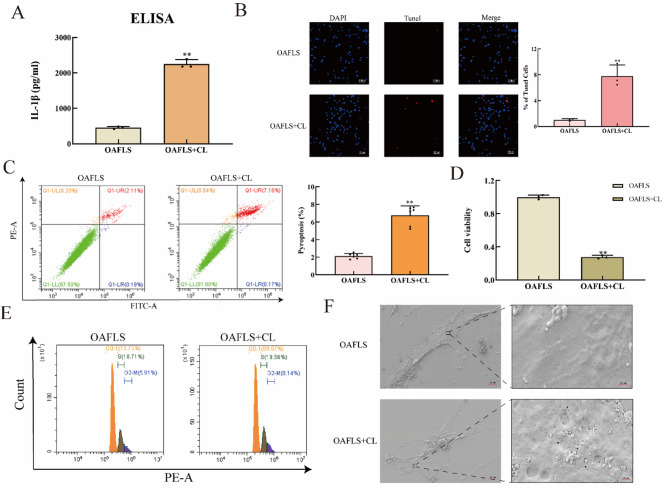
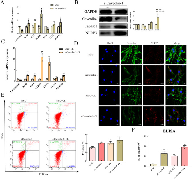
Results: CLs were engulfed by FLSs, which aggravated inflammatory cytokine release and NLRP3 inflammasome-mediated FLS pyroptosis. NLRP3 expression was significantly upregulated in human OA-FLSs and FLSs cocultured with CLs, while CAV1 was downregulated. CAV1 overexpression reversed the inflammatory phenotype in FLSs and simultaneously rescued pyroptosis in CL-pre-treated FLSs. Both synovial hyperplasia and inflammatory infiltration in C57BL/6 mice with DMM surgery were alleviated after intra-articular AAV-CAV1 injection. Moreover, the CL-specific protein LIM-containing lipoma preferred partner (LPP) markedly exacerbated FLS pyroptosis and inflammation.

Conclusions: CLs were endocytosed by FLSs through CAV1, and the CL-specific protein LPP stimulated NLRP3 inflammasome-mediated pyroptosis and synovitis by inhibiting CAV1 expression. Our findings offer a novel therapeutic target for treating synovitis.

Keywords: Caveolin-1; Chondrocyte lysate; Fibroblast-like synoviocyte; LIM-containing lipoma preferred partner; NLRP3.


Conflict of interest statement

Declarations. Ethics approval and consent to participate: All experiments involving animal and patient samples in this study adhered to ethical policies and procedures approved by the Ethics Committee of the First Affiliated Hospital of Sun Yat-Sen University, China. (Approval no. IRB: IIT-2021-667; SYSU-IACUC-2020-000504). Consent for publication: Written informed consent for publication was obtained from all participants. Competing interests: The authors declare no competing interests.

Figures

Fig. 1
Fig. 1
Chondrocyte lysates upregulate the expression of NLRP3 via endocytosis by FLSs. (A). Quantitative PCR analysis of NLRP3 in primary FLSs from normal articular (NA) tissue and OA tissue (n = 6). (B) WB analysis of NLRP3 in synovium obtained from individuals with NA and from patients with OA (n = 3). (C) Representative coimmunofluorescence images of NLRP3 (red fluorescence) and vimentin (green fluorescence), a marker of FLSs, in synovial tissues harvested from NA and OA patients. A scale bar is presented in each image for precision. (D) Immunostaining of NLRP3 (green light) in primary FLSs isolated from NA and OA patients. (E) qRT‒PCR for NLRP3, IL-1β, IL-18, TNF-α, TLR4 and MMP13 in FLSs with or without CL pretreatment. (F) WB analysis of NLRP3, caspase-1, GSDMD, IL-1β, and IL-18 in FLSs with and without CL stimulation. (G) Representative images of coimmunostaining for NLRP3 (green fluorescence) and CLs (red fluorescence) in FLSs with and without CL pretreatment. All the data are presented as the means ± SEMs. Paired t tests and one-way analysis of variance (ANOVA) were used for statistical analysis. * p < 0.05, ** p < 0.01, ***p < 0.001. FLSs, fibroblast-like synoviocytes; NA, normal articular; OA, osteoarthritis
Fig. 2
Fig. 2
Chondrocyte lysates promote the secretion of inflammatory cytokines by enhancing pyroptosis in FLSs. (A) ELISA of the protein levels of IL-1β in the cell supernatants of primary FLSs with or without CL treatment. (B). Representative images of TUNEL-stained primary FLSs with or without CL treatment and quantification of the percentage of TdT-positive cells (n = 3). A scale bar is presented in each image for precision. (C). The pyroptotic rates of FLSs with or without CL pretreatment were detected via flow cytometry via Annexin V-FITC/PI double staining and quantified via FACS analysis. (D). The viability of FLSs with and without CL pretreatment was quantified via the CCK-8 assay. (E). The effect of CLs on the cell cycle of FLSs was detected by flow cytometry and PI staining. (F). Representative SEM images focused on the perforation of the cellular membrane on FLSs with and without CL pretreatment. All the data are presented as the means ± SEMs. Paired t tests and one-way analysis of variance (ANOVA) were used for statistical analysis. * p < 0.05, ** p < 0.01, ***p < 0.001. A scale bar is presented in each image for precision. FLSs, fibroblast-like synoviocytes;
Fig. 3
Fig. 3
The endocytosis of chondrocyte lysates by FLSs is regulated through CAV1, and CLs downregulate the expression of CAV1 in FLSs. (A). qRT‒PCR analysis of CAV1 in FLSs with and without CL pretreatment (n = 6). (B). WB and quantification of CAV1 levels in FLSs with or without CL pretreatment (n = 3). (C). Representative coimmunofluorescence images of CAV1 (green fluorescence) and CLs (red fluorescence). (D). Representative coimmunostaining images for CAV1 and NLRP3 in FLSs with or without CL pretreatment after transfection with si-CAV1 or a control. (E). WB and quantitative analysis of ERK and p-ERK levels in FLSs with or without CL pretreatment. (F). WB and quantitative analysis of CAV1, ERK and p-ERK levels in CL-stimulated FLSs with or without hypaphorine treatment (n = 3). All the data are presented as the means ± SEMs. Paired t tests and one-way analysis of variance (ANOVA) were used for statistical analysis. * p < 0.05, ** p < 0.01, ***p < 0.001. FLSs, fibroblast-like synoviocytes; NA, normal articular; OA, osteoarthritis
Fig. 4
Fig. 4
Silencing CAV1 in FLSs in vitro promotes NLRP3 inflammasome-mediated pyroptosis and is associated with synovitis. (A). qRT‒PCR of CAV1, IL-1β, IL-18, NLRP3, TNF-α, TLR4, and MMP13 in FLSs transfected with si-CAV1 for 2 days or transfected with the control (n = 6). (B). WB and quantification of CAV1, caspase-1 and NLRP3 levels after transfection with si-CAV1 for 3 days or after transfection with the control (n = 3). (C). qRT‒PCR analysis of CAV1, IL-1β, IL-18, NLRP3, TNF-α, TLR4, and MMP13 in CL-stimulated FLSs after transfection with si-CAV1 for 2 days or the control (n = 3). (D). Representative images of coimmunostaining for CAV1 and NLRP3 in FLSs with or without CL pretreatment after transfection with si-CAV1 or the control. (E). The rates of pyroptosis were analysed via flow cytometry via Annexin V-FITC/PI double staining, and the number of pyroptotic cells was determined via FACS. (F). ELISA was used to detect the concentration of IL-1β in the cell supernatants of FLSs treated with CLs and si-CAV1 or the control (n = 3). All the data are presented as the means ± SEMs. Paired t tests and one-way analysis of variance (ANOVA) were used for statistical analysis. * p < 0.05, ** p < 0.01, ***p < 0.001
Fig. 5
Fig. 5
CAV1 overexpression in FLSs alleviates synovitis in a DMM-induced OA model through the regulation of NLRP3 inflammasome-mediated pyroptosis. (A). Representative images of HE-stained knee joints from C57BL/6J (wild-type) DMM mice and NLRP3 knockout DMM mice with or without intra-articular injection of AAV-CAV1 (oeCAV1) and quantification of the synovitis score (n = 5). (B). Representative images of coimmunofluorescence of CAV1 (red fluorescence) and vimentin (green fluorescence) in knee joints from C57BL/6J (wild-type) DMM mice with or without intra-articular injection of AAV-CAV1 (n = 5). (C). Representative images of NLRP3 (red fluorescence) and vimentin (green fluorescence) in knee joints from C57BL/6J (wild-type) DMM mice with or without intra-articular injection of AAV-CAV1 (n = 5). (D). Representative images of coimmunofluorescence of CAV1 (red fluorescence) and vimentin (green fluorescence) in the knee joints of NLRP3 knockout mice with or without intra-articular injection of AAV-CAV1 (n = 5). All the data are presented as the means ± SEMs. Paired t tests and one-way analysis of variance (ANOVA) were used for statistical analysis. * p < 0.05, ** p < 0.01, ***p < 0.001. HE, haematoxylin‒eosin; DMM, destabilization of the medial meniscus; AAV, adeno-associated virus. KO, knockout
Fig. 6
Fig. 6
LPP in chondrocyte lysates promotes NLRP3 inflammasome-mediated pyroptosis in FLSs. (A). Venn diagram of screened proteins detected by LC‒MS from CLs and FLSs isolated from 3 different groups (Ligand A/B/C) of OA patients with CL stimulation. (B). Co-IP analysis of LPP pulled down from NLRP3 in FLSs with or without CL pretreatment. (C). qRT‒PCR analysis of CAV1, IL-1β, IL-18, NLRP3, TNF-α, TLR4, and MMP13 levels in cells with or without recombinant LPP protein treatment (n = 6). (D). WB and quantification of CAV1, NLRP3, and LPP levels in FLSs with and without LPP treatment. (E). The pyroptotic rates of FLSs with and without recombinant LPP protein treatment were quantified via flow cytometry with double Annexin V-FITC/PI staining (n = 3). (F). Representative images of the coimmunofluorescence of CAV1 (green fluorescence) and NLRP3 (red fluorescence) with and without LPP treatment are shown. (G). Schematic representation of the mechanisms by which CLs regulate synovitis. All the data are presented as the means ± SEMs. Paired t tests and one-way analysis of variance (ANOVA) were used for statistical analysis. * p < 0.05, ** p < 0.01, ***p < 0.001. LC‒MS

Similar articles

References

    1. Yao Q, Wu X, Tao C, Gong W, Chen M, Qu M, Zhong Y, He T, Chen S, Xiao G. Osteoarthritis: pathogenic signaling pathways and therapeutic targets. Signal Transduct Target Ther. 2023;8(1):56. - PMC - PubMed
    1. Oppenheim JJ, Yang D. Alarmins: chemotactic activators of immune responses. Curr Opin Immunol. 2005;17(4):359–65. - PubMed
    1. Millerand M, Berenbaum F, Jacques C. Danger signals and inflammaging in osteoarthritis. Clin Exp Rheumatol. 2019;37(Suppl 120):48–56. - PubMed
    1. Nefla M, Holzinger D, Berenbaum F, Jacques C. The danger from within: alarmins in arthritis. Nat Rev Rheumatol. 2016;12(11):669–83. - PubMed
    1. Huang J, Xie Y, Sun X, Zeh HJ 3rd, Kang R, Lotze MT, Tang D. DAMPs, ageing, and cancer: the ‘DAMP hypothesis’. Ageing Res Rev. 2015;24(Pt A):3–16. - PMC - PubMed

MeSH terms

LinkOut - more resources