Randomized Controlled Trial
. 2025 Aug;24(8):e70108.
doi: 10.1111/acel.70108. Epub 2025 May 15.

Quercetin Reduces Vascular Senescence and Inflammation in Symptomatic Male but Not Female Coronary Artery Disease Patients

Affiliations
Randomized Controlled Trial

Quercetin Reduces Vascular Senescence and Inflammation in Symptomatic Male but Not Female Coronary Artery Disease Patients

Pauline Mury et al. Aging Cell. 2025 Aug.

Abstract

Recent studies suggest that vascular senescence and its associated inflammation fuel the inflammaging to favor atherogenesis; whether these pathways can be therapeutically targeted in coronary artery disease (CAD) patients remains unknown. In a randomized, double-blind trial, 97 patients (78 men) undergoing coronary artery bypass graft surgery were treated with either quercetin (500 mg twice daily, 47 patients) or placebo (50 patients) for two days pre-surgery through hospital discharge. Primary outcomes were reduced inflammation and improved endothelial function ex vivo. Exploratory analyses included plasma proteomics and single-nuclei RNA sequencing of internal thoracic artery (ITA) samples. Quercetin treatment showed a trend toward reduced C-reactive protein at discharge (p = 0.073) and differentially modulated circulating inflammatory protein expression between men and women, with a pro-inflammatory effect of quercetin in females. Endothelial acetylcholine-induced relaxation improved significantly with quercetin (p = 0.049), with effects in men (p = 0.043) but not in women (p = 0.852). ITA transcriptomics revealed the overexpression of senescence and inflammaging pathways in male vascular cells, which quercetin reversed. In female cells, quercetin had minimal endothelial benefit and increased inflammaging in fibroblasts. In male cells, a candidate target of quercetin involves interactions between the receptor PLAUR and its ligands PLAU and SERPINE1. Post-operative atrial fibrillation incidence was significantly lower with quercetin, representing 4% of the patients compared to 18% in the placebo group (p = 0.033). In conclusion, short-term quercetin treatment effectively targeted vascular senescence in male CAD patients, improving inflammatory and functional outcomes. However, these benefits were not observed in female patients. Trial Registration: https://clinicaltrials.gov, NCT04907253.

Keywords: inflammaging; cellular senescence; coronary artery disease; endothelium‐dependent relaxation; sex‐dimorphism; snRNA‐seq; systemic inflammatory proteomic.


Conflict of interest statement

The authors declare no conflicts of interest.

Figures

FIGURE 1
FIGURE 1
Impact of quercetin on systemic inflammatory biomarkers. (A) Flow‐chart of the primary endpoint of the study (impact of quercetin on systemic inflammation) at each timepoint of hs‐CRP measurement (created using BioRender). (B) Mean plasma hs‐CRP levels measured at baseline (pre‐treatment), post‐operative day (POD) 1, POD4 and at hospital discharge (POD7) after a coronary by‐pass graft surgery, in the intent‐to‐treat (ITT; 97 patients) population. Data are mean ± SEM of 50 placebo patients vs. 47 quercetin patients. ANCOVA was performed (effect of Group p = 0.824, Time p < 0.0001, Group × Time p = 0.025). (C) Mean plasma hs‐CRP levels of 39 men placebo patients vs. 11 women placebo patients and of 39 men quercetin patients vs. 8 women quercetin patients. *p < 0.05 vs. men placebo. (D) Mean plasma hs‐CRP levels of 39 men quercetin vs. 39 men placebo (left panel) and 8 women quercetin vs. 11 women placebo (right panel). (E) Flowchart of the proteomic analysis (Olink Proteomics) at POD4 (created using BioRender). (F) VolcanoPlot visualization of 384 inflammation‐related biomarkers in placebo vs. quercetin patients illustrating any differentially expressed proteins (DEP) between placebo and quercetin groups in the ITT population (proteins are considered significantly differently expressed when false discovery rate (FDR) < 0.05 and Log2 fold change (L2FC) > 0.25). (G) All DEP of the Inflammation panel between women placebo and men placebo groups (left), and comparison between women quercetin and men quercetin (right) (proteins are considered significantly differently expressed when FDR < 0.05 and L2FC > 0.25).
FIGURE 2
FIGURE 2
Impact of quercetin on endothelial relaxant function to acetylcholine. (A) Flow‐chart of the vascular endothelial function analysis performed the day of the surgery, after 2 days of exposure to quercetin (created using BioRender). (B) Endothelial‐dependent relaxation to ACh was assessed ex vivo on pre‐constricted arterial segments of the ITT population made available by the surgeons. EC50, half maximal effective concentration of ACh; Emax, maximal effect of ACh. Values are expressed as mean ± SEM of n = 34 quercetin patients and n = 44 placebo patients arterial rings. A Mann–Whitney test was used to compare groups. *p < 0.05 vs. placebo. (C) Values are expressed as mean ± SEM of n = 28 men quercetin patients and n = 35 men placebo patients arterial rings. A Mann–Whitney test was used to compare groups. *p < 0.05 vs. men placebo. (D) Values are expressed as mean ± SEM of n = 6 women quercetin patients and n = 9 women placebo patients arterial rings. A Mann–Whitney test was used to compare groups. *p < 0.05 vs. women placebo.
FIGURE 3
FIGURE 3
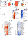
Sex‐dependent effect of quercetin treatment on single‐nuclei RNA sequencing. (A) Flow‐chart of the transcriptomic analysis using single‐nuclei RNA sequencing, performed on 3 samples per group collected the day of the surgery, after 2 days of quercetin treatment (created using BioRender). (B) Uniform Manifold Approximation and Projection (UMAP) plot, in 2 dimensions (UMAP‐1 and UMAP‐2), of annotated cell‐types clustered based on their specific gene expression, present in ITA graft arteries. (C) Proportion (%) of the 5 cell‐types (EC: Endothelial cells; SMC: Smooth muscle cells; FIB: Fibroblasts; IMM: Immune cells; PER: Pericytes) in men (M) and women (W) arteries, in placebo and quercetin groups. (D) UMAP plot colored by sample in men and women in placebo and quercetin groups. (E) Cell type prioritization representation of AUGUR predictive score, reflecting the effect of quercetin on the transcriptomic response in each arterial cell type, in men cells (brown circles) and in women cells (green circles); the higher the score (AUC), the higher the impact of quercetin. (F) Heatmap showing Gene Set Variation Analysis (GSVA) analysis of selected senescence‐related genes in the 5 cell types between men placebo and women placebo (F) and between men and women treated with quercetin (G). Rows represent gene‐related pathways (log2 fold change), and columns represent cell‐types. The color of each cell in the matrix represents the expression level of a specific pathway: Red corresponds to higher expression in men, and blue represents higher expression in women. On the heatmap, the dendrogram shows the hierarchical clustering of gene pathways based on the similarity of their expression profiles. H.eatmap showing GSVA analysis of Hallmark collection in the 5 cell types between men placebo and women placebo (H) and between men and women treated with quercetin (I).
FIGURE 4
FIGURE 4
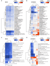
Pathway enrichment analysis reveals a uniform response to quercetin in male vascular cells and a cell type‐dependent response in female cells. (A) Heatmap showing GSVA analysis of Hallmark collection in the 5 cell types between men placebo and men quercetin. (B) Heatmap showing GSVA analysis of Hallmark collection in the 5 cell types between women placebo and women quercetin. (C) Heatmap showing GSVA analysis of selected senescence‐related gene sets in the 5 cell types between men placebo and men quercetin. (D) Heatmap showing GSVA analysis of selected senescence‐related gene sets in the 5 cell types between women placebo and women quercetin. Red and blue squares represent up‐regulated and down‐regulated pathways by quercetin, respectively.
FIGURE 5
FIGURE 5
Identification of perturbed cell‐to‐cell interactions on the cell types the most affected by quercetin. (A) Differential expression and perturbation scores of ligands and receptors in women patients between placebo and quercetin groups. The left panel shows significant log2 fold changes (L2FC) of ligands in endothelial (EC) and smooth muscle cells (SMC), and the middle panel presents the significant L2FC for corresponding receptors in SMC. The right panel illustrates the perturbation scores, highlighting significant interactions among pathways between EC and SMC, and within SMC. (B) Differential expression and perturbation scores of ligands and receptors in men patients between placebo and quercetin groups. The left panel shows significant L2FC of ligands in all cell types of the wall, and the middle panel presents the L2FC for corresponding receptors. The right panel illustrates the perturbation scores, highlighting significant interactions among pathways in paracrine (cell‐to‐cell) and autocrine (one cell type) environment. (C) Circos Plot showing differential interactions illustrated in B (ligand up‐regulated by quercetin in the upper half, and receptors up‐regulated by quercetin in the bottom half) among the 5 male cell types, with connecting lines representing significant communication links in the signaling network between cell types.

References

    1. Amor, C. , Feucht J., Leibold J., et al. 2020. “Senolytic CAR T Cells Reverse Senescence‐Associated Pathologies.” Nature 583, no. 7814: 127–132. 10.1038/s41586-020-2403-9. - DOI - PMC - PubMed
    1. Bloom, S. I. , Islam M. T., Lesniewski L. A., and Donato A. J.. 2023. “Mechanisms and Consequences of Endothelial Cell Senescence.” Nature Reviews. Cardiology 20, no. 1: 38–51. 10.1038/s41569-022-00739-0. - DOI - PMC - PubMed
    1. Budamagunta, V. , Kumar A., Rani A., et al. 2023. “Effect of Peripheral Cellular Senescence on Brain Aging and Cognitive Decline.” Aging Cell 22, no. 5: e13817. 10.1111/acel.13817. - DOI - PMC - PubMed
    1. Caldemeyer, L. , Dugan M., Edwards J., and Akard L.. 2016. “Long‐Term Side Effects of Tyrosine Kinase Inhibitors in Chronic Myeloid Leukemia.” Current Hematologic Malignancy Reports 11, no. 2: 71–79. 10.1007/s11899-016-0309-2. - DOI - PubMed
    1. Celermajer, D. S. , Sorensen K. E., Spiegelhalter D. J., Georgakopoulos D., Robinson J., and Deanfield J. E.. 1994. “Aging Is Associated With Endothelial Dysfunction in Healthy Men Years Before the Age‐Related Decline in Women.” Journal of the American College of Cardiology 24, no. 2: 471–476. 10.1016/0735-1097(94)90305-0. - DOI - PubMed

Publication types

Associated data