. 2025 May;55(5):e202451644.
doi: 10.1002/eji.202451644.

The Human Bone Marrow May Offer an IL-15-Dependent Survival Niche for EOMES+ Tr1-Like Cells

Affiliations

The Human Bone Marrow May Offer an IL-15-Dependent Survival Niche for EOMES+ Tr1-Like Cells

Nadia Pulvirenti et al. Eur J Immunol. 2025 May.

Abstract

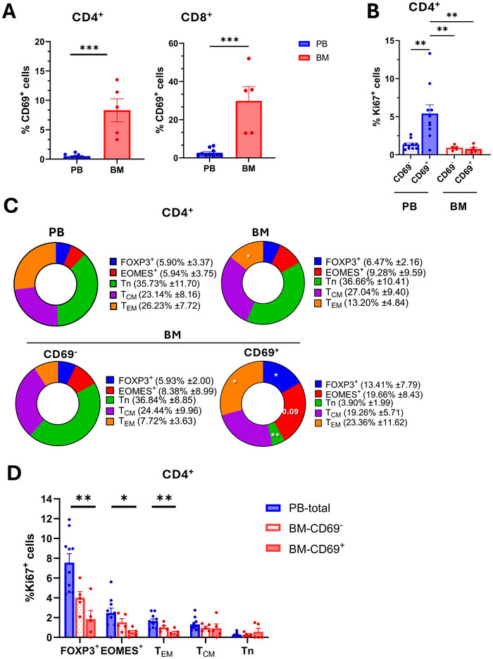
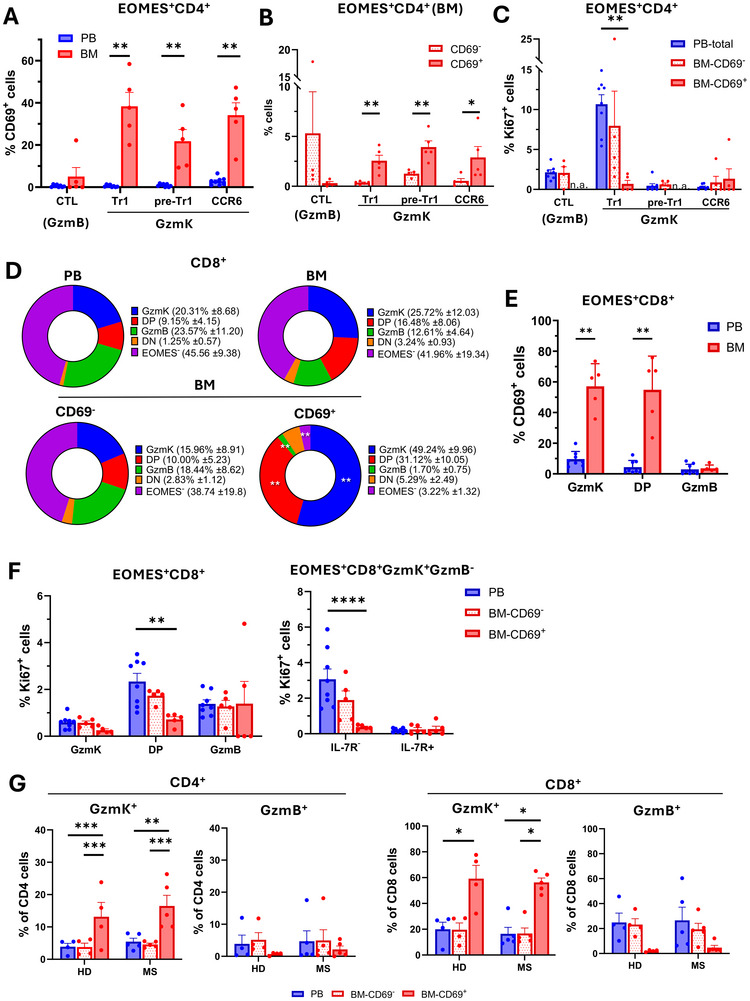
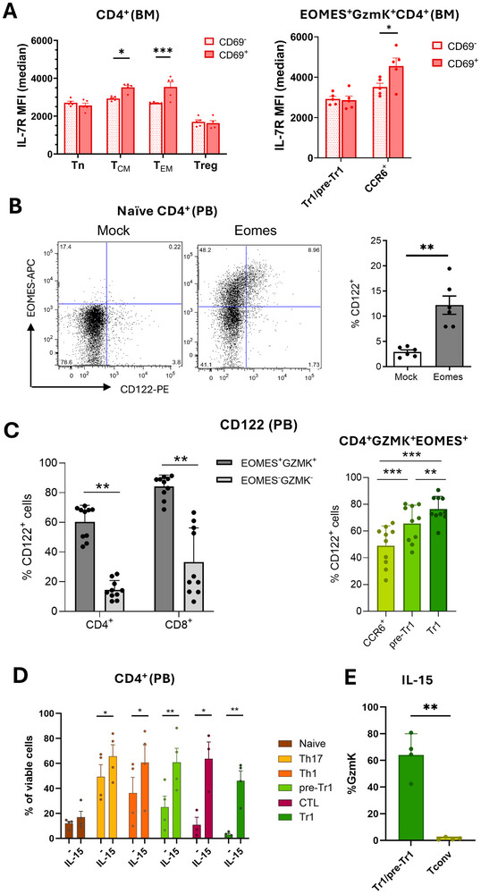
Maintenance of memory T-cells in the bone marrow and systemically depends on the homeostatic cytokines IL-7 and IL-15. An immunological memory may also exist for regulatory T-cells. EOMES+type-1 regulatory (Tr1)-like cells have a rapid in vivo turnover, but whether they are short-lived effector cells or are maintained long-term has not been investigated. EOMES+Tr1-like cells expressing GzmK were enriched among CD69+Ki67-T-cells in the bone marrow of healthy donors, suggesting that they became quiescent and bone marrow-resident. Conversely, CD4+GzmB+ effector T-cells were excluded from the bone marrow-resident fraction. The dichotomy between GzmK+ and GzmB+T-cells was observed both in healthy individuals and in multiple sclerosis patients, and also among CD8+T-cells. Intriguingly, bone marrow-resident CD4+ memory T-cells expressed increased levels of IL-7Rα, while EOMES+Tr1-like cells were consistently IL-7Rαlo. However, EOMES+Tr1-like cells expressed the IL-2/15Rβ chain, and the latter was induced upon forced expression of EOMES in primary human CD4+ T-cells. Finally, IL-15 rescued EOMES+Tr1-enriched populations from death by neglect but was not required for CD4+ memory T-cell survival. These findings suggest that the bone marrow may provide a survival niche for EOMES+Tr1-like cells. The different IL-7 and IL-15 receptor expression patterns of CD4+ memory T-cells and EOMES+Tr1-like cells suggest furthermore that they compete for different homeostatic niches.

Keywords: CD69; bone marrow; memory T lymphocytes; regulatory T‐cells.


Conflict of interest statement

The authors declare no conflicts of interest.

Figures

FIGURE 1
FIGURE 1
EOMES+ and FOXP3+CD4+T‐cells in the bone marrow upregulate CD69 and become quiescent. (A) Percentages of CD69+ cells among CD4+ and CD8+T‐cells in the blood (PB, n = 11) and in the bone marrow (BM, n = 5). (B) Percentages of Ki67+ cells among CD69+ and CD69CD4+T‐cells in the blood and the bone marrow. (C) Upper pie charts illustrate the contributions of naïve, central memory, and effector memory cells as well as FOXP3+ and EOMES+ cells to the CD4+T‐cell compartment in the blood and the bone marrow. Lower pie charts show the contributions of the same subsets to the CD69+ and CD69 compartments in the bone marrow. Statistical tendencies (p‐values >0.05) are reported as numbers in the pie charts. (D) Ki67 expression in the CD4+T‐cell subsets analyzed in (C) in peripheral blood and in the CD69+ and CD69 fractions in the bone marrow.
FIGURE 2
FIGURE 2
Bone marrow‐resident CD4+ and CD8+T‐cells are strongly enriched for GzmK‐expressing subsets, whereas GzmB+ effector CTL are largely excluded. (A) Percentages of CD69+ cells among CD4+EOMES+T‐cell subsets in the blood and in the bone marrow (BM). (B) Frequencies of CD4+EOMES+T‐cell subsets among CD69+ and CD69CD4+T‐cells in the bone marrow. (C) Ki67 expression in CD4+ EOMES+T‐cell subsets in peripheral blood and the CD69+ and CD69 fractions in the bone marrow. (D) The upper pie charts show the contributions of EOMES+ subsets classified according to the expression of GzmK and/or GzmB, as well as EOMES cells, to the CD8+T‐cell compartments in the blood and the bone marrow. Lower pie charts show the contributions of the same subsets to the CD69+ and CD69 compartments in the bone marrow. (E) Percentages of CD69+ cells among CD8+EOMES+T‐cell subsets in the blood and the bone marrow. (F) Ki67 expression in CD8+EOMES+T‐cell subsets in peripheral blood and in the CD69+ and CD69 fractions in the bone marrow. The left panel shows CD8+EOMES+T‐cell subsets expressing GzmK and/or GzmB (DP: double‐positive: GzmK+GzmB+), the right panel CD8+EOMES+GzmK+GzmBT‐cell cells stratified according to IL‐7Rα expression. (G) Percentages of GzmK+ and GzmB+T‐cell subsets in paired samples of peripheral blood (PB), and in the CD69+ and CD69 fractions of the bone marrow (BM) in four healthy donors (HD) and in five multiple sclerosis patients (MS). Upper histograms show CD4+T‐cells and lower histograms CD8+T‐cells.
FIGURE 3
FIGURE 3
IL‐7RloEOMES+Tr1‐like cells express high levels of IL‐2/15Rβ and are rescued from death by neglect by IL‐15. (A) IL‐7Rα expression levels on CD4+T‐cell subsets in the CD69+ and CD69 compartments of the bone marrow. The upper panel shows naïve, central, and effector memory cells as well as FOXP3+Treg and the lower panel shows CD4+EOMES+GzmK+ subsets. Representative histogram overlays are shown in Figure S4A. (B) Naive CD4+T‐cells were activated with anti‐CD3 and anti‐CD28 antibodies, IL‐2 and IL‐12, and transduced with lentiviral vectors coding for GFP (Mock) or for GFP and Eomesodermin (Eomes). Left: Representative Dot plots of cells analyzed for EOMES and CD122 (IL‐2/15Rβ) expression. Mean percentage of CD122 expression (n = 6). (C) Percentages of CD122+ cells in the indicated T‐cell subsets. The gating strategy is shown in Figure S5. (D) CD4+T‐cell subsets were enriched by cell sorting according to the gating strategy reported in Figure S6. They were cultured for 4 days in the absence or presence of IL‐15, and the mean percentage of viable cells was reported (n = 4). (E) Pre‐Tr1/Tr1‐cell‐enriched populations and CD4+CCR5CD27+ Tconv control cells were cultured for 4 days with IL‐15, and GzmK expression of viable cells was analyzed.

References

    1. Sprent J. and Surh C. D., “T Cell Memory,” Annual Review of Immunology 20 (2002): 551–579. - PubMed
    1. Manz R. A., Thiel A., and Radbruch A., “Lifetime of Plasma Cells in the Bone Marrow [letter],” Nature 388 (1997): 133–134. - PubMed
    1. Lenz D. C., Kurz S. K., Lemmens E., et al., “IL‐7 Regulates Basal Homeostatic Proliferation of Antiviral CD4+T Cell Memory,” PNAS 101 (2004): 9357–9362. - PMC - PubMed
    1. Purton J. F., Tan J. T., Rubinstein M. P., Kim D. M., Sprent J., and Surh C. D., “Antiviral CD4+ Memory T Cells Are IL‐15 Dependent,” Journal of Experimental Medicine 204 (2007): 951–961. - PMC - PubMed
    1. Zhang X., Sun S., Hwang I., Tough D. F., and Sprent J., “Potent and Selective Stimulation of Memory‐phenotype CD8+ T Cells in Vivo by IL‐15,” Immunity 8 (1998): 591–599. - PubMed

MeSH terms

LinkOut - more resources