Novel fascial mapping of muscle spindles distribution: insights from a murine model study
- PMID: 40376118
- PMCID: PMC12078280
- DOI: 10.3389/fphys.2025.1571500
Novel fascial mapping of muscle spindles distribution: insights from a murine model study
Abstract
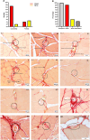
Muscle spindles (MSs) are essential for proprioception and motor control. The precise distribution and localization of MSs have been the focus of major research efforts to provide a foundation for understanding their roles in various diseases and motor dysfunctions. However, there are currently disagreements on the distribution patterns of MSs, and these discrepancies hinder the advancement of novel physical therapy techniques based on MS functionality. In this study, we present an innovative fascia-based distribution pattern for MSs. Using the rat quadriceps femoris muscle as the target, serial sections of the muscle were meticulously prepared following tissue sampling, fixation, and embedding. Furthermore, four additional rat gastrocnemius and eight human muscles were processed and cut into non-successive sections by the above method. The MSs were identified and characterized using Sirius Red staining, and their locations, quantities, associated structures, and basic parameters were documented via microscopy. Our findings demonstrate that the MSs are primarily located within the fascial layers and predominantly within the perimysium; the MS capsule is structurally continuous with the perimysium and forms multiple connections in different orientations. This study demonstrates that MSs are influenced by not only changes in muscle length but also alterations in the fascia tension or state, which may have more significant impacts. Furthermore, both nerves and vessels were observed near or within the capsule of the MS but were not always presented. In some sections, no microscopically distinguishable vessels or nerve fibers were observed around the MSs. This study proposes a novel fascia-based distribution model for MSs by highlighting that MSs are embedded within the fascial matrix and that the fascia may serve as a key structural marker for locating MSs. Additionally, the structural continuity of the fascia with the MS capsule suggests its role as a potential mediator in MS functions. The present study challenges the traditional concepts of MS distribution by introducing a more refined and efficient approach for studying MSs through the fascial perspective, thereby representing a significant advancement.
Keywords: distribution; fascia; motor control; muscle spindle; perimysium; proprioception.
Copyright © 2025 Sun, Petrelli, Fede, Biz, Incendi, Porzionato, Pirri, Zhao and Stecco.
Conflict of interest statement
The authors declare that the research was conducted in the absence of any commercial or financial relationships that could be construed as a potential conflict of interest.
Figures


Similar articles
-
Lower Genitourinary Trauma.2023 May 8. In: StatPearls [Internet]. Treasure Island (FL): StatPearls Publishing; 2025 Jan–. 2023 May 8. In: StatPearls [Internet]. Treasure Island (FL): StatPearls Publishing; 2025 Jan–. PMID: 32491459 Free Books & Documents.
-
Age-Related Alterations of Hyaluronan and Collagen in Extracellular Matrix of the Muscle Spindles.J Clin Med. 2021 Dec 24;11(1):86. doi: 10.3390/jcm11010086. J Clin Med. 2021. PMID: 35011824 Free PMC article.
-
Anatomy, Fascia Layers.2023 Jul 24. In: StatPearls [Internet]. Treasure Island (FL): StatPearls Publishing; 2025 Jan–. 2023 Jul 24. In: StatPearls [Internet]. Treasure Island (FL): StatPearls Publishing; 2025 Jan–. PMID: 30252294 Free Books & Documents.
-
Quantity and Distribution of Muscle Spindles in Animal and Human Muscles.Int J Mol Sci. 2024 Jul 3;25(13):7320. doi: 10.3390/ijms25137320. Int J Mol Sci. 2024. PMID: 39000428 Free PMC article. Review.
-
Role of fascial connectivity in musculoskeletal dysfunctions: A narrative review.J Bodyw Mov Ther. 2020 Oct;24(4):423-431. doi: 10.1016/j.jbmt.2020.07.020. Epub 2020 Jul 30. J Bodyw Mov Ther. 2020. PMID: 33218543 Review.
References
-
- Banks R. W., Barker D. (2004). “The muscle spindle,” in Myology. Editors Engel A. G., Franzini-Armstrong C. 3rd edn (New York, NY, USA: McGraw-Hill; ), 489–509.
LinkOut - more resources
Full Text Sources

