. 2025 May 15:29:0207.
doi: 10.34133/bmr.0207. eCollection 2025.

Sono-Gas-Mediated Precise Stiffness Remodeling for Triple-Negative Breast Cancer Mechanical Immunotherapy

Affiliations

Sono-Gas-Mediated Precise Stiffness Remodeling for Triple-Negative Breast Cancer Mechanical Immunotherapy

Yaqin Hu et al. Biomater Res. .

Abstract

Triple-negative breast cancer (TNBC) is a highly invasive cancer, and its poor therapeutic outcomes are often associated with the mechanical properties of the tumor microenvironment, which is characterized by altered extracellular matrix (ECM) flexibility and increased stiffness. Herein, a mechanical immunomodulator, namely, red blood cell membrane-IR780-L-arginine nanoparticles (R-I-LA NPs), was designed to precisely regulate the stiffness of the ECM for mechanical immunotherapy of TNBC. In tumor cells, the low-intensity focused ultrasound activates R-I-LA NPs to produce reactive nitrogen species, which damages tumor cells and remodels the stiffness of ECM. Meanwhile, the softened ECM can normalize the tumor vasculature to alleviate hypoxia and increase the production of reactive oxygen species, thereby enhancing the efficacy of sonodynamic therapy and stimulating immunogenic cell death. Additionally, R-I-LA NPs stimulate the immune system and suppress pulmonary metastasis. Overall, this study offers a distinctive "sono-gas-mediated mechanical immunity" strategy for ECM regulation, potentially overcoming current TNBC therapy limitations.


Conflict of interest statement

Competing interests: The authors declare that they have no competing interests.

Figures

Fig. 1.
Fig. 1.
(A) The preparation process of R-I-LA NPs. (B) The sono-activated biomimetic nanoparticles (R-I-LA NPs) generate reactive nitrogen species (RNS) through a cascade reaction activated using low-intensity focused ultrasound (LIFU). RNS effectively remodels the tumor mechanical microenvironment (TMM) by reducing extracellular type I collagen, which improves the effectiveness of sonodynamic therapy (SDT) and strengthens the immune response against TNBC.
Fig. 2.
Fig. 2.
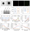
Characterization of R-I-LA NPs. (A) TEM images of I-LA NPs and R-I-LA NPs. (B) CLSM images of R-I-LA NPs. (C) Western blotting analysis of CD47. (D) Size distribution of I-LA NPs and R-I-LA NPs. (E) Zeta potential of I-LA NPs and R-I-LA NPs. (F) Stability of R-I-LA NPs based on DLS and PDI. (G) UV–vis–NIR absorbance spectra of different NPs (free IR780, R-LA NPs, R-I NPs, and R-I-LA NPs). (H) UV–vis–NIR absorbance spectra of free IR780 at elevated concentrations. (I) SOSG fluorescence intensity of R-I-LA NPs during LIFU irradiation. (J) LIFU irradiation time-dependent singlet oxygen (1O2) yield of different NPs (R-LA NPs, R-I NPs, and R-I-LA NPs). (K) UV–vis absorption of the R-I-LA NPs solution after adding Griess reagent during LIFU irradiation. The experiments were repeated thrice independently. ANOVA with Tukey’s post-hoc test.
Fig. 3.
Fig. 3.
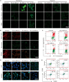
Evaluation of intracellular uptake. (A) CLSM images of DiI-labeled different NPs (LA NPs, I-LA NPs, and R-I-LA NPs) colocalized with 4T1 cells. (B) FCM analysis of 4T1 cells after coincubation with different NPs for various times. (C) CLSM images of Raw264.7 mouse macrophage cells treated with I-LA NPs or R-I-LA NPs. (D) CLSM images of DiI-labeled R-LA NPs or R-I-LA NPs colocalized with Mito-Tracker green in 4T1 cells. The experiments were repeated thrice independently. ANOVA with Tukey’s post-hoc test.
Fig. 4.
Fig. 4.
Therapeutic efficacy of R-I-LA NPs in vitro. (A) Cell viability of 4T1 cells after different treatments. (B) CLSM images of cells stained with Calcein-AM/PI staining after different treatments. (C) Apoptosis quantified by FCM analysis. The experiments were repeated thrice independently. ANOVA with Tukey’s post-hoc test. **P < 0.01 and ****P < 0.0001.
Fig. 5.
Fig. 5.
SDT capacity and induction of ICD by R-I-LA NPs in vitro. (A) DCFH-DA probe for evaluating the ROS level; DAF-FM DA probe for the detection of NO generation; RNS probe for the detection of RNS generation. (B) CLSM images and (C) FCM analysis of JC-1-stained 4T1 cells after different treatments. (D) CLSM images of intracellular CRT and HGMB1expression of 4T1 cells after different treatments. (E) FCM analysis of matured DCs (CD11c+CD80+CD86+) after different treatments. The experiments were repeated thrice independently. ANOVA with Tukey’s post-hoc test.
Fig. 6.
Fig. 6.
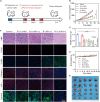
FL/PAI imaging ability of R-I-LA NPs. (A) In vivo FL images of 4T1 tumor-bearing mice at different time points and (B) the corresponding FL intensities of tumors. (C) FL images and (D) corresponding quantitative analysis of isolated organs and tumors. (E) In vitro PAI images and PAI values of R-I-LA NPs at different concentrations. (F) In vivo PAI images of 4T1 tumor-bearing mice at different time points and (G) the corresponding PAI intensities of tumors. The experiments were repeated thrice independently. ANOVA with Tukey’s post-hoc test. *P < 0.05, **P < 0.01, and ***P < 0.001.
Fig. 7.
Fig. 7.
Antitumor efficacy of R-I-LA NPs in vivo. (A) Treatment schedule for the R-I-LA NPs. (B) Tumor growth curves. (C) Tumor weights. (D) Body weight changes. (E) Photographs of tumors dissected from mice. (F) H&E, PCNA, TUNEL, Collagen I, HIF-α, and hypoxia staining of tumors. The experiments were repeated thrice independently. ANOVA with Tukey’s post-hoc test. *P < 0.05, **P < 0.01, and ****P < 0.0001.
Fig. 8.
Fig. 8.
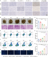
Evaluation of anticancer immune responses caused by R-I-LA NPs. (A) 3-NT, MMP-1, and MMP-2 staining of tumors. (B) Bouin’s trichrome fixed lung tissue and H&E staining images and (C) the number of pulmonary nodules after receiving different treatments. (D) Representative FCM plots of matured DCs in spleens and (E) corresponding quantitative analysis. (F) Representative FCM plots of CD8+ T cells in spleens and (G) corresponding quantitative analysis. (H) Immunofluorescence images of CD8+ T cells in tumors after different treatments. The experiments were repeated thrice independently. ANOVA with Tukey’s post-hoc test. **P < 0.01 and ****P < 0.0001.

References

    1. Tarantino P, Corti C, Schmid P, Cortes J, Mittendorf EA, Rugo H, Tolaney SM, Bianchini G, Andrè F, Curigliano G. Immunotherapy for early triple negative breast cancer: Research agenda for the next decade. NPJ Breast Cancer. 2022;8(1):23. - PMC - PubMed
    1. Leon-Ferre RA, Goetz MP. Advances in systemic therapies for triple negative breast cancer. BMJ. 2023;381: Article e071674. - PubMed
    1. Lakshmanan VK, Jindal S, Packirisamy G, Ojha S, Lian S, Kaushik A, Alzarooni A, Metwally YAF, Thyagarajan SP, Do Jung Y, et al. . Nanomedicine-based cancer immunotherapy: Recent trends and future perspectives. Cancer Gene Ther. 2021;28(9):911–923. - PubMed
    1. Nguyen HM, Paulishak W, Oladejo M, Wood L. Dynamic tumor microenvironment, molecular heterogeneity, and distinct immunologic portrait of triple-negative breast cancer: An impact on classification and treatment approaches. Breast Cancer. 2023;30(2):167–186. - PubMed
    1. Wang Y, Goliwas KF, Severino PE, Hough KP, Van Vessem D, Wang H, Tousif S, Koomullil RP, Frost AR, Ponnazhagan S, et al. . Mechanical strain induces phenotypic changes in breast cancer cells and promotes immunosuppression in the tumor microenvironment. Lab Investig. 2020;100(12):1503–1516. - PMC - PubMed

LinkOut - more resources