Frontal Lobe Cysticercosis
- PMID: 40376341
- PMCID: PMC12081067
- DOI: 10.7759/cureus.82321
Frontal Lobe Cysticercosis
Abstract
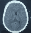
Cysticercosis, a brain infection caused by the parasite Taenia solium, presents considerable health challenges, especially in tropical and developing areas, where it contributes significantly to the incidence of epilepsy. The lifecycle of this parasite involves humans as the primary hosts and pigs as the intermediate hosts. In this case study, a 12-year-old girl exhibited unusual symptoms, such as fainting episodes and a decline in academic performance. Although she adhered to a vegetarian diet, her contact with pigs resulted in an infection, confirmed by a CT scan that revealed a cyst in the right frontal lobe containing a hyperdense scolex. The treatment regimen included valproic acid, prednisolone, and albendazole, which successfully resolved her seizures over a follow-up period of 24 months. This case underscores the diagnostic challenges associated with cysticercosis and highlights the necessity of identifying atypical cognitive symptoms related to this infection, particularly in populations at risk of exposure.
Keywords: albendazole; attention-deficit; cysticercosis; frontal lobe; taenia sodium.
Copyright © 2025, Singh et al.
Conflict of interest statement
Human subjects: Consent for treatment and open access publication was obtained or waived by all participants in this study. Conflicts of interest: In compliance with the ICMJE uniform disclosure form, all authors declare the following: Payment/services info: All authors have declared that no financial support was received from any organization for the submitted work. Financial relationships: All authors have declared that they have no financial relationships at present or within the previous three years with any organizations that might have an interest in the submitted work. Other relationships: All authors have declared that there are no other relationships or activities that could appear to have influenced the submitted work.
Figures


References
-
- Taeniasis/cysticercosis. [ Mar; 2025 ]. 2022. https://www.who.int/news-room/fact-sheets/detail/taeniasis-cysticercosis https://www.who.int/news-room/fact-sheets/detail/taeniasis-cysticercosis
-
- Neurocysticercosis in infants and toddlers: report of seven cases and review of published patients. Del Brutto OH. Pediatr Neurol. 2013;48:432–435. - PubMed
-
- Revised diagnostic criteria for neurocysticercosis. Del Brutto OH, Nash TE, White AC Jr, et al. J Neurol Sci. 2017;372:202–210. - PubMed
Publication types
LinkOut - more resources
Full Text Sources
