

A Rare Case of Lipidized Fibrous Histiocytoma of the Oral Cavity
- PMID: 40376352
- PMCID: PMC12078029
- DOI: 10.7759/cureus.82265
A Rare Case of Lipidized Fibrous Histiocytoma of the Oral Cavity
Abstract
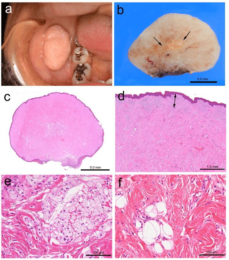
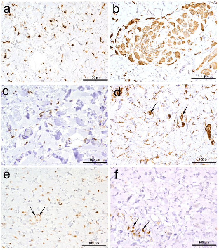
Benign fibrous histiocytoma (FH) is a common cutaneous tumor that rarely occurs in the oral cavity. Lipidized FH is an uncommon variant characterized by abundant foam cells in collagenized stroma. To our knowledge, only a single case of oral lipidized FH affecting the tongue has been reported. We examine a case of lipidized FH in the buccal mucosa of a patient presenting with a gradually enlarging, pedunculated lesion measuring up to 26 mm. A histological examination revealed a well-marginated but unencapsulated tumor with a Grenz zone, hyalinized stroma, and central foam cell aggregation. Immunohistochemically, the spindle and foam cells were CD68-positive, with focal Factor XIIIa positivity and negative bcl-2 staining. The lesion was completely excised, and no recurrence was observed. To our knowledge, this is the second reported case of oral lipidized FH and the first case in the buccal mucosa. Given its rarity and histological overlap with other xanthomatous lesions, accurate diagnosis is crucial with immunohistochemistry. Complete excision appears to be curative; however, a long-term follow-up is recommended, considering the subtype of fibrous histiocytoma developing in the orofacial region.
Keywords: cd68; factor xiiia; foam cells; lipidized fibrous histiocytoma; oral cavity.
Copyright © 2025, Shimada et al.
Conflict of interest statement
Human subjects: Consent for treatment and open access publication was obtained or waived by all participants in this study. Ethics Committee of Matsumoto Dental University issued approval 0294. Conflicts of interest: In compliance with the ICMJE uniform disclosure form, all authors declare the following: Payment/services info: All authors have declared that no financial support was received from any organization for the submitted work. Financial relationships: All authors have declared that they have no financial relationships at present or within the previous three years with any organizations that might have an interest in the submitted work. Other relationships: All authors have declared that there are no other relationships or activities that could appear to have influenced the submitted work.
Figures
References
-
- WHO Classification of Tumours Editorial Board. Skin Tumors. Lyon: International Agency for Research on Cancer; 2025. WHO Classification of Tumours Editorial Board: Skin tumors [Internet; beta version ahead of print]. [cited 20th February 2025. 5th ed.
-
- Benign fibrous histiocytoma of the oral and maxillofacial region: A systematic review. Kirschnick LB, Schuch LF, Silveira FM, et al. Oral Surg Oral Med Oral Pathol Oral Radiol. 2022;133:0–56. - PubMed
-
- Signet-ring cell dermatofibroma. Garrido-Ruiz MC, Carrillo R, Enguita AB, Peralto JL. Am J Dermatopathol. 2009;31:84–87. - PubMed
-
- Cutaneous fibrohistiocytic tumours - an update. Luzar B, Calonje E. Histopathology. 2010;56:148–165. - PubMed
Publication types
LinkOut - more resources
Full Text Sources
Miscellaneous