An evaluation of distinct adeno-associated virus vector strategies for driving transgene expression in spinal inhibitory neurons of the rat
- PMID: 40376611
- PMCID: PMC12078132
- DOI: 10.3389/fnins.2025.1558581
An evaluation of distinct adeno-associated virus vector strategies for driving transgene expression in spinal inhibitory neurons of the rat
Abstract
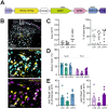
The spinal cord dorsal horn (DH) is essential for processing and transmitting nociceptive information. Its neuronal subpopulations exhibit significant heterogeneity in morphology and intrinsic properties, forming complex circuits that remain only partially understood. Under physiological and pathological conditions, inhibitory interneurons in the DH are of particular interest. These neurons modulate and refine pain-related signals entering the central nervous system. The ability to selectively target these inhibitory interneurons is key to investigating the underlying circuitry and mechanisms of pain processing, as well as to understand the specific role of inhibitory signaling within these processes. We employed a viral vector approach to deliver a fluorescent reporter protein specifically to inhibitory interneurons in the rat spinal cord. Using adeno-associated virus (AAV) vectors designed to express enhanced green fluorescent protein (EGFP) under the control of various promoters, we targeted distinct subtypes of spinal inhibitory interneurons. Through immunostaining, in situ hybridization, and confocal imaging, we evaluated the specificity and efficacy of these promoters. Our findings revealed that the promoter/vector combinations used did not achieve the desired specificity for targeting distinct interneuron populations in the DH. Despite these limitations, this work provides valuable insights into the potential and challenges of designing AAV-based approaches for selective neuronal targeting. These results emphasize the need for further refinement of promoter designs to achieve precise and reliable expression in specific spinal interneuron subtypes. Addressing these challenges will be crucial for advancing our understanding of spinal nociceptive circuits and developing targeted therapeutic approaches for pain syndromes.
Keywords: adeno-associated virus vectors; dorsal horn; inhibitory interneurons; intraparenchymal injection; nociception; rat; spinal cord.
Copyright © 2025 Klinger, Siegert, Holzinger, Trofimova, Ada and Drdla-Schutting.
Conflict of interest statement
The authors declare that the research was conducted in the absence of any commercial or financial relationships that could be construed as a potential conflict of interest.
Figures


Similar articles
-
Differential Activation of Pain Circuitry Neuron Populations in a Mouse Model of Spinal Cord Injury-Induced Neuropathic Pain.J Neurosci. 2022 Apr 13;42(15):3271-3289. doi: 10.1523/JNEUROSCI.1596-21.2022. Epub 2022 Mar 7. J Neurosci. 2022. PMID: 35256528 Free PMC article.
-
Neonatal Injury Evokes Persistent Deficits in Dynorphin Inhibitory Circuits within the Adult Mouse Superficial Dorsal Horn.J Neurosci. 2020 May 13;40(20):3882-3895. doi: 10.1523/JNEUROSCI.0029-20.2020. Epub 2020 Apr 14. J Neurosci. 2020. PMID: 32291327 Free PMC article.
-
Network model of nociceptive processing in the superficial spinal dorsal horn reveals mechanisms of hyperalgesia, allodynia, and spinal cord stimulation.J Neurophysiol. 2023 Nov 1;130(5):1103-1117. doi: 10.1152/jn.00186.2023. Epub 2023 Sep 20. J Neurophysiol. 2023. PMID: 37727912
-
Transgenic Mouse Models for the Tracing of “Pain” Pathways.In: Kruger L, Light AR, editors. Translational Pain Research: From Mouse to Man. Boca Raton (FL): CRC Press/Taylor & Francis; 2010. Chapter 7. In: Kruger L, Light AR, editors. Translational Pain Research: From Mouse to Man. Boca Raton (FL): CRC Press/Taylor & Francis; 2010. Chapter 7. PMID: 21882471 Free Books & Documents. Review.
-
PKCγ interneurons, a gateway to pathological pain in the dorsal horn.J Neural Transm (Vienna). 2020 Apr;127(4):527-540. doi: 10.1007/s00702-020-02162-6. Epub 2020 Feb 27. J Neural Transm (Vienna). 2020. PMID: 32108249 Review.
References
LinkOut - more resources
Full Text Sources

