

Dose-dependent changes in global brain activity and functional connectivity following exposure to psilocybin: a BOLD MRI study in awake rats
- PMID: 40376612
- PMCID: PMC12078138
- DOI: 10.3389/fnins.2025.1554049
Dose-dependent changes in global brain activity and functional connectivity following exposure to psilocybin: a BOLD MRI study in awake rats
Abstract
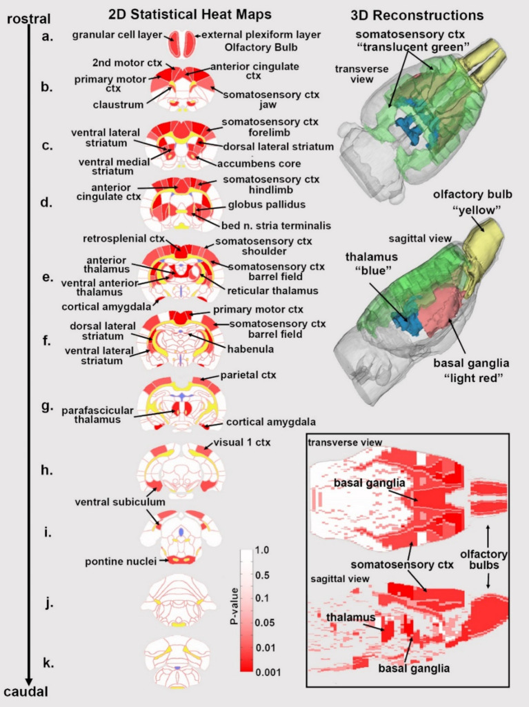
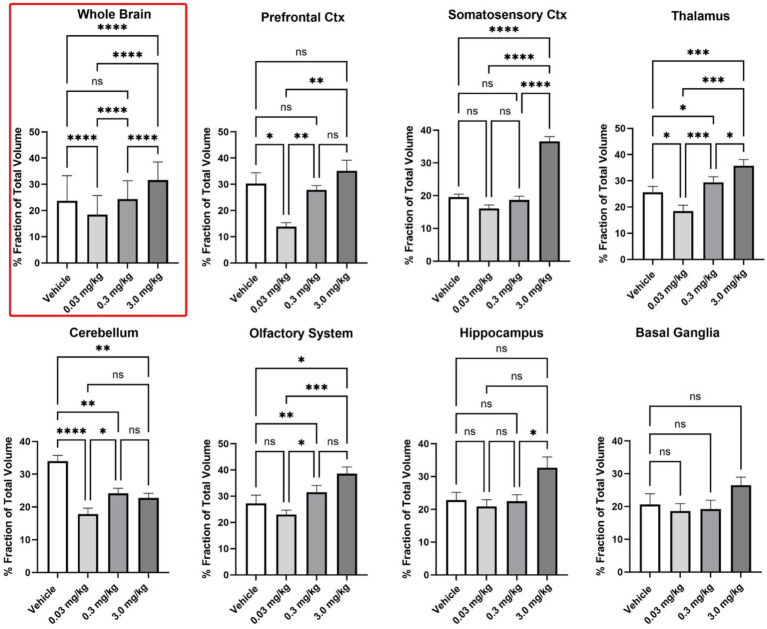
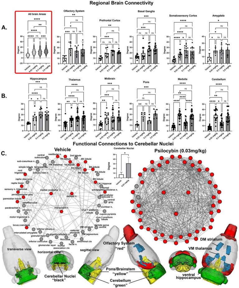
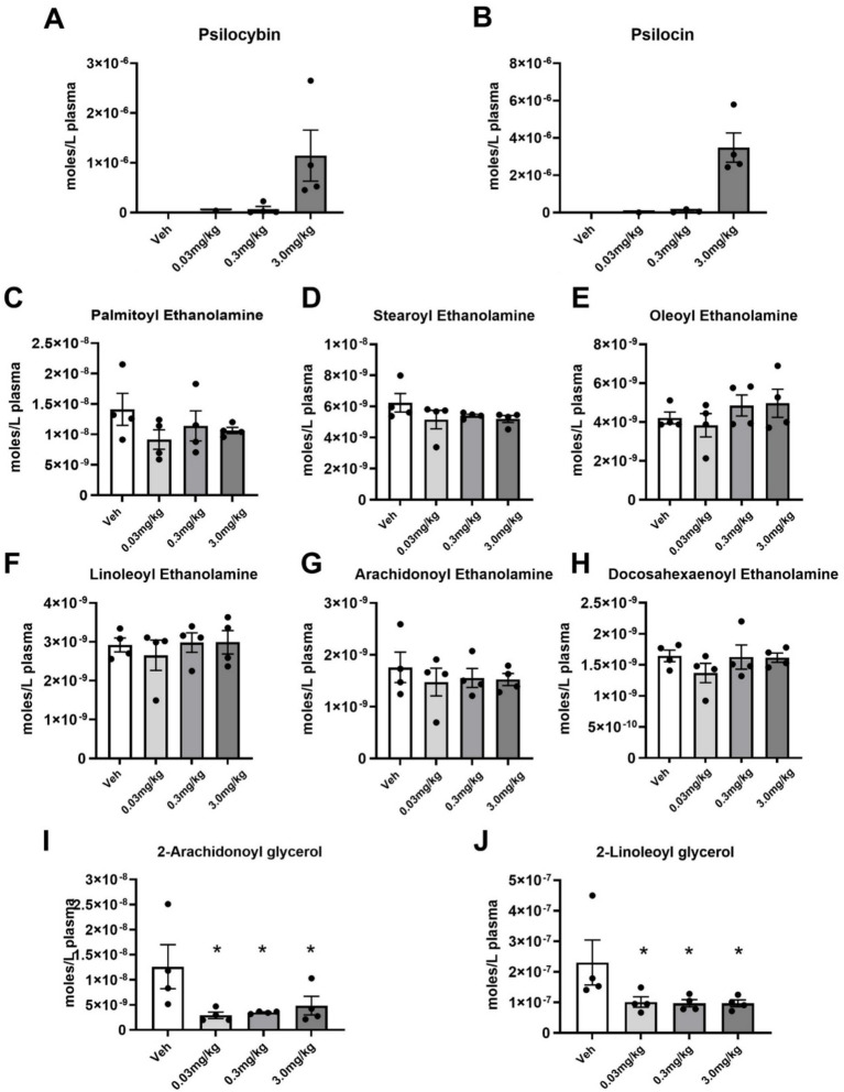
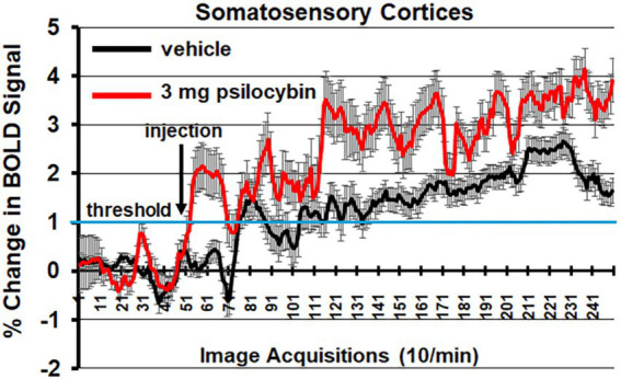
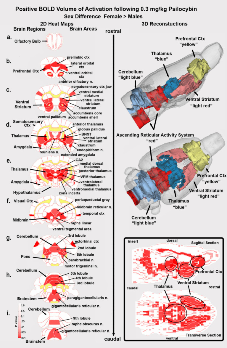
Psilocybin is a hallucinogen with complex neurobiological and behavioral effects. This is the first study to use MRI to follow functional changes in brain activity in response to different doses of psilocybin in fully awake, drug naive rats. We hypothesized that psilocybin would show a dose-dependent increase in activity in the prefrontal cortex and thalamus, while decreasing hippocampal activity. Female and male rats were given IP injections of vehicle or psilocybin in doses of 0.03 mg/kg, 0.3 mg/kg, and 3.0 mg/kg while fully awake during the imaging session. These levels were validated by measuring psilocybin and its metabolite, psilocin. Changes in BOLD signal were recorded over a 20 min window. Data for resting state functional connectivity were collected approximately 35 min post injection. All data were registered to rat 3D MRI atlas with 169 brain areas providing site-specific changes in global brain activity and changes in functional connectivity. Treatment with psilocybin resulted in a significant dose-dependent increase in positive BOLD signal. The areas most affected by the acute presentation of psilocybin were the somatosensory cortex, basal ganglia and thalamus. Males and females showed different sensitivity to psilocybin dose, with females exhibiting greater activation than males at 0.3 mg/kg, especially in thalamic and basal ganglia regions. There was a significant dose-dependent global increase in functional connectivity, highlighted by hyperconnectivity to the cerebellum. Brain areas hypothesized to be involved in loss of sensory filtering and organization of sensory motor stimuli, such as the cortico-striato-thalamo-cortical circuit and the claustrum, showed increased activation at higher doses of psilocybin. Indeed, the general neuroanatomical circuitry associated with the psychedelic experience was affected but the direction of the BOLD signal and pattern of activity between neural networks was inconsistent with the human literature.
Keywords: 5-HT2A receptor; BOLD resting state functional connectivity; cerebellar nuclei; hyperconnectivity; psilocin.
Copyright © 2025 Fuini, Chang, Ortiz, Nasseef, Edwards, Latta, Gonzalez, Woodward, Axe, Maheswari, Cavallaro, Bradshaw, Kulkarni and Ferris.
Conflict of interest statement
CF and PK have a financial interest in Ekam Imaging. The remaining authors declare that the research was conducted in the absence of any commercial or financial relationships that could be construed as a potential conflict of interest.
Figures
Similar articles
-
Dose-dependent LSD effects on cortical/thalamic and cerebellar activity: brain oxygen level-dependent fMRI study in awake rats.Brain Commun. 2024 Jun 4;6(3):fcae194. doi: 10.1093/braincomms/fcae194. eCollection 2024. Brain Commun. 2024. PMID: 38863575 Free PMC article.
-
Dose-dependent effects of GAT107, a novel allosteric agonist-positive allosteric modulator (ago-PAM) for the α7 nicotinic cholinergic receptor: a BOLD phMRI and connectivity study on awake rats.Front Neurosci. 2023 Jun 23;17:1196786. doi: 10.3389/fnins.2023.1196786. eCollection 2023. Front Neurosci. 2023. PMID: 37424993 Free PMC article.
-
Psilocybin acutely alters the functional connectivity of the claustrum with brain networks that support perception, memory, and attention.Neuroimage. 2020 Sep;218:116980. doi: 10.1016/j.neuroimage.2020.116980. Epub 2020 May 23. Neuroimage. 2020. PMID: 32454209 Free PMC article.
-
Effects of (-)-MBP, a novel 5-HT2C agonist and 5-HT2A/2B antagonist/inverse agonist on brain activity: A phMRI study on awake mice.Pharmacol Res Perspect. 2023 Oct;11(5):e01144. doi: 10.1002/prp2.1144. Pharmacol Res Perspect. 2023. PMID: 37837184 Free PMC article.
-
Psilocybin induces spatially constrained alterations in thalamic functional organizaton and connectivity.Neuroimage. 2022 Oct 15;260:119434. doi: 10.1016/j.neuroimage.2022.119434. Epub 2022 Jul 2. Neuroimage. 2022. PMID: 35792293 Free PMC article.
References
LinkOut - more resources
Full Text Sources