

Comparison of Fascin Expression in Oral Verrucous Carcinoma and Oral Squamous Cell Carcinoma
- PMID: 40376627
- PMCID: PMC12081144
- DOI: 10.1155/ijod/5530533
Comparison of Fascin Expression in Oral Verrucous Carcinoma and Oral Squamous Cell Carcinoma
Abstract
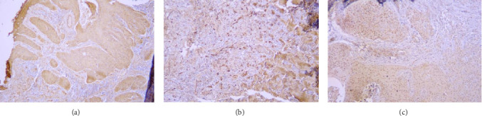
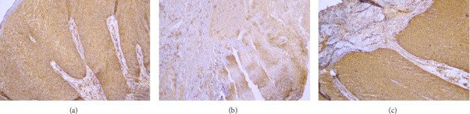
Introduction: One of the diagnostic problems of pathology is to differentiate between oral verrucous carcinoma (OVC) and oral squamous cell carcinoma (OSCC). Fascin increases the invasion of normal and neoplastic cells by stabilizing cytoplasmic filamentous actin. The present study aimed to investigate the expression of fascin in OSCC and OVC. Methods: This descriptive-analytical cross-sectional was conducted on 25 blocks of OSCC, 22 blocks of OVC, and 10 blocks of healthy mucosa as a control group. After immunohistochemical staining, samples were observed by two maxillofacial pathologists simultaneously, and the percentage of stained cells, intensity of staining, and the location of stained cells were obtained. Results: There was no significant difference in the gender (p=0.123) and age (p=0.276) distribution of participants in the groups. There was a significant difference in the distribution of the involved area in the patients of the studied groups (p < 0.001). There was a significant difference in the intensity of staining and the percentage of stained cells between the studied groups (p < 0.001). Conclusions: The percentage and intensity of staining were higher in the OSCC, OVC and, control groups, respectively. It seems that Fascin expression has an important role in predicting OVC and OSCC.
Keywords: fascin; immunohistochemistry; oral squamous cell carcinoma; pathology; verrucous carcinoma.
Copyright © 2025 Nima Mohammadi et al. International Journal of Dentistry published by John Wiley & Sons Ltd.
Conflict of interest statement
The authors declare no conflicts of interest.
Figures
Similar articles
-
Differential Expression of EZH2 and H3K27me3 in Oral Verrucous Carcinoma and Oral Verrucous Hyperplasia.Head Neck Pathol. 2021 Jun;15(2):408-415. doi: 10.1007/s12105-020-01209-0. Epub 2020 Jul 27. Head Neck Pathol. 2021. PMID: 32720035 Free PMC article.
-
Expression of Matrix Metalloproteinase-10 at Invasive Front of Squamous Cell Carcinoma and Verrucous Carcinoma in the Oral Cavity.Asian Pac J Cancer Prev. 2015;16(15):6609-13. doi: 10.7314/apjcp.2015.16.15.6609. Asian Pac J Cancer Prev. 2015. PMID: 26434883
-
Comparison of E-Cadherin Expression in Oral Verrucous Carcinoma and Normal Oral Mucosa: An Immunohistochemical Study.Cureus. 2024 Nov 27;16(11):e74634. doi: 10.7759/cureus.74634. eCollection 2024 Nov. Cureus. 2024. PMID: 39735013 Free PMC article.
-
Oral Proliferative Verrucous Leukoplakia: Progression to Malignancy and Clinical Implications. Systematic Review and Meta-Analysis.Cancers (Basel). 2021 Aug 13;13(16):4085. doi: 10.3390/cancers13164085. Cancers (Basel). 2021. PMID: 34439238 Free PMC article. Review.
-
Oral verrucous carcinoma: From multifactorial etiology to diverse treatment regimens (Review).Int J Oncol. 2016 Jul;49(1):59-73. doi: 10.3892/ijo.2016.3501. Epub 2016 Apr 26. Int J Oncol. 2016. PMID: 27121637 Review.
References
-
- Neville B. W., Damm D. D., Chi A. C., Allen C. M. Oral and Maxillofacial Pathology . Elsevier Health Sciences; 2015. pp. 394–423.
-
- WHO Classification of Tumours Editorial Board. Head and Neck Tumours (WHO Classification of Tumours Series) 5th. Lyon (France): International Agency for Research on Cancer; 2022.
LinkOut - more resources
Full Text Sources
