Review
. 2025 Dec;22(1):1-10.
doi: 10.1080/15476286.2025.2504727. Epub 2025 May 16.

Translation elongation: measurements and applications

Affiliations
Review

Translation elongation: measurements and applications

Leslie Watkins et al. RNA Biol. 2025 Dec.

Abstract

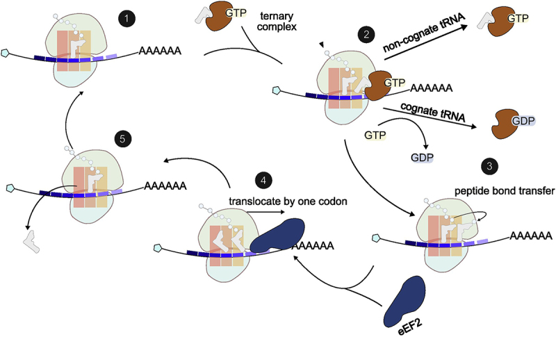
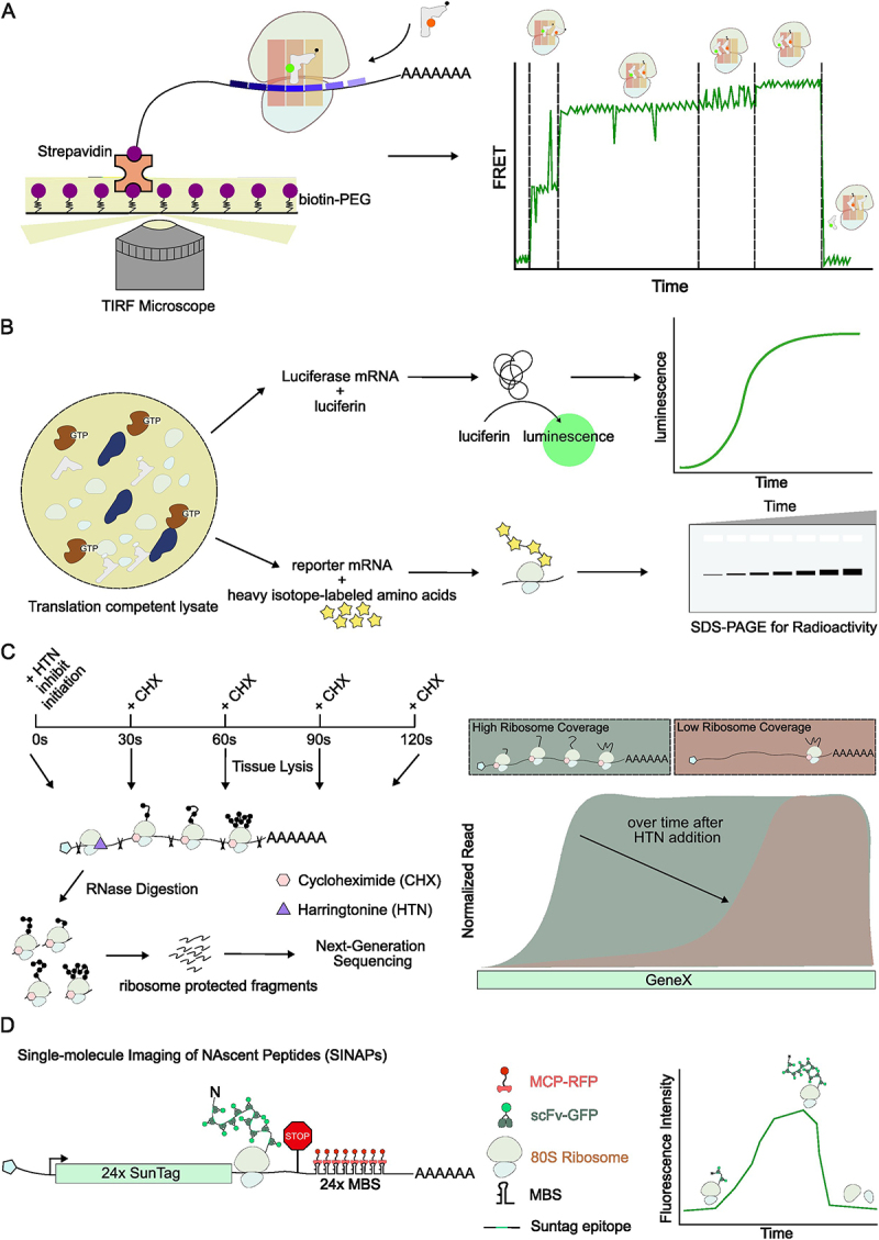
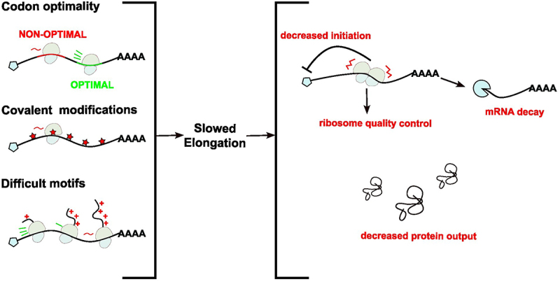
Translation converts genetic information in mRNAs into functional proteins. This process occurs in four major steps: initiation, elongation, termination and ribosome recycling; each of which profoundly impacts mRNA stability and protein yield. Over recent decades, regulatory mechanisms governing these aspects of translation have been identified. In this review, we focus on the elongation phase, reviewing the experimental methods used to measure elongation rates and discussing how the measurements shed light on the factors that regulate elongation and ultimately gene expression.

Keywords: RNA modification; Translation elongation; codon optimality; ribosome; ribosome profiling; single molecule.


Conflict of interest statement

No potential conflict of interest was reported by the author(s).

Figures

Figure 1.
Figure 1.
Overview of translation elongation cycles. Structural basis of eukaryotic elongation is extensively reviewed elsewhere [15–18]. Briefly, an elongation cycle starts with a charged aminoacyl-tRNA positioned at the P site of 80S ribosomes. A G-protein eEF1A delivers an aminoacyl-tRNA to the A-site of ribosomes. Upon cognate codon-anti-codon pairing, GTP-hydrolysis triggers the release of eEF1A from ribosomes, locking tRNA into the A-site. Next, the peptidyl transferase centre (PTC) catalyzes the peptide bond formation, transferring the nascent peptide from the P-site tRNA to the A-site tRNA. This process induces ribosome rotation, forming a hybrid state of E/P and P/A state tRNA. eEF2, a GTPase, binds to the rotated ribosome and translocates the ribosome forward by one codon, shifting tRNAs from site A to P and site P to E. eEF2 restores the ribosome to its original conformation, making the A-site available for the next tRNA.
Figure 2.
Figure 2.
Methods for measuring elongation rates. (A). single-molecule FRET to measure a single elongation cycle. Aminoacyl tRNAs are fluorescently labelled and applied to ribosomes in translation-competent cell lysates. Distinct FRET states correspond to ribosome mechanics. This method measures elongation rate by measuring the time duration of FRET state cycles. (B). Translation competent cell lysate. mRNA templates are added to the lysate to initiate translation. Luciferase mRNAs can be used to probe elongation rate by measuring luminescence signal over time. To measure elongation rate of mRNAs lack of fluorescence or bioluminescence, mRNA templates are added to lysate with heavy isotope-labelled amino acids, and newly synthesized peptides can be visualized on SDS-PAGE. Accumulation of radioactive signal over time reports translation rate. (C). Ribosome profiling. This method determines gene-specific elongation rates by using harringtonine (HTN) to block initiation, followed by cycloheximide (CHX) to freeze elongating ribosomes at different time points. Sequencing ribosome-protected fragments maps ribosome positions on transcripts. The loss of ribosome coverage on transcripts over time reveals elongation rates. High sequencing depth is required for low-expression transcripts. The exact time course of CHX treatment may need optimization depending on gene targets. (D). Live-cell translation imaging. Single-molecule imaging of nascent peptides (SINAPs) utilizes an array of genetically encoded epitope tags and single chain variant fragments conjugated with green fluorescent proteins (scFv-GFP) to visualize nascent peptides, whereas single mRNAs are visualized via repeats of MS2 binding sites (MBS) in their 3’UTR and MS2 coat proteins fused to red fluorescent proteins (MCP-RFP). This method measures elongation rates by tracking either ribosome dwell time on a transcript or the dynamics of nascent peptide signal.
Figure 3.
Figure 3.
Factors affecting elongation rates. Non-optimal codons (red) may be translated slower than optimal codons (green). m1Ψ modified bases (red) may cause elongation speed to slow compared to unmodified ones. Besides mRNA sequence, peptides encoding polybasic residues cause elongation to slow and even stall. As a consequence of slow elongation, ribosomes can stall and collide. These collisions are cleared by the RQC pathway. Ultimately, slow elongation leads to decreased initiation, mRNA decay and decreased protein output.

Similar articles

References

    1. Merrick WC, Pavitt GD.. Protein synthesis initiation in eukaryotic cells. Cold Spring Harb Perspect Biol. 2018;10(12):a033092. doi: 10.1101/cshperspect.a033092 - DOI - PMC - PubMed
    1. Hinnebusch AG. Structural insights into the mechanism of scanning and start codon recognition in eukaryotic translation initiation. Trends Biochem Sci. 2017;42(8):589–611. doi: 10.1016/j.tibs.2017.03.004 - DOI - PubMed
    1. Hinnebusch AG, Lorsch JR. The mechanism of eukaryotic translation initiation: new insights and challenges. Cold Spring Harb Perspect Biol. 2012;4(10):a011544–a011544. doi: 10.1101/cshperspect.a011544 - DOI - PMC - PubMed
    1. Sonenberg N, Hinnebusch AG. Regulation of translation initiation in eukaryotes: mechanisms and biological targets. Cell. 2009;136(4):731–745. doi: 10.1016/j.cell.2009.01.042 - DOI - PMC - PubMed
    1. Brito Querido J, Díaz-López I, Ramakrishnan V. The molecular basis of translation initiation and its regulation in eukaryotes. Nat Rev Mol Cell Biol. 2024;25(3):168–186. doi: 10.1038/s41580-023-00624-9 - DOI - PubMed

LinkOut - more resources