. 2025 May 10;53(9):gkaf426.
doi: 10.1093/nar/gkaf426.

During recombinase-mediated homology recognition RecQ helicases inhibit formation of toxic long-lived D-loops that could promote genomic instability

Affiliations

During recombinase-mediated homology recognition RecQ helicases inhibit formation of toxic long-lived D-loops that could promote genomic instability

Claudia Danilowicz et al. Nucleic Acids Res. .

Abstract

Mutations in RecQ family helicases underlie human genetic disorders associated with genomic instability and cancer predisposition, but questions remain about how properly functioning RecQ reduces these deleterious effects. Importantly, some of the deleterious effects may result from incorrect repair of DNA double-strand breaks (DSBs) by recombinase proteins. Displacement loops (D-loops) are three-strand intermediates formed by recombinases during repair of DSB. RecQ helicases might enhance genome stability by disassembling incorrect recombinase-mediated D-loops formed between mismatched sequences and/or between short regions of accidental homology. We used bulk FRET and gel electrophoresis assays to probe the effects of RecQ family helicases in the context of ongoing recombinase-mediated D-loop formation. We found that RecQ does not differentially promote disassembly of short D-loops or D-loops that include mismatched base pairs. Thus, RecQ does not reduce genomic instability by discriminating against incorrect D-loops. In contrast, our results suggest that RecQ intervenes during D-loop formation to limit the length of recombinase-mediated D-loops. Without that intervention, D-loops can become so long that they do not spontaneously reverse. We suggest that RecQ prevents undesirable long-lived connections between chromosomes that could compromise chromosome metabolism and/or segregation and promote genomic instability.


Conflict of interest statement

None declared.

Figures

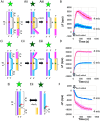
Graphical Abstract
Graphical Abstract
Figure 1.
Figure 1.
Effect of RecQ on D-loops formed by homologous recombination by RecA for matched or mismatched invading strands. (A) The nucleoprotein filament is formed by combining RecA (blue ovals) and a 98 nt ssDNA (Supplementary Table S6). The orange line indicates the 79 nt sequence region at the 3′ end of the ssDNA that is homologous to the dsDNA. The magenta line at the 5′ end indicates the 19 bases that are heterologous to the dsDNA. The purple and light blue lines indicate the complementary and displaced strands in the 180-bp dsDNA (Supplementary Table S1), respectively. The base pairing in the homologous region is indicated in gray, whereas the base pairing in the flanking heterologous tails is indicated in yellow. The red circle indicates the rhodamine label. Dark green and bright green stars indicate quenched fluorescein and unquenched fluorescein, respectively. (i) Initial conditions. (ii) Strand exchange forms a D-loop in which the complementary and invading strands are base-paired, and the displaced strand is not base-paired. The fluorescein label is not quenched. (iii) ATP hydrolysis drives unbinding of RecA, forming a RecA free three-strand product in which the fluorescein is not quenched. (iv) The heteroduplex reverses with or without the help of RecQ, restoring the homoduplex dsDNA in which the fluorescein is quenched. (v) A new nucleoprotein filament can form on the invading strand. (B) ΔF (calculated as the difference between each emission value and the heterologous emission at 1800 s) versus time curves for a completely heterologous invading strand, an invading strand with 79 contiguous homologous bases at the 3′ end, or an invading strand in which the 79 nt at the 3′ end include mismatches (mis) positioned 36 nt from the 3′ end: two grouped mismatches (two mis-grouped), three grouped mismatches (three mis-grouped), and four grouped mismatches (four mis-grouped). No RecQ was present initially. The arrow indicates the time when RecQ was added. The curves represent averages of at least two independent runs. (C) ΔF versus time curves for a completely heterologous invading strand or an invading strand with single mismatches evenly distributed (mis dis) along the 79-nt invading strand: four mismatches distributed (four mis dis), which is one mismatch every 17 bp, and 13 mismatches distributed (13 mis dis), which is 1 mismatch every 6 bp. (D) RatioRecQ, the fractional decrease in emission that occurs in the first 250 s after adding RecQ as a function of the number of mismatches (see text for description). The black curve shows the results for the grouped mismatches [curves shown in panel (B)]. The red triangle shows the result for four distributed mismatches [purple curve in panel (C)].
Figure 2.
Figure 2.
Effect of WRN on D-loops formed by homologous recombination for matched or mismatched invading strands. (A) ΔF versus time curves for homologous recombination performed by DMC1, which is a eukaryotic homolog of RecA. WRN was added at 800 s, as indicated by the arrow. The results for a completely heterologous invading strand and for an invading strand with 79 contiguous homologous bases at the 3′ end (Supplementary Table S6) are shown. Schematics are the same as in Fig. 1A and B. (B) Same as A except the homologous recombination was performed by RecA. The result for a filament with four grouped mismatches is also shown. The conditions are the same as those for Fig. 1B, except in Fig. 1 RecQ helicase was used. The curves represent averages of at least two independent runs. The lighter regions surrounding the curves indicate the range for the runs.
Figure 3
Figure 3
Effect of RecQ on D-loop-like structures and flapped DNAs A. dsDNA is formed by combining two 90-nucleotide ssDNAs (Supplementary Table S3). (A) (i) The two ssDNAs include 50 mismatched bases flanked on both sides by 20-bp matched dsDNA. The mismatched bases in the blue strand are shown in gray. The annealed ssDNA forms a dsDNA with a 50-bp ssDNA bubble. One of the strands has a rhodamine label (red circle) in the bubble. That strand can base pair with the 50 sequence-matched bases (orange line) at the 3′ end of a fluorescein (green star) labeled ssDNA that also includes a 25-nt 5′ tail (magenta line). (ii) When the ssDNA base pairs with the rhodamine labeled strand, fluorescein emission is quenched. (iii) Adding RecQ increases fluorescein emission if the helicase unwinds and separates the rhodamine- and fluorescein-labeled strands. (B) ΔF versus time curves when the 50 nt in the ssDNA are completely sequence matched (0 mis) or contain four mismatches (4 mis) for the structure shown in panel (A). The arrows indicate the time when RecQ was added. (C) Same as A, but the ssDNA has a 25-nt heterologous tail at the 3′ end. (D) ΔF versus time curves when the 50 nt in the ssDNA are completely sequence-matched (0 mis) or contain four mismatches (4 mis) for the structure shown in panel (C). The arrows indicate the time when RecQ was added. (E) (i) Flapped dsDNA where fluorescein label is bright if strands are annealed. The light blue strand includes a 14-nt 5′ tail shown in gray that is heterologous to the purple strand. The red arrow indicates the position of the mismatch in the structure where the mismatch is near the fluorophore. (ii) If RecQ unwinds and separates the fluorescein-labeled strand, emission decreases in the fluorescein-labeled ssDNA product. We cannot detect whether the orange strand separates, but previous work suggests that it would [1]. (F) ΔF versus time curves when the fluorescein-labeled strand is sequence matched (0 mis) or contains four mismatches (4 mis) in a flapped DNA. For B, D, and F, the curves represent averages of at least two independent runs. The lighter regions surrounding the curves indicate the ranges for the runs.
Figure 4.
Figure 4.
Effect of RecQ helicases on D-loops formed by homologous recombination for different N values. (A) Schematic of interactions between a 180 bp dsDNA (Supplementary Table S1) labeled with rhodamine (red circle) and fluorescein (green star) and filaments with homologous length N = 82, 100, and 140 (Supplementary Table S7) and total length L = 98, 100, and 140. (B) ΔF versus time curves for a completely heterologous invading strand and invading strands with N = 82, N = 100, or N = 140. The gray arrow indicates the time when the RecQ was added. The curves represent averages of at least two independent runs. The lighter regions surrounding the curves indicate the ranges for the runs. (C) Schematic of the interaction between a 562-bp dsDNA labeled with rhodamine (red circle) and fluorescein (green star) (Supplementary Table S2) and filaments with N = 320 (Supplementary Table S7). One filament included only 320 nt, but the other two filaments were 370 nt long and included 50 heterologous nt at either the 3′ or 5′ end of the invading strand. (D) ΔF versus time curves for filaments with N = 320 with or without heterologous tails and for a heterologous filament (Supplementary Table S6).
Figure 5.
Figure 5.
Effect of RecQ helicase on D-loops formed by homologous recombination for different N values. (A) ΔF versus time curves for a completely heterologous invading strand or a filament with N = 50. Strand exchange curve without RecQ or with RecQ added initially. (B) Same as A but for N = 82. (C) Same as B except for N = 140. (D) Same as C except for N = 320. Schematics are shown in Fig. 4.
Figure 6.
Figure 6.
RecQ could block formation of long strand exchange products (A) initial invading strand nucleoprotein filament and fluorophore-labeled dsDNA as in Fig. 1A. (B) RecA-bound D-loop established at the 5′ end of the invading strand. (C) RecA-bound D-loop extends toward the 3′ end. (D) RecA begins to unbind at the 5′ end, creating a protein-free three-strand product. (E) RecQ binds in the RecA-free region. (F) RecQ begins to reverse the RecA free product at the 5′ end, but the D-loop has finally reached the 3′ end. (G) RecQ continues to shorten the D-loop.

Similar articles

References

    1. Harmon FG, Kowalczykowski SC RecQ helicase, in concert with RecA and SSB proteins, initiates and disrupts DNA recombination. Genes Dev. 1998; 12:1134–44.10.1101/gad.12.8.1134. - DOI - PMC - PubMed
    1. Croteau DL, Popuri V, Opresko PL et al. . Human RecQ helicases in DNA repair, recombination, and replication. Annu Rev Biochem. 2014; 83:519–52.10.1146/annurev-biochem-060713-035428. - DOI - PMC - PubMed
    1. Mohaghegh P, Hickson ID DNA helicase deficiencies associated with cancer predisposition and premature ageing disorders. Hum Mol Genet. 2001; 10:741–6.10.1093/hmg/10.7.741. - DOI - PubMed
    1. Lu H, Davis AJ Human RecQ helicases in DNA double-strand break repair. Front Cell Dev Biol. 2021; 9:640755.10.3389/fcell.2021.640755. - DOI - PMC - PubMed
    1. Karow JK, Wu L, Hickson ID RecQ family helicases: roles in cancer and aging. Curr Opin Genet Dev. 2000; 10:32–8.10.1016/S0959-437X(99)00039-8. - DOI - PubMed

LinkOut - more resources