. 2025 Jun 17;10(6):e0047325.
doi: 10.1128/msystems.00473-25. Epub 2025 May 16.

Light and polyphosphate kinase 2 cooperatively regulate the production of zero-valent sulfur in a deep-sea bacterium

Affiliations

Light and polyphosphate kinase 2 cooperatively regulate the production of zero-valent sulfur in a deep-sea bacterium

Tianhang Zhang et al. mSystems. .

Abstract

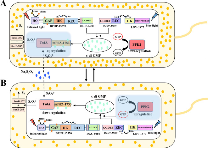
It is well established that different wavelengths of light exist in various deep-sea environments, and many deep-sea microorganisms have evolved specialized mechanisms for sensing and utilizing light energy. Our previous research found that blue light promotes zero-valent sulfur (ZVS) production in Erythrobacter flavus 21-3, a bacterium isolated from a deep-sea cold seep. Given that long-wavelength light is more prevalent in deep-sea environments, the present study investigates the mechanism by which E. flavus 21-3 senses infrared light (wavelength 940 nm) and regulates ZVS production. We found that the bacteriophytochrome BPHP-15570 is responsible for sensing infrared light, which induces autophosphorylation of BPHP-15570, activating the diguanylate cyclase DGC-0450 for c-di-GMP biosynthesis. Subsequently, the PilZ domain-containing protein mPilZ-1753 binds to c-di-GMP, triggering a well-established ZVS production pathway involving thiosulfate dehydrogenase (TsdA) and two homologs of thiosulfohydrolases (SoxB). Notably, polyphosphate kinase 2 (PPK2) is recruited to compete for GTP, the direct precursor of c-di-GMP biosynthesis. This competition downregulates ZVS production as well as other important metabolic processes. This negative regulatory pathway helps the bacterium avoid excessive ZVS accumulation, which could be toxic to bacterial growth. Overall, E. flavus 21-3 has evolved a sophisticated regulatory pathway to sense both blue and infrared light, triggering ZVS production. Our study provides a valuable model for understanding light utilization and its coupling with sulfur cycling in deep-sea environments.IMPORTANCEIt is widely believed that deep-sea ecosystems operate independently of light, relying primarily on chemical energy. However, the discovery of non-photosynthetic bacteria in various deep-sea environments that can sense and utilize light has challenged this assumption. In a recent study, we found that blue light significantly promotes the production of zero-valent sulfur (ZVS) in the deep-sea bacterium Erythrobacter flavus 21-3. Given that long-wavelength light is more prevalent in deep-sea environments, we investigated whether infrared light also plays a role in regulating ZVS production in E. flavus 21-3. Our results indicate that infrared light does promote ZVS formation in this bacterium. We identified PPK2 as a negative regulator, maintaining intracellular ZVS at safe levels to prevent toxicity due to excessive accumulation. Overall, our study offers a valuable model for exploring how light is utilized and its interaction with microbial sulfur cycling in the extreme conditions of the deep sea.

Keywords: c-di-GMP; deep-sea bacterium; infrared light; polyphosphate kinase 2; zero-valent sulfur.


Conflict of interest statement

The authors declare no conflict of interest.

Figures

Fig 1
Fig 1
A bacteriophytochrome (BPHP-15570) responds to infrared light and triggers the production of ZVS in the deep-sea bacterium E. flavus 21-3. (A) Comparison of ZVS production in E. flavus 21-3 cultured under blue light, infrared light, or dark conditions for 1, 2, 3, and 4 days (N = 3 biological replicates). (B) Comparison of the growth of E. flavus 21-3 cultured under blue light, infrared light, or dark conditions for 1, 2, 3, and 4 days (N = 3 biological replicates; n.s. indicates no significant difference). (C) Sequence alignment of D0Y83-RS15570 and other homologs. The conserved binding sites (Cys in the GAF domain and His in the HK domain) are marked with asterisks. The numbers on the left indicate the accession numbers of the corresponding proteins. The blue bar chart shows the frequency of base occurrence. (D) Absorption spectrum analysis of BPHP-15570 after infrared light illumination. Black arrows indicate the absorption peaks corresponding to infrared light. The inset shows the SDS-PAGE analysis of purified BPHP-15570 from E. coli BL21 (DE3) cells (M, protein marker). (E) Infrared light sensing assay of BPHP-15570 in E. coli BL21 (DE3) cells. E. coli BL21 (DE3) cells co-expressing BPHP-15570 and heme oxygenase displayed the red color on Congo red-containing agar plates (50 µg/mL) under infrared light illumination.
Fig 2
Fig 2
The bacteriophytochrome BPHP-15570 activates the diguanylate cyclase DGC-0450 for c-di-GMP biosynthesis. (A) Comparison of c-di-GMP yields in E. flavus 21-3 cultured under infrared light and dark conditions for 12, 24, and 36 hours (N = 3 biological replicates). (B) Proteomic analysis revealed the expression levels of five GGDEF-domain-containing proteins in E. flavus 21-3, which were significantly up-regulated under infrared light compared to dark conditions. (C) Detection of interaction between BPHP-15570 and the predicted GGDEF-domain-containing proteins. Assays were performed on MacConkey plates with 1% maltose and X-gal-LB indicator plates (LB broth with 40 µg/mL X-gal) with 0.5 mM IPTG. E. coli BTH101 strains were transformed with T18 and T25 fusion constructs to exclude self-activation. Positive control: E. coli BTH101 with pKNT25-Zip and pCH363-Zip plasmids. Negative control: E. coli BTH101 with empty plasmids. (D) Comparison of intracellular c-di-GMP concentrations in E. coli BL21(DE3) with and without the overexpression of DGC-0450. The c-di-GMP concentration in E. coli BL21(DE3) transformed with the empty vector was normalized as the control (N = 3 biological replicates). (E) Comparison of intracellular c-di-GMP concentrations in E. coli BL21(DE3) co-expressing heme oxygenase (HO), BPHP-15570, and DGC-0450 under infrared light and dark conditions (N = 3 biological replicates). (F) Comparison of intracellular c-di-GMP concentrations in E. coli BL21(DE3) co-expressing BPHP-15570 mutant (with His524 mutated to Gly) and heme oxygenase (HO) after 24 hours of cultivation under infrared light and dark conditions (N = 3 biological replicates; n.s. indicates no significant difference). (G) Comparison of intracellular c-di-GMP concentrations in E. coli BL21(DE3) co-expressing BPHP-15570 mutant (with Asp742 mutated to Gly) and heme oxygenase (HO) after 24 hours of cultivation under infrared light and dark conditions (N = 3 biological replicates; n.s. indicates no significant difference).
Fig 3
Fig 3
Proposed model for ZVS production pathway triggered by infrared light in E. flavus 21-3. In the presence of infrared light, BPHP-15570 senses the light signal with the assistance of heme oxygenase (HO), which induces autophosphorylation and activates the diguanylate cyclase DGC-0450, leading to the production of c-di-GMP. The generated c-di-GMP then binds to mPilZ-1753, triggering its interaction with thiosulfate dehydrogenase (TsdA). This interaction facilitates the conversion of thiosulfate to tetrathionate. Finally, tetrathionate is further transformed into ZVS by thiosulfohydrolases SoxB-277 and SoxB-285. HO: heme oxygenase; TsdA: thiosulfate dehydrogenase; SoxB: thiosulfohydrolase; PPK2: polyphosphate kinase 2.
Fig 4
Fig 4
Downregulation of ZVS formation by polyphosphate kinase 2 (PPK2) through decreased c-di-GMP biosynthesis. (A) Comparison of intracellular GTP concentrations in wild-type and mutant Δppk2 strains of E. flavus 21-3 cultured under infrared light and dark conditions for 12, 24, and 36 hours (N = 3 biological replicates; n.s. indicates no significant difference). (B) Comparison of intracellular c-di-GMP concentrations in wild-type and mutant Δppk2 strains of E. flavus 21-3 cultured under infrared light and dark conditions for 12, 24, and 36 hours (N = 3 biological replicates; n.s. indicates no significant difference). (C) Comparison of ZVS concentrations produced by wild-type and mutant Δppk2 strains of E. flavus 21-3 cultured under infrared light and dark conditions for 1, 2, 3, and 4 days (N = 3 biological replicates). (D) Comparison of growth levels of wild-type and mutant Δppk2 strains of E. flavus 21-3 cultured under infrared light and dark conditions for 1, 2, 3, and 4 days (N = 3 biological replicates; n.s. indicates no significant difference). (E) Qualitative analysis of biofilm formation in wild-type and mutant Δppk2 strains of E. flavus 21-3 cultured under infrared light and dark conditions (N = 3 biological replicates). (F) Quantitative analysis of biofilm formation as shown in panel E. (G) Proteomic analysis of the expression of GGDEF-domain-containing proteins, as well as flagellar assembly and chemotaxis-related proteins, in wild-type and mutant Δppk2 strains of E. flavus 21-3 cultured under infrared light.
Fig 5
Fig 5
Downregulation of c-di-GMP production by polyphosphate kinase 2 (PPK2) in E. coli BL21 (DE3) and P. aeruginosa PAO1. (A) Comparison of intracellular c-di-GMP concentrations in E. coli BL21 (DE3) overexpressing PPK2 and the control. The control strain consists of E. coli BL21 (DE3) overexpressing the empty vector pET28a (+) (N = 3 biological replicates). (B) Comparison of intracellular GTP concentrations in E. coli BL21 (DE3) overexpressing PPK2 and the control. The control strain consists of E. coli BL21 (DE3) overexpressing the empty vector pET28a (+) (N = 3 biological replicates). (C) Comparison of intracellular c-di-GMP concentrations in P. aeruginosa PAO1 overexpressing PPK2 and the control. The control strain consists of P. aeruginosa PAO1 overexpressing the empty vector pUCP18-Gm (N = 3 biological replicates). (D) Comparison of intracellular GTP concentrations in P. aeruginosa PAO1 overexpressing PPK2 and the control. The control strain consists of P. aeruginosa PAO1 overexpressing the empty vector pUCP18-Gm (N = 3 biological replicates). (E) Qualitative analysis of biofilm formation in P. aeruginosa PAO1 overexpressing PPK2 and the control. The control strain consists of P. aeruginosa PAO1 overexpressing the empty vector pUCP18-Gm. (F) Quantitative analysis of biofilm formation as shown in panel E (N = 3 biological replicates).
Fig 6
Fig 6
Proposed model for the cooperative regulation of ZVS production by light and polyphosphate kinase in E. flavus 21-3. (A) In the presence of light (infrared or blue) and thiosulfate, E. flavus 21-3 activates light sensors BPHP-15570 and LOV-1477, which, respectively, detect infrared and blue light. These light sensors then interact with the diguanylate cyclases DGC-0450 and DGC-2902, stimulating their activity to produce c-di-GMP. Concurrently, the expression of PPK2 is negatively regulated. As a result, c-di-GMP accumulates and binds to mPilZ-1753, promoting the ZVS production pathway via TsdA and SoxB. (B) To prevent the harmful effects of excessive ZVS production, E. flavus 21-3 increases the expression of PPK2, which consumes intracellular GTP and reduces c-di-GMP generation. This, in turn, downregulates the downstream production of ZVS. HO: heme oxygenase; TsdA: thiosulfate dehydrogenase; SoxB: thiosulfohydrolase; PPK2: polyphosphate kinase 2.

Similar articles

References

    1. van der Horst MA, Key J, Hellingwerf KJ. 2007. Photosensing in chemotrophic, non-phototrophic bacteria: let there be light sensing too. Trends Microbiol 15:554–562. doi: 10.1016/j.tim.2007.09.009 - DOI - PubMed
    1. Kushkevych I, Procházka J, Gajdács M, Rittmann SK-MR, Vítězová M. 2021. Molecular physiology of anaerobic phototrophic purple and green sulfur bacteria. Int J Mol Sci 22:6398. doi: 10.3390/ijms22126398 - DOI - PMC - PubMed
    1. Cai RN, He WY, Zhang J, Liu R, Yin ZY, Zhang X, Sun CM. 2023. Blue light promotes zero-valent sulfur production in a deep-sea bacterium. EMBO J 42:e112514. doi: 10.15252/embj.2022112514 - DOI - PMC - PubMed
    1. Wiltbank LB, Kehoe DM. 2019. Diverse light responses of cyanobacteria mediated by phytochrome superfamily photoreceptors. Nat Rev Microbiol 17:37–50. doi: 10.1038/s41579-018-0110-4 - DOI - PubMed
    1. Braatsch S, Klug G. 2004. Blue light perception in bacteria. Photosynth Res 79:45–57. doi: 10.1023/B:PRES.0000011924.89742.f9 - DOI - PubMed

MeSH terms

Substances

LinkOut - more resources