Review
. 2025 Jun 9;65(11):5785-5800.
doi: 10.1021/acs.jcim.5c00462. Epub 2025 May 16.

Entabolons: How Metabolites Modify the Biochemical Function of Proteins and Cause the Correlated Behavior of Proteins in Pathways

Affiliations
Review

Entabolons: How Metabolites Modify the Biochemical Function of Proteins and Cause the Correlated Behavior of Proteins in Pathways

Jeffrey Skolnick et al. J Chem Inf Model. .

Abstract

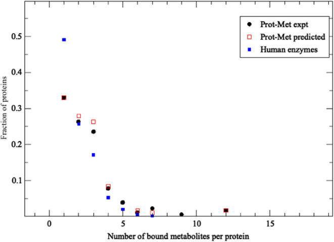
Although there are over 100,000 distinct human metabolites, their biological significance is often not fully appreciated. Metabolites can reshape the protein pockets to which they bind by COLIG formation, thereby influencing enzyme kinetics and altering the monomer-multimer equilibrium in protein complexes. Binding a common metabolite to a set of protein monomers or multimers results in metabolic entanglements that couple the conformational states and functions of nonhomologous, nonphysically interacting proteins that bind the same metabolite. These shared metabolites might provide the collective behavior responsible for protein pathway formation. Proteins whose binding and functional behavior is modified by a set of metabolites are termed an "entabolon"─a portmanteau of metabolic entanglement and metabolon. 55%-60% (22%-24%) of pairs of nonenzymatic proteins that likely bind the same metabolite have a p-value that they are in the same pathway, which is <0.05 (0.0005). Interestingly, the most populated pairs of proteins common to multiple pathways bind ancient metabolites. Similarly, we suggest how metabolites can possibly activate, terminate, or preclude transcription and other nucleic acid functions and may facilitate or inhibit the binding of nucleic acids to proteins, thereby influencing transcription and translation processes. Consequently, metabolites likely play a critical role in the organization and function of biological systems.


Figures

1
1
Fraction of proteins with the given number of bound types of metabolites versus the number of bound metabolites. Prot-Met data set results are in black circles, the results of LIGMAP predictions are in red squares, and LIGMAP predictions for human enzymes with PDB structures are in blue squares.
2
2
LIGMAP’s results are shown in the colored circles. The precision versus recall curves are in black, and the precision versus NPV value (defined in Table ) are in red. The random results are shown in the squares. The precision versus recall curves are in blue and the precision versus NPV value (defined in Table ) are in green.
3
3
Pocket remodeling associated with native pairs of COLIGs, at least one of which is a human metabolite. The crystal structure is colored green. (A-D) Both ligands (red and yellow) are native; (E–H) the native metabolite is colored red, and the predicted COLIG metabolite is colored yellow. The right-hand structure in each panel shows the COLIGs without the protein. (A) COLIG formed by GLC (red), alpha-d-glucopyranose and ADP (yellow), and adenosine 5′ phosphate in 2r4uA, a glycogen synthase. (B) COLIG formed by enzyme reactants UDP (red), uridine 5′-diphosphate, and LCN (yellow), 1,5-anhydro-d-arabino-hex-1-enitol, buried in the interior of 3s28A, sucrose synthase-1. (C) A COLIG involved in bond ligation is shown for 3guhA, a glycogen synthase, whose COLIG is ADP, adenosine-5′-diphosphate (red), and ASO (yellow), 4-[(11beta,17beta)-17-methoxy-17-(methoxymethyl)-3-oxoestra-4,9-dien-11-yl] benzaldehyde oxime. (D) GDP (red) (guanosine 5′-diphosphate) and B12 (yellow) (cobalamin) form a surface COLIG in 3o0oA, a ribonucleotide reductase. (E) Remodeled pocket in 1a9tA, a purine nucleotide phosphorylase, whose ligands are XAN (yellow) and R1P (red), 1-O-phosphono-alpha-d-ribofuranose. (F) Remodeled pocket in 2dorA, a dihydroorotate dehydrogenase, whose predicted and native ligands are DOR (yellow), (4s)-2,6-dioxohexahydropyrimidine-4-carboxylic acid, and FMN (red), flavin mononucleotide. (G) Remodeled pocket in 1e2fA, a human thymidylate kinase, whose native ligand TMP (red), thymidine 5′ phosphate, has a binding surface created by ATP (yellow). (H) Remodeled pocket in 1tgyA, uridine phosphorylase, whose native ligand R1P’s (red), 1-O-phosphono-alpha-d-ribofuranose, binding is partly stabilized by TDR (yellow), thymine.
4
4
Pocket remodeling associated with native pairs of COLIGs, at least one of which is a human metabolite. Nonpolar residues are white, basic residues are blue, acid residues are red, and polar residues are green. The ligands are shown in a licorice representation, colored by the atom type with oxygen in red, nitrogen in blue, carbon in cyan, and hydrogen in white. In Figures 4A–D, both ligands are native; in Figures 4E–H, native and predicted ligands are shown. (A) COLIG formed by GLC, alpha-d-glucopyranose and ADP, adenosine 5′ phosphate in 2r4uA, a glycogen synthase. (B) COLIG formed by enzyme reactants UDP, uridine 5′-diphosphate, and LCN, 1,5-anhydro-d-arabino-hex-1-enitol, buried in the interior of 3s28A, sucrose synthase-1. (C) A COLIG involved in bond ligation is shown for 3guhA, a glycogen synthase, whose COLIG is ADP (adenosine-5′-diphosphate) and ASO (4-[(11beta,17beta)-17-methoxy-17-(methoxymethyl)-3-oxoestra-4,9-dien-11-yl] benzaldehyde oxime). (D) GDP (guanosine 5′-diphosphate) and B12 (cobalamin) form a surface COLIG in 3o0oA, a ribonucleotide reductase. (E) Remodeled pocket in 1a9tA, a purine nucleotide phosphorylase, whose ligands are XAN and R1P (1-O-phosphono-alpha-d-ribofuranose). (F) Remodeled pocket in 2dorA, a dihydroorotate dehydrogenase, whose predicted and native ligands are DOR ((4s)-2,6-dioxohexahydropyrimidine-4-carboxylic acid) and FMN (flavin mononucleotide). (G) Remodeled pocket in 1e2fA, a human thymidylate kinase, whose native ligand TMP, thymidine 5′ phosphate, has a binding surface created by ATP. (H) Remodeled pocket in 1tgyA, uridine phosphorylase, whose native ligand R1P’s, 1-O-phosphono-alpha-D-ribofuranose, binding is partly stabilized by TDR, thymine. The figures were generated by VMD.
5
5
Examples of ligand pinning COLIGs in native PDB structures. The native protein structure is colored green. (A–D) Both ligands (red and yellow) are native; (E–H) the native metabolite is colored red, and the predicted metabolite is colored yellow. The right-hand structures in each panel show the COLIGS with their native structure removed. (A) HEM (yellow), protoporphyrin IX pins HSO (red), l-histidinol in1h3jA, a peroxidase. (B) NAD (yellow), nicotinamide-adenine-dinucleotide, pins IMP, inosinic acid (red) in 6ua2A, human inosine monophosphate dehydrogenase. (C) Example of intertwined pocket remodeling/ligand pinning, by CLA, chlorophyll A (red), and DDG, digalactosyl diacyl glycerol (yellow) in 5h2fC, part of photosystem II. (D) FMN, flavin mononucleotide (red), is totally pinned by ORO (yellow), orotic acid in 6fmdA, human dihydroorotate dehydrogenase. (E) Native ligand FAD (red) forms a COLIG with MLI (yellow), malonate ion that partially blocks its exit from 1bwkA, old yellow enzyme. (F) The native ligand, UDP (red), uridine-5′-diphosphate, is partially pinned by GLC (yellow), alpha-d-glucopyranose in1k4vA, beta-galactoside-alpha-1,3-galactosyltransfera. (G) The native ligand, FMN (red), flavin adenine dinucleotide, is pinned by FMN (yellow), flavin mononucleotides in 3r6wA, an azoreductase. (H) The native ligand TDR (red), thymine, is pinned to the side of the pocket by R1P, 1-O-phosphono-alpha-d-ribofuranose, in 4txm, a uridine phosphatase. The figures were generated by VMD.
6
6
Examples of ligand pinning COLIGs in native PDB structures. Nonpolar residues are white, basic residues are blue, acid residues are red, and polar residues are green. The small molecule ligands are shown in a licorice representation, colored by atom type in the protein with oxygen in red, nitrogen in blue, carbon in cyan, and hydrogen in white. (A–D) Both ligands are native; (E–H) native and predicted metabolites are shown. (A) HEM, protoporphyrin IX, pins HSO, l-histidinol in1h3jA, a peroxidase. (B) NAD, nicotinamide-adenine-dinucleotide, pins IMP, inosinic acid in 6ua2A, human inosine monophosphate dehydrogenase. (C) Example of intertwined pocket remodeling/ligand pinning, by CLA, chlorophyll A, and DDG, digalactosyl diacyl glycerol, in 5h2fC, part of photosystem II. (D) FMN, flavin mononucleotide, is totally pinned by ORO, orotic acid, in 6fmdA, human dihydroorotate dehydrogenase. (E) Native ligand FAD forms a COLIG with MLI, malonate ion, that partially blocks its exit from 1bwkA 6, old yellow enzyme. (F) The native ligand, UDP, uridine-5′-diphosphate, is partially pinned by GLC, alpha-d-glucopyranose in1k4vA, beta-galactoside-alpha-1,3-galactosyltransfera. (G) The native ligand, FMN, flavin adenine dinucleotide, is pinned by FMN, flavin mononucleotides in 3r6wA, an azoreductase. (H) The native ligand TDR, thymine, is pinned to the side of the pocket by R1P, 1-O-phosphono-alpha-d-ribofuranose, in 4txm, a uridine phosphatase. The figures were generated by VMD.
7
7
Examples of “metabolic entanglement.” (A) Metabolite (dark blue) binding to the pocket adjacent to the protein–protein interfaces stabilizes the dimer relative to its dissociated monomers. This shifts the monomer–dimer equilibrium. (B) Binding of the metabolite (dark blue) induces an allosteric transition in the dimer. (C) The “adj-vs-adj” case considers situations where the metabolite (dark blue) binds adjacent to the protein–protein interface in two distinct dimers. Both molecules remain in the dimeric state, but the presence and absence metabolite binding induces an allosteric transition within the pair of dimers. (D) The “inter-vs-inter” case occurs when the binding of a metabolite (dark blue) to an interfacial pocket in a monomer precludes dimer formation. Dissociation of the metabolite from the light blue monomer allows it to bind to an interfacial pocket in another monomeric (orange) protein. This induces dissociation of the left-hand (yellow/orange) dimer and association of the light blue and white monomers to form a dimer. (E) The “adj-vs-inter” case involves the shift of equilibrium of the yellow/orange dimer containing the interface adjacent metabolite (dark blue) to the interfacial pocket in one of the chains of the light blue/white dimer. This dissociates the light blue/white dimer and might cause an allosteric transition within the yellow/orange dimer.
8
8
Examples how small molecules/metabolites can assist with DNA–protein binding and DNA ligation. The protein is shown in pink, and the DNA and predicted ligands from LIGMAP are shown in a licorice representation, colored by atom type, with oxygen in red, nitrogen in blue, carbon in cyan, and hydrogen in white. (A) 3khhA DNA bound to Y-family polymerase 4 whose binding is possibly assisted by 24 ligands including metabolites DCP, 2’-deoxycytidine-5′-triphosphate; DTP, 2’-deoxyadenosine 5′-triphosphate; and ACT. (B) 3t3fA, the large fragment of Taq DNA polymerase, is predicted to bind 31 ligands including DCP. (C) 6cvrA is the structure of aprataxin that protects genome integrity and fixes abortive DNA ligation arising during ribonucleotide and base excision DNA repair associated with cytotoxic adenylated DNA strand breaks. Five examples of the bound AMP are also shown and are part of the DNA ligation process.

Similar articles

References

    1. Milo R.. What is the total number of protein molecules per cell volume? A call to rethink some published values. Bioessays. 2013;35(12):1050–1055. doi: 10.1002/bies.201300066. - DOI - PMC - PubMed
    1. Bennett B. D., Kimball E. H., Gao M., Osterhout R., Van Dien S. J., Rabinowitz J. D.. Absolute metabolite concentrations and implied enzyme active site occupancy in Escherichia coli. Nat. Chem. Biol. 2009;5(8):593–599. doi: 10.1038/nchembio.186. - DOI - PMC - PubMed
    1. Bionumbers What are the concentrations of free metabolites in cells?. 2024. https://book.bionumbers.org/what-are-the-concentrations-of-free-metaboli....
    1. Goodman, L. S. ; Brunton, L. L. ; Chabner, B. ; Knollmann, B. R. C. . Goodman & Gilman’s pharmacological basis of therapeutics; McGraw-Hill, 2011.
    1. Wishart D. S., Guo A., Oler E., Wang F., Anjum A., Peters H., Dizon R., Sayeeda Z., Tian S., Lee B. L.. et al. HMDB 5.0: the Human Metabolome Database for 2022. Nucleic Acids Res. 2022;50(D1):D622–D631. doi: 10.1093/nar/gkab1062. - DOI - PMC - PubMed

LinkOut - more resources