. 2025 May 16;11(20):eadv1286.
doi: 10.1126/sciadv.adv1286. Epub 2025 May 16.

Hsc70-4: An unanticipated mediator of dsRNA internalization in Drosophila

Affiliations

Hsc70-4: An unanticipated mediator of dsRNA internalization in Drosophila

Sabrina J Fletcher et al. Sci Adv. .

Abstract

The small interfering RNA pathway is the primary antiviral defense mechanism in invertebrates and plants. This systemic mechanism relies on the recognition, transport, and internalization of double-stranded RNA (dsRNA). Our aim was to identify cell surface proteins that bind extracellular dsRNA and mediate its internalization in Drosophila cells. We used coimmunoprecipitation coupled with proteomics analysis and found that silencing heat shock cognate protein 70-4 (Hsc70-4), a constitutively expressed heat shock protein, impairs dsRNA internalization. Unexpectedly, despite lacking a predicted transmembrane domain, Hsc70-4 localizes to the cell membrane via lipid interactions. Antibody blocking experiments revealed an extracellular domain on Hsc70-4 that is essential for dsRNA internalization. Intriguingly, this dsRNA-specific binding capacity of Hsc70-4 functions independently of its chaperone activity. These findings not only highlight Hsc70-4 as a previously uncharacterized and essential component in the dsRNA internalization process but also offer promising insights for advancing RNA interference-based technologies to combat pests and vector-borne diseases.


Figures
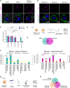
Fig. 1.
Fig. 1.. S2 cell model and proteomic approaches to identify cell surface dsRNA binding proteins.
(A) Internalization of dsRNA-Cy3 evaluated by fluorescence confocal imaging of single Z-sections. dsRNA soaking was done for 40 min. Actin is in green, and nuclei are in blue. Dynasore was added for 1 hour before dsRNA soaking. (B) Specificity of nucleic acid internalization by S2Xpress cells. dsRNA-Cy3 (dsFLuc), dsDNA-Cy3 (FLuc), or siRNA-Cy3 (siGAPDH) (magenta) was added during soaking. Staining is as in (A). (C) Silencing of firefly luciferase by internalized dsRNA. Cells were cotransfected with plasmids expressing firefly and Renilla luciferase followed by soaking with dsRNA targeting firefly luciferase (dsFLuc) or GFP (dsCtl). As positive controls, dsRNAs and plasmids were cotransfected. Luciferase activity was measured 24 hours after induction. Firefly luciferase values were normalized to Renilla luciferase values. The histogram shows the mean + SD firefly/Renilla ratio relative to soaking with dsCtl (Soak. dsCtl) from three independent experiments (n = 8 to 9). Welch’s ANOVA was used to detect significant differences compared to Soak. dsCtl. (D) Protocol used to purify cell surface proteins from S2naive and S2Xpress cells. (E) Venn diagram showing proteins identified from S2naive and S2Xpress cells in (D). (F) Cellular component analysis of the proteins identified by the protocol in (D). Statistical analysis was done with FunRich (57) (hypergeometric test). (G) Protocol used to purify dsRNA binding proteins from S2Xpress cells by IP. Dynasore was used to prevent internalization of dsRNA-Cy3. (H) Venn diagram showing proteins identified in S2naive and S2express cells using the different proteomic protocols. Venn diagrams were done with FunRich (57). Protocol schemes were created with BioRender.com. Scale bars represent 5 μm. ALU, arbitrary luciferase units. *P < 0.05; **P < 0.01; ****P < 0.0001.
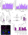
Fig. 2.
Fig. 2.. High-content screen of selected candidates.
(A) Candidate genes were silenced in S2Xpress cells by transfection with gene-specific dsRNA (dsRNA target) or nonspecific dsRNA (Ctl−, dsCtl, Dynasore) followed by soaking with nonspecific dsRNA-Cy3 (dsFLuc) and analyzed with the Opera Phenix High-Content microscope. Cy3 intensity, number of Cy3 spots per cell, and mean spot area were quantified on single Z-sections with Columbus software. Histograms show the means + SD of spots per cell and mean spot area. ANOVA with Dunnett’s post hoc tests was used to detect significant differences compared to dsCtl (Ctl− and dsCtl, n = 8; dsCandidates and Dynasore, n = 3; dsHmu, n = 2). P values are indicated in fig. S2B. a.u., arbitrary units. (B) High-content analysis to determine the effects of silencing Hsc70-4 on dsRNA internalization. Experiments and analyses were performed as in (A). Data are from three independent experiments (Ctl−, n = 24; Dynasore, n = 9; dsCtl, n = 23; dsHsc70-4, n = 23) (Welch’s ANOVAs with Dunnett’s T3 post hoc). (C) Immunoblot and quantification of anti-Hsc70-4 signal in S2Xpress cells transfected with dsRNA targeting Hsc70-4 (dsHsc70-4) compared to the control (dsCtl). Tubulin was used as a control. Histograms show the means + SD normalized to tubulin (unpaired t test, n = 9). (D) Confocal imaging of single Z-sections to confirm the findings shown in (B). S2Xpress cells were transfected with dsHsc70-4 or dsCtl for 72 hours followed by soaking of dsRNA-Cy3 (magenta). Actin is in green, and nuclei are in blue. Scale bars represent 5 μM. (E) Effect of down-regulation of Hsc70-4 on silencing by luciferase assay. Cells were transfected with dsHsc70-4, dsCtl, or dsAgo2 (as a positive control of effect on silencing). Experiments were done as in Fig. 1C. The histogram shows the mean + SD firefly/Renilla ratio relative to Soak. dsCtl from two independent experiments (n = 6). ANOVA with Dunnett’s post hoc tests was used to detect significant differences compared to dsCtl. *P < 0.05; **P < 0.01; ***P < 0.001; ****P < 0.0001. ns, not significant.
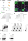
Fig. 3.
Fig. 3.. Cell surface localization of Hsc70-4 and binding to dsRNA.
(A) Schematic representation of immunofluorescence staining performed with anti-Hsc70-4 on S2 cells under permeabilized and nonpermeabilized conditions to detect Hsc70-4 localization. The cartoon was created using BioRender.com. (B) Fluorescence confocal images of single Z-sections of S2naive and S2Xpress under permeabilized and nonpermeabilized conditions. Hsc70-4 is in green, actin is in magenta, and nuclei are in blue. (C and D) PIP Strip membranes were used to detect the binding of Drosophila recombinant protein (rHsc70-4) or its human ortholog (rHsc70) to various lipids. (E) EMSA assay testing binding of rHsc70-4 (0.25, 0.5, 1, 2, 3, 4, and 5 μM) to dsRNA-Cy3 (dsCat, 0.76 nM). The electrophoretic shift of dsRNA-Cy3 was evaluated by native PAGE. A mobility shift of dsRNA-Cy3 incubated with rHsc70-4 confirms binding. (F) In a competition assay, unlabeled dsRNA and Cy3-labeled dsRNA were incubated with rHsc70-4 (0.25, 0.5, and 1 μM) in a 10:1 unlabeled:labeled dsRNA ratio. An increase in the mobility of Cy3-labeled dsRNA confirmed that unlabeled dsRNA displaced the labeled dsRNA. (G) EMSA using a dsRNA with a different sequence (dsRNA-2). (H) EMSA of dsDNA-Cy3 (Cat, 0.76 nM) with rHsc70-4 (0.25, 0.5, 1, and 2 μM) as in (E). (I) EMSA of siRNA-Cy3 (siGAPDH, 0.76 nM) with rHsc70-4 (0.5, 1, and 2 μM) as in (E). All EMSA experiments were performed at least twice, giving similar results. The first lane in all gels corresponds to Cy3-labeled nucleic acid (dsRNA, dsDNA, or siRNA) incubated without rHsc70-4. Scale bars represent 5 μm.
Fig. 4.
Fig. 4.. Effect of nucleotide substrates on rHsc70-4 binding to dsRNA and JG-98 treatment on silencing of luciferase.
EMSAs testing the binding of rHsc70-4 (2 μM) to dsRNA-Cy3 (dsCat, 0.76 nM) in the presence of (A) ATP-γ-S, (B) AMP-PNP, or (C) ADP. Concentrations of 1, 2.5, 5, and 10 mM of each substrate were used. The electrophoretic shift of dsRNA-Cy3 was assessed by native PAGE. All EMSA experiments were performed at least three times, giving similar results. The first lane in all gels corresponds to dsRNA-Cy3 incubated without rHsc70-4. The last lane in all gels corresponds to dsRNA-Cy3 incubated without rHsc70-4 and with each substrate (10 mM). (D) Effect of pretreatment of S2Xpress cells with a JG-98 inhibitor on silencing of luciferase. Experiments were performed as in Fig. 1C. After transfection with luciferase-expressing plasmids, cells were incubated with JG-98 (0.125, 0.25, 0.5, and 1 μM) or DMSO (1 μM). Then, dsFluc or dsCtl was added to the media. Last, luciferase activity was measured. As positive and negative controls of RNAi silencing, dsFluc and dsCtl were cotransfected, respectively. The histogram shows the mean + SD firefly/Renilla ratio relative to Soak. dsCtl from three independent experiments (n = 9). ANOVA followed by Tukey’s post hoc test was used to detect significant differences between treatments. **P < 0.01; ***P < 0.001; ****P < 0.0001.
Fig. 5.
Fig. 5.. Blocking of dsRNA internalization with an anti-Hsc70-4 antibody.
(A) Effect of pretreatment of S2Xpress cells with an anti-Hsc70-4 antibody on silencing of luciferase. Experiments were performed as in Fig. 1C. After transfection with luciferase-expressing plasmids, cells were incubated with anti-Hsc70-4 (40, 60, and 80 ng/μl) or an unrelated IgG antibody (80 ng/μl) as a control for 1 hour. Cells were then washed with PBS, and dsFluc or dsCtl was added to the media. Last, luciferase activity was measured. For positive and negative controls of RNAi silencing, dsFluc and dsCtl were cotransfected, respectively. As a positive control of inhibition of RNAi silencing, dsAgo2 was cotransfected. The histogram shows the mean + SD Firefly/Renilla ratio relative to Soak. dsCtl from four independent experiments (n = 12). ANOVA followed by Tukey’s post hoc test was used to detect significant differences between treatments. (B) Blocking of dsRNA-Cy3 internalization evaluated by fluorescence confocal imaging of single Z-sections. S2Xpress cells were incubated with anti-Hsc70-4 (80 ng/μl) or control IgG (80 ng/μl) for 1 hour. After washing with PBS, dsRNA-Cy3 was added to the growth medium for 40 min. dsRNA is shown in magenta, actin is shown in green, and nuclei are shown in blue. (C) Proposed model for dsRNA internalization. Our data suggest that Hsc70-4 acts as a co-receptor with a cell surface partner to bind and internalize extracellular dsRNA. The model was created using BioRender.com. Scale bars represent 5 μm. *P < 0.05; ****P < 0.0001.

References

    1. Riera Romo M., Pérez-Martínez D., Castillo Ferrer C., Innate immunity in vertebrates: An overview. Immunology 148, 125–139 (2016). - PMC - PubMed
    1. Ferrandon D., Imler J. L., Hetru C., Hoffmann J. A., The Drosophila systemic immune response: Sensing and signalling during bacterial and fungal infections. Nat. Rev. Immunol. 7, 862–874 (2007). - PubMed
    1. Grant M., Lamb C., Systemic immunity. Curr. Opin. Plant Biol. 9, 414–420 (2006). - PubMed
    1. Karlikow M., Goic B., Saleh M. C., RNAi and antiviral defense in Drosophila: Setting up a systemic immune response. Dev. Comp. Immunol. 42, 85–92 (2014). - PubMed
    1. Kemp C., Mueller S., Goto A., Barbier V., Paro S., Bonnay F., Dostert C., Troxler L., Hetru C., Meignin C., Pfeffer S., Hoffmann J. A., Imler J.-L., Broad RNA interference-mediated antiviral immunity and virus-specific inducible responses in Drosophila. J. Immunol. 190, 650–658 (2013). - PMC - PubMed

MeSH terms

LinkOut - more resources