. 2025 May 16;11(20):eadu6632.
doi: 10.1126/sciadv.adu6632. Epub 2025 May 16.

Dual DNA demethylation mechanisms implement epigenetic memory driven by the pioneer factor PAX7

Affiliations

Dual DNA demethylation mechanisms implement epigenetic memory driven by the pioneer factor PAX7

Juliette Harris et al. Sci Adv. .

Abstract

Pioneer transcription factors have the unique ability to open chromatin at enhancers to implement new cell fates. They also provide epigenetic memory through demethylation of enhancer DNA, but the underlying mechanisms remain unclear. We now show that the pioneer paired box 7 (PAX7) triggers DNA demethylation using two replication-dependent mechanisms, including direct PAX7 interaction with the E3 ubiquitin-protein ligase (UHRF1)-DNA methyltransferase 1 (DNMT1) complex that is responsible for DNA methylation maintenance. PAX7 binds to UHRF1 and prevents its interaction with DNMT1, thus blocking activation of its enzyme activity. The ten-eleven translocation DNA dioxygenase (TET) DNA demethylases also contribute to the replication-dependent loss of DNA methylation. Thus, PAX7 hijacks UHRF1 to block activation of DNMT1 after replication, leading to loss of DNA methylation by dilution, and the process is assisted by the action of TET demethylases.


Figures
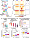
Fig. 1.
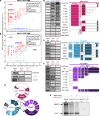
Fig. 1.. PAX7 triggers progressive and localized DNA demethylation.
(A) Current scheme for PAX7 pioneer action (6) indicating steps of enhancer chromatin opening (20). Step 2 is dependent on passage through replication and sensitive to mimosine blockade. (B) Scatter plots representing levels (%) of CpG dinucleotide methylation derived from WGBS performed on control AtT-20 (Neo) and PAX7-expressing AtT-20 (PAX7) cells. For representation, the 8450 CpGs present within the Pioneer enhancer subset (n = 876) were used together with subsets of the same number of randomly selected CpGs from the other types of enhancers centered on PAX7 peak summit. Data are shown for Constitutive, Pioneer, and Resistant enhancers, as defined in (9, 19). (C) Average profiles of normalized ATAC-seq data for Neo (blue) and PAX7 cells (red) at indicated enhancer subsets. (D) Violin plots showing CpG methylation (%) at Pioneer enhancers derived from ChIP-bis-seq performed on AtT-20 expressing ER-PAX7 induced with Tam for 30 min (′) or 9 days (d) and for 10 days followed by 20 days without Tam. P values from the t test are shown. ns, not significant. (E) Violin plots showing CpG methylation (%) at Pioneer enhancers derived from ChIP-bis-seq performed on AtT-20 expressing ER-PAX7 induced with Tam for the indicated times. The data include CpGs within a ±150-bp window around PAX7 peaks (sequencing coverage ≥10 reads per CpG in all samples) that showed ≥ 10% of absolute demethylation between 30 min and 9 days of Tam treatments. (F) Time course of relative PAX7 recruitment (FlagM2 ChIP-seq), chromatin opening (ATAC-seq), and DNA demethylation [sites from (D)]. Standard errors of the means are shown as black bars. h, hours. (G to I) Box plots of normalized signals at Pioneer enhancers for H3K4me1 ChIP-seq (G) (±250 bp), ATAC-seq (H) (±50 bp), and 5mC determined by WGBS (I) (±150 bp) performed on ER-PAX7 cells treated or not with Tam and/or mimosine (Mim), as indicated. P values were calculated by the t test.
Fig. 2.
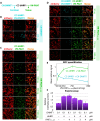
Fig. 2.. Identification of candidates for DNA demethylation.
(A) Scatter plot of protein enrichment determined by RIME (21) after immunoprecipitation using a Flag-M2 antibody in control Neo AtT-20 (blue) or PAX7 AtT-20 cells (red). (B) Same as (A), immunoprecipitation using a Flag-M2 antibody in AtT-20 cells expressing Flag-PAX7 (red) compared to TPIT immunoprecipitation in AtT-20 cells (green). (C) Pull-down assay of 35S-labeled UHRF1 and DNMT1 binding to MBP-PAX7. MBP-βGAL is used as a control. (D) UHRF1 and DNMT1 binding to PAX7 was assessed by pull-down assays using indicated MBP-PAX7 chimeras. WT, wild type. (E and F) PAX7 binding to indicated deletions of UHRF1 (E) and DNMT1 (F) was assessed in pull-down assays. (G) Summary of interactions between PAX7, UHRF1, and DNMT1. (H) Order-of-addition pull-down experiment performed with either MBP-PAX7 or MBP-βGal. 35S-labeled UHRF1 or DNMT1 was added first as indicated.
Fig. 3.
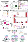
Fig. 3.. PAX7 binding to UHRF1 displaces UHRF1:DNMT1 interaction and prevents DNMT1 activation.
(A) Schema of fluorescence complementation between CC-UHRF1 and CN-DNMT1 or VN-PAX735–273. (B) BiFC between CC-UHRF1 and CN-DNMT1 (blue fluorescence) cotransfected with mCherry (red fluorescence) as a transfection control. (C) BiFC of CC-UHRF1 with increasing amounts (nanograms of plasmid per dish) of VN-PAX735–273 (green fluorescence), cotransfected with mCherry. (D) Bicolor BiFC between CC-UHRF1 and CN-DNMT1 (blue fluorescence) is competed by increasing concentrations of VN-PAX735–273 (green fluorescence). (E) Quantification of fluorescence signals (±SEM) in (D). (F) Relative DNA methylase activity of DNMT1 assessed with or without indicated concentrations of UHRF1, PAX7, or both.
Fig. 4.
Fig. 4.. Two mechanisms implement PAX7-driven DNA demethylation.
(A) Box plot of normalized signals at PAX7 Pioneer enhancers for 5hmC ChIP-seq (±50 bp) after 0- or 48-hour Tam activation with/without mimosine cell cycle arrest. (B) TET3 recruitment (ChIP-qPCR) at control and PAX7 Pioneer enhancers [from (20)] before/after Tam activation for 72 hours. (C) Time course of 5hmC enrichment assessed by ChIP-seq (peak ±50 bp) at Pioneer enhancers compared to PAX7 recruitment (ChIP-seq), chromatin gain of accessibility (ATAC-seq), and DNA demethylation (WGBS). (D) Modified CpG dinucleotides predicted to occur after DNA replication (left) and following oxidation by TET enzymes (right). (E) Methylation patterns determined by nanopore sequencing of unamplified genomic DNA duplexes at control CpGs that are not demethylated (<1% change of absolute DNA methylation between Neo and PAX7 cells at PAX7 peak summits ±250 bp). (F) Methylation patterns at CpGs within Pioneer sites (PAX7 summits ±250 bp) that show demethylation (≥10%) after PAX7 activation. Significant changes compared to Tam at 0 hours (P values ≤10−5) are shown in (E) and (F) by black lines above relevant bars. P values are provided in table S2. (G) Relative change of occurrence for indicated CpG methylation patterns [(F) above] compared to Tam at 0 hours. (H) Box plots showing CpG methylation (%) before and after PAX7 activation at Pioneer enhancers that are positive (black) or not (pink) for 5hmC in ChIP-Seq at any time point assessed in (C). (I) Model for PAX7-driven enhancer DNA demethylation. Blockade of the UHRF1-DNMT1 DNA methylation maintenance system will lead to progressive CpG demethylation through dilution at each replication cycle (left). A postreplication TET action will lead to partial CpG methylation maintenance by hydroxymethylation of hemimethylated CpGs (center) that are poor substrates for UHRF1 and DNMT1 (35, 59). The combined PAX7-dependent blockade of UHRF1 activation together with TET action would ensure efficient enhancer demethylation (right).

References

    1. Smith Z. D., Meissner A., DNA methylation: Roles in mammalian development. Nat. Rev. Genet. 14, 204–220 (2013). - PubMed
    1. Bird A. P., CpG-rich islands and the function of DNA methylation. Nature 321, 209–213 (1986). - PubMed
    1. Pacis A., Mailhot-Léonard F., Tailleux L., Randolph H. E., Yotova V., Dumaine A., Grenier J. C., Barreiro L. B., Gene activation precedes DNA demethylation in response to infection in human dendritic cells. Proc. Natl. Acad. Sci. U.S.A. 116, 6938–6943 (2019). - PMC - PubMed
    1. Jadhav U., Cavazza A., Banerjee K. K., Xie H., O’Neill N. K., Saenz-Vash V., Herbert Z., Madha S., Orkin S. H., Zhai H., Shivdasani R. A., Extensive recovery of embryonic enhancer and gene memory stored in hypomethylated enhancer DNA. Mol. Cell 74, 542–554.e5 (2019). - PMC - PubMed
    1. Orlanski S., Labi V., Reizel Y., Spiro A., Lichtenstein M., Levin-Klein R., Koralov S. B., Skversky Y., Rajewsky K., Cedar H., Bergman Y., Tissue-specific DNA demethylation is required for proper B-cell differentiation and function. Proc. Natl. Acad. Sci. U.S.A. 113, 5018–5023 (2016). - PMC - PubMed

MeSH terms

Substances

LinkOut - more resources