. 2025 May 16;11(20):eado3440.
doi: 10.1126/sciadv.ado3440. Epub 2025 May 16.

Ppp1r3b is a metabolic switch that shifts hepatic energy storage from lipid to glycogen

Affiliations

Ppp1r3b is a metabolic switch that shifts hepatic energy storage from lipid to glycogen

Kate Townsend Creasy et al. Sci Adv. .

Abstract

The PPP1R3B gene, encoding PPP1R3B protein, is critical for liver glycogen synthesis and maintaining blood glucose levels. Genetic variants affecting PPP1R3B expression are associated with several metabolic traits and liver disease, but the precise mechanisms are not fully understood. We studied the effects of both Ppp1r3b overexpression and deletion in mice and cell models and found that both changes in Ppp1r3b expression result in dysregulated metabolism and liver damage, with overexpression increasing liver glycogen stores, while deletion resulted in higher liver lipid accumulation. These patterns were confirmed in humans where variants increasing PPP1R3B expression had lower liver fat and decreased plasma lipids, whereas putative loss-of-function variants were associated with increased liver fat and elevated plasma lipids. These findings support that PPP1R3B is a crucial regulator of hepatic metabolism beyond glycogen synthesis and that genetic variants affecting PPP1R3B expression levels influence if hepatic energy is stored as glycogen or triglycerides.


Figures

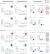
Fig. 1.
Fig. 1.. Mice with hepatocyte Ppp1r3b deletion develop metabolic dysfunction contributing to hepatic steatosis with age, while Ppp1r3b-overexpressing mice rapidly increase glycogen synthesis and storage contributing to liver damage with aging.
Metabolic phenotypes of 4-month-aged mice. (A to D) Four-hour–fasted metabolites in Ppp1r3bf/f (black, n = 9), Ppp1r3bΔhep (pink, n = 8), and Ppp1r3bhepOE (green, n = 8) mice. (A) Blood glucose. (B) Plasma insulin. (C) Blood lactate. (D) Blood ketones. (E to L) Characteristics of overnight (16 hours)–fasted Ppp1r3bf/f (black, n = 7), Ppp1r3bΔhep (pink, n = 6), and Ppp1r3bhepOE (green, n = 6) mice. (E) Liver-to-body weight ratio. (F) Hepatic glycogen content. (G) Hepatic TG content. (H) Fasted plasma ALT measurements. [(I) to (L)] Paraffin-embedded liver sections sectioned and stained with (I) hematoxylin and eosin (H&E), (J) PAS to detect polysaccharide content, (K) immunohistochemistry with PLIN2 antibodies to indicate lipid droplets, and (L) Picro-Sirius Red to detect collagen deposition and liver lesions. Group average presented as bar + SD with individual animals represented as shapes. n.s., not significant.
Fig. 2.
Fig. 2.. Ppp1r3bΔhep mice develop glucose intolerance and steatosis with aging or sucrose diet challenge, while Ppp1r3bOE mice have increased liver glycogen and reduced TG content.
(A to E) Metabolic tests performed on fasted 6- to 9-month-aged Ppp1r3bf/f (black, n = 6) and Ppp1r3bΔhep (pink, n = 8) mice maintained on chow diet. (A) Intraperitoneal (ip) GTT [area under the curve (AUC) inset]. (B) ITT, AUC inset. (C) Hepatic glycogen content. (D) hepatic TG content. (E) Oil Red O staining. (F to I) Metabolic tests performed on fasted 6- to 9-month-aged Ppp1r3bf/f (black, n = 8) and Ppp1r3bhepOE (green, n = 6) mice maintained on chow diet. (F) Intraperitoneal GTT, AUC inset. (G) ITT, AUC inset. (H) Hepatic glycogen content. (I) Hepatic TG content. (J to N) Metabolic tests performed on fasted Ppp1r3bf/f (black, n = 5) and Ppp1r3bΔhep (pink, n = 7) mice on HSD (65%) for 12 weeks. (J) Intraperitoneal GTT, AUC inset. (K) ITT, AUC inset. (L) Hepatic glycogen content. (M) hepatic TG content. (N) Oil Red O staining. (O to S) Metabolic tests performed on fasted Ppp1r3bf/f (black, n = 8) and Ppp1r3bhepOE (green, n = 8) on HSD for 12 weeks. (O) Intraperitoneal GTT, AUC inset. (P) ITT, AUC inset. (Q) Hepatic glycogen content. (R) Hepatic TG content. (S) Oil Red O staining. Group average presented as bar + SD with individual animals represented as shapes. n.d., none detected.
Fig. 3.
Fig. 3.. Changes in hepatocyte Ppp1r3b expression alter glucose and lipid metabolism.
Young (8 weeks old) mice characterized for metabolic changes in response to hepatic deletion or overexpression of Ppp1r3b. (A to D) Metabolic phenotypes of 8-week-old Ppp1r3bf/f (black, n = 6), Ppp1r3bΔhep (pink, n = 6), and Ppp1r3bhepOE (green, n = 6) mice. Four-hour–fasted blood (A) glucose, (B) insulin, (C) lactate, and (D) ketones. (E to I) Characteristics of overnight (16 hours) fasted Ppp1r3bf/f (black, n = 6), Ppp1r3bΔhep (pink, n = 6), and Ppp1r3bhepOE (green, n = 6) mice. (E) Intraperitoneal GTT, AUC inset. (F) Liver-to-body weight ratio. (G) Hepatic glycogen content. (H) Hepatic TG content. (I) Fasted plasma ALT measurements. Group average presented as bar + SD with individual animals represented as shapes.
Fig. 4.
Fig. 4.. Altered hepatocyte Ppp1r3b expression effects on glucose and lipid metabolism.
(A to F) Lipid accumulation pathways: (A) ad libitum–fed plasma NEFAs. (B) Plasma NEFAs after 4 hours of fasting (Ppp1r3bf/f (black, n = 9), Ppp1r3bΔhep (pink, n = 8), and Ppp1r3bhepOE (green, n = 8) mice. (C) Liver mRNA expression levels of lipid transporter Cd36 in Ppp1r3bf/f (black, n = 6), Ppp1r3bΔhep (pink, n = 6), and Ppp1r3bhepOE (green, n = 6) mice. (D) Oxidation of 14C-glucose measured in primary hepatocytes isolated from mice fasted overnight (16 hours). (E) Incorporation of 14C-glucose from media into cellular TG in primary hepatocytes. (F) In vivo DNL assessed by D2O incorporation into newly synthesized liver palmitate in Ppp1r3bf/f (black, n = 6), Ppp1r3bΔhep (pink, n = 6), and Ppp1r3bhepOE (green, n = 6) mice. (G to K) Lipid utilization pathways: (G) in vivo TG secretion as total plasma concentration and (H) rate of secretion in Ppp1r3bf/f (black, n = 5), Ppp1r3bΔhep (pink, n = 5), and Ppp1r3bhepOE (green, n = 6) mice. (I) Oxidation of 14C-oleic acid in primary hepatocytes isolated from mice fasted overnight (16 hours). (J) Incorporation of 14C-oleic acid from media into cellular TG in primary hepatocytes. (K) Seahorse metabolic substrate oxidation assay of primary hepatocytes isolated from fasted Ppp1r3bf/f (black, n = 3), Ppp1r3bΔhep (pink, n = 3), and Ppp1r3bhepOE (green, n = 3) mice, tested in either starvation media or supplemented with glucose, palmitate, or both. Oxygen consumption rate (OCR) normalized to microgram protein per well is graphed as an indication of substrate utilization for ATP production. Group average presented as bar + SD with individual animals represented as shapes.
Fig. 5.
Fig. 5.. Characteristics of LOC157273/PPP1R3B variant carriers corroborate mouse Ppp1r3bΔhep and Ppp1r3bhepOE phenotypes.
(A) Analysis of PMBB participants with whole-exome sequencing and CT-derived hepatic fat quantitation yielded a small cohort of individuals with heterozygous PPP1R3B pLOF mutations (n = 14) as well as carriers of common SNPs in LOC157273 (n = 41,754). Beta indicates the direction of effect (>0 indicates increased trait metric, <0 indicates decreased trait metric), SE, standard error; significant P values indicated by bold italic font. (B and C) Plasma metabolomics data from UK Biobank participants with LOC157273 rs4841132-A SNPs [(B) n = 17] and PPP1R3B pLOF variants [(C) n = 25]. Horizontal line indicates Bonferroni-corrected P value with significant metabolites above the line. Vertical line indicates direction of metabolite plasma concentration (>0 indicates increased metabolite; <0 indicates decreased metabolite). (D) Model of the effects of altered Ppp1r3b expression on liver metabolism. Left, with WT Ppp1r3b expression, postprandial glucose is mainly stored as hepatic glycogen with excess glucose stored as TG. In early fasting, glycogen is converted to free glucose to maintain blood glucose levels and meet extrahepatic energy demands, with lipid utilization and TG secretion as VLDL when glycogen is low. Middle, with deletion of hepatocyte Ppp1r3b expression, postprandial glycogen synthesis is impaired and exogenous glucose is shunted to lipogenesis and stored as hepatic TG. During fasting, stored TG is oxidized to produce ketones, and lipid is secreted as VLDL. Right, overexpression of hepatocyte Ppp1r3b permits enhanced glycogenesis with increased glycogen stores to maintain blood glucose during fasting.

References

    1. Younossi Z. M., Stepanova M., Ong J., Trimble G., AlQahtani S., Younossi I., Ahmed A., Racila A., Henry L., Nonalcoholic steatohepatitis is the most rapidly increasing indication for liver transplantation in the United States. Clin. Gastroenterol. Hepatol. 19, 580–589.e5 (2021). - PubMed
    1. Godoy-Matos A. F., Silva Júnior W. S., Valerio C. M., NAFLD as a continuum: From obesity to metabolic syndrome and diabetes. Diabetol. Metab. Syndr. 12, 60 (2020). - PMC - PubMed
    1. Sookoian S., Pirola C. J., Genetic predisposition in nonalcoholic fatty liver disease. Clin. Mol. Hepatol. 23, 1–12 (2017). - PMC - PubMed
    1. Manning A. K., Hivert M.-F., Scott R. A., Grimsby J. L., Bouatia-Naji N., Chen H., Rybin D., Liu C.-T., Bielak L. F., Prokopenko I., Amin N., Barnes D., Cadby G., Hottenga J.-J., Ingelsson E., Jackson A. U., Johnson T., Kanoni S., Ladenvall C., Lagou V., Lahti J., Lecoeur C., Liu Y., Martinez-Larrad M. T., Montasser M. E., Navarro P., Perry J. R. B., Rasmussen-Torvik L. J., Salo P., Sattar N., Shungin D., Strawbridge R. J., Tanaka T., van Duijn C. M., An P., de Andrade M., Andrews J. S., Aspelund T., Atalay M., Aulchenko Y., Balkau B., Bandinelli S., Beckmann J. S., Beilby J. P., Bellis C., Bergman R. N., Blangero J., Boban M., Boehnke M., Boerwinkle E., Bonnycastle L. L., Boomsma D. I., Borecki I. B., Böttcher Y., Bouchard C., Brunner E., Budimir D., Campbell H., Carlson O., Chines P. S., Clarke R., Collins F. S., Corbatón-Anchuelo A., Couper D., de Faire U., Dedoussis G. V., Deloukas P., Dimitriou M., Egan J. M., Eiriksdottir G., Erdos M. R., Eriksson J. G., Eury E., Ferrucci L., Ford I., Forouhi N. G., Fox C. S., Franzosi M. G., Franks P. W., Frayling T. M., Froguel P., Galan P., de Geus E., Gigante B., Glazer N. L., Goel A., Groop L., Gudnason V., Hallmans G., Hamsten A., Hansson O., Harris T. B., Hayward C., Heath S., Hercberg S., Hicks A. A., Hingorani A., Hofman A., Hui J., Hung J., Jarvelin M.-R., Jhun M. A., Johnson P. C. D., Wouter Jukema J., Jula A., Kao W. H., Kaprio J., Kardia S. L. R., Keinanen-Kiukaanniemi S., Kivimaki M., Kolcic I., Kovacs P., Kumari M., Kuusisto J., Kyvik K. O., Laakso M., Lakka T., Lannfelt L., Lathrop G. M., Launer L. J., Leander K., Li G., Lind L., Lindstrom J., Lobbens S., Loos R. J. F., Luan J., Lyssenko V., Mägi R., Magnusson P. K. E., Marmot M., Meneton P., Mohlke K. L., Mooser V., Morken M. A., Miljkovic I., Narisu N., O’Connell J., Ong K. K., Oostra B. A., Palmer L. J., Palotie A., Pankow J. S., Peden J. F., Pedersen N. L., Pehlic M., Peltonen L., Penninx B., Pericic M., Perola M., Perusse L., Peyser P. A., Polasek O., Pramstaller P. P., Province M. A., Räikkönen K., Rauramaa R., Rehnberg E., Rice K., Rotter J. I., Rudan I., Ruokonen A., Saaristo T., Sabater-Lleal M., Salomaa V., Savage D. B., Saxena R., Schwarz P., Seedorf U., Sennblad B., Serrano-Rios M., Shuldiner A. R., Sijbrands E. J. G., Siscovick D. S., Smit J. H., Small K. S., Smith N. L., Smith A. V., Stančáková A., Stirrups K., Stumvoll M., Sun Y. V., Swift A. J., Tönjes A., Tuomilehto J., Trompet S., Uitterlinden A. G., Uusitupa M., Vikström M., Vitart V., Vohl M.-C., Voight B. F., Vollenweider P., Waeber G., Waterworth D. M., Watkins H., Wheeler E., Widen E., Wild S. H., Willems S. M., Willemsen G., Wilson J. F., Witteman J. C. M., Wright A. F., Yaghootkar H., Zelenika D., Zemunik T., Zgaga L., DIAbetes Genetics Replication And Meta-analysis (DIAGRAM) Consortium, The Multiple Tissue Human Expression Resource (MUTHER) Consortium, Wareham N. J., McCarthy M. I., Barroso I., Watanabe R. M., Florez J. C., Dupuis J., Meigs J. B., Langenberg C., A genome-wide approach accounting for body mass index identifies genetic variants influencing fasting glycemic traits and insulin resistance. Nat. Genet. 44, 659–669 (2012). - PMC - PubMed
    1. Tin A., Balakrishnan P., Beaty T. H., Boerwinkle E., Hoogeveen R. C., Young J. H., Kao W. H. L., GCKR and PPP1R3B identified as genome-wide significant loci for plasma lactate: The Atherosclerosis Risk in Communities (ARIC) study. Diabet. Med. 33, 968–975 (2016). - PMC - PubMed

LinkOut - more resources