. 2025 Jul;24(7):100993.
doi: 10.1016/j.mcpro.2025.100993. Epub 2025 May 14.

Native Top-Down Analysis of Membrane Protein Complexes Directly From In Vitro and Native Membranes

Affiliations

Native Top-Down Analysis of Membrane Protein Complexes Directly From In Vitro and Native Membranes

Wonhyeuk Jung et al. Mol Cell Proteomics. 2025 Jul.

Abstract

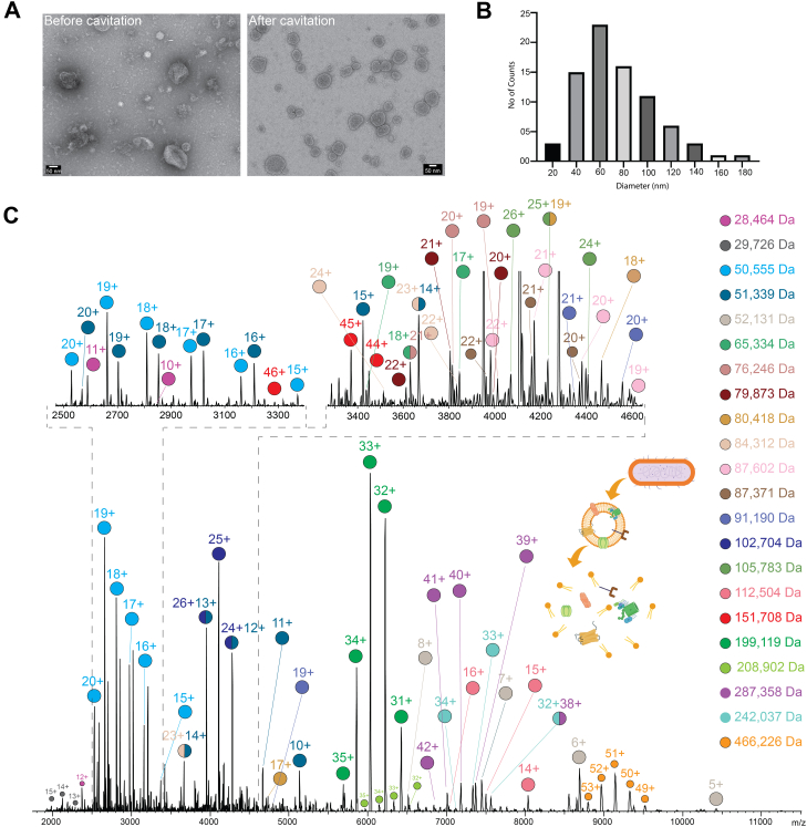
Macromolecular organization of proteins and lipids in cellular membranes is fundamental to cell functionality. Recent advances in native mass spectrometry (nMS) have established it as a key analytical tool for capturing these associations. This typically necessitates the extraction of target membrane proteins (MPs) from their physiological environments into detergent-like surroundings. In our recent studies using in vitro synthetic liposomes, we discovered that gas phase supercharging can selectively destabilize lipid bilayers and enable MS1 detection of embedded and associated protein-lipid complexes. Here, we further extend and apply this methodology to native cell-derived membrane vesicles. We demonstrate our ability to detect and ID protein complexes and their proteoforms directly from native membranes using supercharger-assisted prequadrupole activation followed by downstream native top-down tandem mass spectrometry, which combines both collision-based and electron capture-based fragmentation approaches. We first demonstrated this approach through native top-down identification of several integral MPs from in vitro membranes. Subsequently, we developed a protocol to produce nMS-ready native membrane vesicles. Applying to Escherichia coli total membranes, we generated nMS-ready vesicles and identified both integral and membrane-associated protein complexes of homomeric and heteromeric nature using our supercharging-enabled native top-down platform. For the heteropentameric β-barrel-assembly machinery (BAM) complex, which includes the integral MP BAM-A, we detected several lipidated proteoforms. For peripheral homodimeric dihydrolipoyl dehydrogenase, we identified bound endogenous metabolite cofactors. Furthermore, using BAM complex, a crucial antibiotic target, we show how this platform could be utilized to study drug binding to MPs directly from their native membranes.

Keywords: drug binding; electron capture fragmentation; membrane protein complex; native top–down mass spectrometry; proteoform analysis.


Conflict of interest statement

Conflict of interest The authors declare no competing interests.

Figures

None
Graphical abstract
Fig. 1
Fig. 1
Native top–down mass spectrometry (nTD-MS) analysis of membrane proteins (MPs) from in vitro liposomes.A, schematic representation of MP analysis from membrane vesicle via nMS. MPs are ablated out of the lipid bilayer by utilizing the front-end activation and are directly detected (1). This is facilitated by supercharging agents such as glycerol, which increases the amount of charge deposited onto the vesicle during the ESI process. The liberated MP complexes are quadrupole isolated for downstream native complex-up (nCU) experiment, where the complex is dissociated into its components (2). Alternatively, the isolated species can be subjected to nTD-MS analysis in which they undergo fragmentation by CID or EChcD (3). Proteoliposomes formed by incorporating VAMP2 (B), semiSWEET (C), and AqpZ (D) were subjected to nTD analysis. The physiological oligomeric states of the respective proteins (VAMP2 [monomer], semiSWEET [dimer], and AqpZ [tetramer]) were quadrupole isolated and fragmented with EChcD approach, and a representative section of the corresponding spectra is shown. The MS2 data were searched against the modified database that includes the total Escherichia coli proteome supplemented with VAMP2, semiSWEET, and AqpZ sequences. The respective P scores are displayed here along with the sequence coverage. The b/y and c/z fragments are highlighted in green and red, respectively. The experimental and theoretical masses of the proteins, in their physiological oligomeric states, are denoted as Exp and Th. Mass, respectively. The HCD voltage and the status of the ECD cell are stated in each MS/MS spectrum. CID, collision-induced dissociation; ECD, electron-capture dissociation; EChcD, electron capture higher-energy collisional dissociation; ESI, electrospray ionization; HCD, higher-energy collisional dissociation; nMS, native mass spectrometry.
Fig. 2
Fig. 2
Proteins ejected out of membrane vesicles analyzed via nMS.A, EM picture of Escherichia coli membrane vesicles before and after N2 cavitation. B, nMS analysis of the cavitated vesicles yields a variety of endogenous proteins/protein complexes ranging from 28 to 466 kDa. nMS, native mass spectrometry. C, the size distribution of the vesicles was measured by manual counting. The vesicles are relatively homogeneous, with 68% of the vesicle’s diameter ranging from 40 to 80 nm, confirming that cavitation generates small vesicles that are homogeneous in size.
Fig. 3
Fig. 3
nTD-MS and complex-up analysis of an endogenous protein from Escherichia coli membrane vesicle.A, the 25+ charge state of the 102 kDa species was isolated via quadrupole and then subjected to nTD-MS analysis with EChcD approach. A representative part of the fragmentation spectra is shown here, and the full annotation is available in Figure 4. B, the MS2 data from (A) were searched against the E. coli membrane proteome database using ProSight Native. As shown, this led to an unambiguous ID of the protein as DLDH with a very confident P score, E value, and >20% sequence coverage. The annotated terminal ion types are marked on the sequence. C, the dimeric endogenous oligomeric state of the protein was further confirmed by subjecting the same precursor to a complex-up analysis by CID. Data show clear ejection of a high charge state unfolded monomers and a low charge state folded monomer, with the endogenous cofactor (FAD, represented in yellow) still bound. The experimental and theoretical masses of the FAD-bound dimers are denoted as Exp and Th. Mass, respectively. The HCD voltage and the status of the ECD cell are stated in each MS/MS spectrum. FAD, flavin adenine dinucleotide. CID, collision-induced dissociation; DLDH, dihydrolipoyl dehydrogenase; ECD, electron-capture dissociation; EChcD, electron capture higher-energy collisional dissociation; HCD, higher-energy collisional dissociation; nTD-MS, native top–down mass spectrometry.
Fig. 4
Fig. 4
Annotated nTD-MS data of dimeric DLDH. 25+ charge state of the dimeric DLDH is quadrupole isolated, subjected to EChcD fragmentation. Sections of annotated EChcD nTD-MS data are shown. Only terminal ions are annotated. Three different neutral losses, NH3 (denoted with #), CONH2 (denoted with ##), and H2O (denoted as ∗), were included. Overall, the data yield 21% sequence coverage. The HCD voltage and the status of the ECD cell are stated in each MS/MS spectrum. DLDH, dihydrolipoyl dehydrogenase; ECD, electron-capture dissociation; EChcD, electron capture higher-energy collisional dissociation; HCD, higher-energy collisional dissociation; nTD-MS, native top–down mass spectrometry.
Fig. 5
Fig. 5
nMS analysis of BAM complex and its ligand from Escherichia coli membrane vesicle.A, complex-up analysis of the 34+ charge state of the 199 kDa protein complex yields the subcomplex BAM–ABCD and ejected high-charged unfolded BAM E and subcomplex BAM ABCE through the ejection of monomeric BAM-D. B, deconvoluted high-resolution nMS spectra of the BAM complex show the presence of different protocorms arising through the S-acylation of BAM subunits. BAM B/C/D/E are all peripheral membrane proteins, where S-acylation has been reported as a mechanism for membrane anchoring. The mass additions observed match that of the most common fatty-acyl lipid chain in E. coli that is used for S-acylation. C, MS1 E. coli membrane vesicles treated with MRL494 (bottom panel). As a control, the top panel shows the same m/z region of the spectra of native vesicles, before the addition of MRL494. The spectra are zoomed in to focus on the BAM complex charge state regions. As seen, clear binding of the BAM complex and its inhibitor MRL494 (represented in red) is observed. D, to further confirm the binding, MRL494 bound a 28+ charge state of BAM complex, which was isolated and subjected to CID activation. This leads to the ejection of MRL494 from the complex with or without carrying a charge, generating both 27+ and 28+ apo BAM complexes. The difference calculated from the precursor to any of the apo-peaks again corresponds to the mass of MRL494. E, the MS2 data assignment for bamE for annotated terminal ion types is marked on the sequence, along with the P score. The HCD voltage and the status of the ECD cell are stated in each MS/MS spectrum. BAM, β-barrel–assembly machinery; CID, collision-induced dissociation; ECD, electron-capture dissociation; HCD, higher-energy collisional dissociation; nMS, native mass spectrometry.

Similar articles

References

    1. Sezgin E., Levental I., Mayor S., Eggeling C. The mystery of membrane organization: composition, regulation and roles of lipid rafts. Nat. Rev. Mol. Cell Biol. 2017;18:361–374. - PMC - PubMed
    1. Andersen O.S., Koeppe R.E. Bilayer thickness and membrane protein function: an energetic perspective. Annu. Rev. Biophys. Biomol. Struct. 2007;36:107–130. - PubMed
    1. Chadda R., Bernhardt N., Kelley E.G., Teixeira S.C., Griffith K., Gil-Ley A., et al. Membrane transporter dimerization driven by differential lipid solvation energetics of dissociated and associated states. eLife. 2021;10:e63288. - PMC - PubMed
    1. Levental I., Lyman E. Regulation of membrane protein structure and function by their lipid nano-environment. Nat. Rev. Mol. Cell Biol. 2023;24:107–122. - PMC - PubMed
    1. Fu Z., MacKinnon R. Structure of the flotillin complex in a native membrane environment. Proc. Natl. Acad. Sci. U. S. A. 2024;121 - PMC - PubMed

MeSH terms

LinkOut - more resources