. 2025 Jun;301(6):110236.
doi: 10.1016/j.jbc.2025.110236. Epub 2025 May 14.

ARV1 is a component of the enzyme initiating glycosylphosphatidylinositol biosynthesis

Affiliations

ARV1 is a component of the enzyme initiating glycosylphosphatidylinositol biosynthesis

TianTian Lu et al. J Biol Chem. 2025 Jun.

Abstract

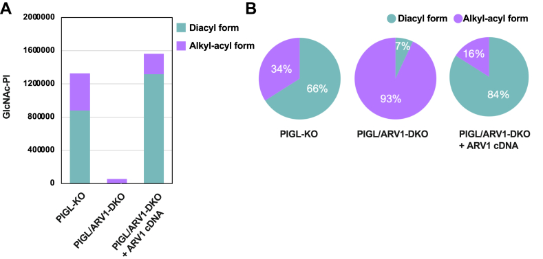
Glycosylphosphatidylinositol (GPI) serves as a membrane anchor of numerous cell surface proteins. It is synthesized in the endoplasmic reticulum from phosphatidylinositol (PI) by stepwise reactions and transferred to the C terminus of the protein. Defects in genes involved in GPI biosynthesis affect the expression of GPI-anchored proteins or their structure, causing the neurological disorder, inherited GPI deficiency. Individuals with ARV1 deficiency have symptoms resembling inherited GPI deficiency, but how ARV1 regulates GPI biosynthesis is poorly understood. Here, we show that ARV1 acts as a component of the enzyme initiating GPI biosynthesis, GPI N-acetylglucosaminyltransferase (GPI-GnT) complex, which forms a ring structure as predicted by AlphaFold3. ARV1 associates with PIGQ, a GPI-GnT component, and ARV1 mutants defective in this association lose their ability to enhance GPI-GnT activity, showing that association with PIGQ is critical for ARV1's function. ARV1-containing GPI-GnT used PI more efficiently than ARV1-less GPI-GnT in an in vitro enzyme assay. Collectively, our results suggest that ARV1 facilitates efficient recruitment of PI to GPI-GnT, thereby playing a critical role in the regulation of GPI-anchored protein expression.

Keywords: AlphaFold; GPI N-acetylglucosaminyltransferase complex; inherited GPI deficiency; lipidomics; phosphatidylinositol.


Conflict of interest statement

Conflicts of interest The authors declare that they have no conflicts of interest with the contents of this article.

Figures

Figure 1
Figure 1
ARV1-deficient human cells exhibit decreased expression of GPI-APs. A, flow cytometry (FACS) analysis of human fibroblasts. WT (gray), ARV1-KO (blue), and ARV1-rescued (purple) or vector-transfected (orange dotted) ARV1-KO cells were stained for GPI-APs. Mean fluorescence intensity (MFI) of CD59, CD73, and CD109 expressed on ARV1-KO fibroblasts decreased to 80%, 25%, and 18% of WT levels, respectively. Gray dotted lines: isotype control staining. B, top panel. FACS analysis of HEK293 cells. FLAER staining and CD59 and DAF levels on ARV1-KO HEK293 cells (blue) were not decreased from those on WT HEK293 cells (gray). B, three bottom panels. FACS analysis of PIGAR119WKI, PIGOR119WKI, and PIGTT183PKI HEK293 cell lines with further KO of ARV1. Expression of GPI-APs was decreased only in PIGAR119WKI cells by further KO of ARV1. C, FACS analysis of PIGAR119WKI/ARV1-KO HEK293 cells, stably expressing FLAG-ARV1 by the tet-On system, showed partial rescue of CD59 and DAF expression. D, Western blotting analysis of FLAG-ARV1 (34 kDa). GAPDH: a loading control. Each FACS analysis was performed at least two times.
Figure 2
Figure 2
ARV1 directly associates only with PIGQ in GPI–GnT complex. A, Upper: Co-IP assay (IP with HA) using PIGQ-KO HEK293 cells. PIGQ-KO cells with (+) or without (−) rescue of GST-PIGQ were transiently transfected with HA-ARV1 and other FLAG-tagged subunits of GPI-GnT and PIGN, as a negative control. Left two panels (Input): Expression of transfected proteins. Right three panels (IP: HA beads): ARV1-bound GPI–GnT complex only through GST-PIGQ. Lower: Schematic of predicted GPI–GnT complex in PIGQ-KO cells. B, Upper: Co-pull-down assay (pull-down with GST) using PIGA-KO HEK293 cells. PIGA-KO cells with (+) or without (−) rescue of FLAG-PIGA were transiently transfected with HA-ARV1, GST-PIGQ, and other FLAG-tagged subunits and PIGN. Left two panels (Input): Expression of transfected proteins. Right three panels (pull-down: glutathione-Sepharose): PIGQ bound to ARV1 and PIGH in the absence of PIGA. Lower: Schematic of predicted GPI–GnT complex in PIGA-KO cells. For (A and B), experiments were performed at least three times.
Figure 3
Figure 3
ARV1 mutants showing reduced association with PIGQ have decreased GPI–GnT enzyme activity.A, a model of human ARV1 and PIGQ complex predicted by AlphaFold2. Candidate amino acids for protein association are shown with their structures. ARV1, orange; PIGQ, khaki; oxygen atom, red; nitrogen atom, blue; hydrogen bond, yellow dotted line. B, domain structure of ARV1 protein. Candidate amino acids for ARV1-PIGQ association are marked. C, the relative association between HA-ARV1 mutants and FLAG-PIGQ calculated from the results of co-IP assay (pull-down with FLAG) (Fig. S2). Band intensities in Western blotting were quantified using ImageJ (version 2.3.0/1.53q). The relative amounts of HA-ARV1 mutants were normalized by FLAG-PIGQ amounts. D and E, relative expression level of GPI-APs in ARV1-rescued cells. HA-ARV1 mutants were transiently expressed in PIGAR119WKI/ARV1-KO HEK293 cells, and the expression levels of CD59 (D) and DAF (E) were analyzed by FACS (Fig. S2). MFI was quantified using FlowJo 10.9.0. F, correlation of GPI-AP expression levels in ARV1 mutant rescued cells and relative association between ARV1 mutants and PIGQ. Pearson r = 0.91 for association versus CD59 expression (p value, 0.0002), Pearson r = 0.87 for association versus DAF expression (p value, 0.001). For (CE), experiments were performed at least three times, analyzed by one sample t test and the error bars are SD. TMD, transmembrane domain.
Figure 4
Figure 4
ARV1-containing GPI-GnT showed stronger activity than ARV1-less-GPI-GnT in in vitro enzyme assays.A, Western blotting of PIGA/ARV1-DKO HEK293 cells stably expressing GST-PIGA (top panel) with or without tet-On system-controlled expression of FLAG-ARV1 (bottom panel). B, Left: High-performance thin-layer chromatography (HPTLC) of butanol-extracted radiolabeled products from enzyme assay using cell lysates. Products from JY25 cells (human WT B-lymphoblasts) and PIGL-KO Chinese hamster ovary (CHO) cells were used as standards for GlcNAc-PI and GlcN-PI (50). Developing solvent, chloroform/methanol/1 M NH4OH = 10:10:3 (v:v:v) (9). Right: Amounts of radioactive products (GlcNAc-PI + GlcN-PI intensities, normalized by phosphatidylcholine levels detected by molybdenum blue reagent). p value, 0.0045 by one sample t test. C, Left: HPTLC of products from enzyme assay using purified ARV1-containing (left half) and ARV1-less (right half) GPI–GnT complexes with PI at three different concentrations. Right: Quantification of GlcNAc-PI intensities. p value, 0.00002 with 0 μM PI; 0.003 for 10 μM PI; 0.01 for 100 μM PI by Student's t test. For (B and C), assays were performed at least three times. The error bars are SD.
Figure 5
Figure 5
Fewer GPI intermediates are produced without ARV1.A, total amounts of GlcNAc-PI with alkyl/acyl PIs (purple) or diacyl PIs (indigo) in PIGL-KO HEK293 cells and PIGL/ARV1-DKO HEK293 cells without or with ARV1 rescue. B, pie charts showing the percentage of two forms of PIs based on (A). Indigo, diacyl form of GlcNAc-PI; purple, alkyl-acyl form of GlcNAc-PI. (A and B) show representative data of two repeated lipidomic analyses.
Figure 6
Figure 6
Structure of GPI-GnT complex predicted by Alphafold3.A, side view (left) and top view (middle) of the full complex, and top view of an ARV1-less complex (right). Location of the ER membrane with cytosolic and luminal orientation are indicated in the side view. Cyan, PIGA; yellow, PIGH; green, PIGQ; magenta, PIGC; orange, PIGP; red, PIGY; blue, ARV1; and purple, DPM2. B, the orientation of PIGA within the GPI–GnT complex with (cyan) or without (pink) PIGY (red) and PIGP (orange). C, the overlaid structures of MshA (green) and PIGA (cyan). Substrate-binding region is expanded with relevant amino acid side chains of PIGA (top) and MshA (bottom) indicated. Conserved amino acids (AA) involved in substrate binding are aligned on the right. Ino-P, inositol-phosphate.

References

    1. Komath S.S., Fujita M., Hart G.W., Ferguson M.A.J., Kinoshita T. In: Glycosylphosphatidylinositol Anchors in Essentials of Glycobiology. 4th Ed. Varki A., Cummings R.D., Esko J.D., Stanley P., Hart G.W., Aebi M., et al., editors. Cold Spring Harbor; Cold Spring Harbor, NY: 2022. pp. 141–154.
    1. Kinoshita T. Biosynthesis and biology of mammalian GPI-anchored proteins. Open Biol. 2020;10 doi: 10.1098/rsob.190290. - DOI - PMC - PubMed
    1. Wang Y., Hirata T., Maeda Y., Murakami Y., Fujita M., Kinoshita T. Free, unlinked glycosylphosphatidylinositols on mammalian cell surfaces revisited. J. Biol. Chem. 2019;294:5038–5049. - PMC - PubMed
    1. Duval R., Nicolas G., Willemetz A., Murakami Y., Mikdar M., Vrignaud C., et al. Inherited glycosylphosphatidylinositol defects cause the rare Emm-negative blood phenotype and developmental disorders. Blood. 2021;137:3660–3669. - PubMed
    1. Miyata T., Takeda J., Iida Y., Yamada N., Inoue N., Takahashi M., et al. The cloning of PIG-A, a component in the early step of GPI-anchor biosynthesis. Science. 1993;259:1318–1320. - PubMed

MeSH terms

Substances

LinkOut - more resources