Review
. 2025 Jul;301(7):110230.
doi: 10.1016/j.jbc.2025.110230. Epub 2025 May 14.

NDRG1 and its family members: More than just metastasis suppressor proteins and targets of thiosemicarbazones

Affiliations
Review

NDRG1 and its family members: More than just metastasis suppressor proteins and targets of thiosemicarbazones

Mahan Gholam Azad et al. J Biol Chem. 2025 Jul.

Abstract

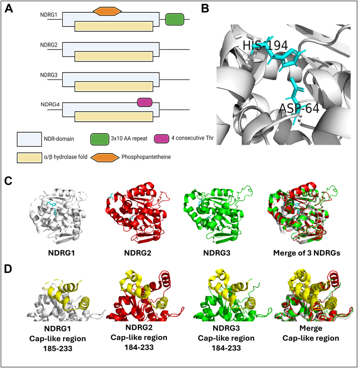
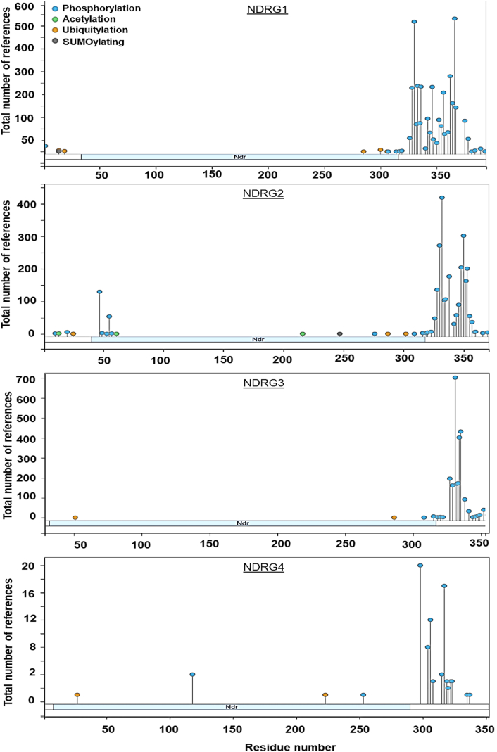
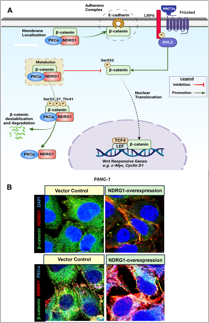
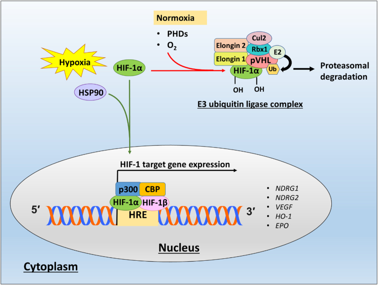
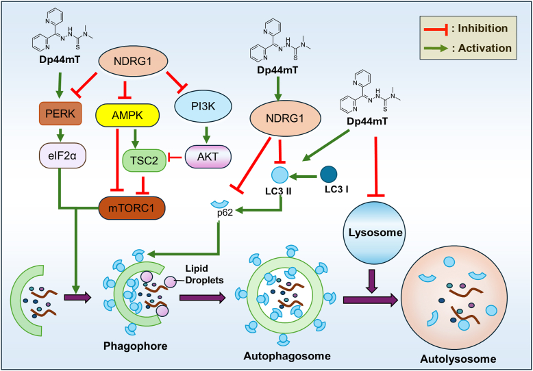
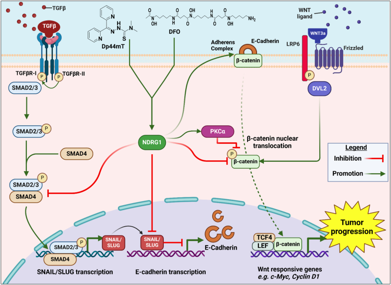
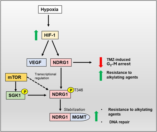
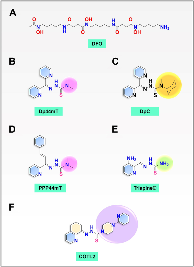
N-Myc downstream regulated gene-1 (NDRG1) and the other three members of this family (NDRG2, 3, and 4) play various functional roles in the cellular stress response, differentiation, migration, and development. These proteins are involved in regulating key signaling proteins and pathways that are often dysregulated in cancer, such as EGFR, PI3K/AKT, c-Met, and the Wnt pathway. NDRG1 is the primary, well-examined member of the NDRG family and is generally characterized as a metastasis suppressor that inhibits the first step in metastasis, the epithelial-mesenchymal transition. While NDRG1 is well-studied, emerging evidence suggests NDRG2, NDRG3, and NDRG4 also play significant roles in modulating oncogenic signaling and cellular homeostasis. NDRG family members are regulated by multiple mechanisms, including transcriptional control by hypoxia-inducible factors, p53, and Myc, as well as post-translational modifications such as phosphorylation, ubiquitination, and acetylation. Pharmacological targeting of the NDRG family is a therapeutic strategy against cancer. For instance, di-2-pyridylketone 4,4-dimethyl-3-thiosemicarbazone (Dp44mT) and di-2-pyridylketone-4-cyclohexyl-4-methyl-3-thiosemicarbazone (DpC) have been extensively shown to upregulate NDRG1 expression, leading to metastasis suppression and inhibition of tumor growth in multiple cancer models. Similarly, targeting NDRG2 demonstrates its pro-apoptotic and anti-proliferative effects, particularly in glioblastoma and colorectal cancer. This review provides a comprehensive analysis of the structural features, regulatory mechanisms, and biological functions of the NDRG family and their roles in cancer and neurodegenerative diseases. Additionally, NDRG1-4 are explored as therapeutic targets in oncology, focusing on recent advances in anti-cancer agents that induce the expression of these proteins. Implications for future research and clinical applications are also discussed.

Keywords: NDRG1; metastasis; metastasis suppression; thiosemicarbazone.


Conflict of interest statement

Conflict of interest The authors declare that they have no conflicts of interest with the contents of this article.

Figures

Figure 1
Figure 1
Structure of NDRG family members.A, schematic of the overall structure of the NDRG1, 2, 3, and 4 proteins and the domains they contain (31, 59). The domains are: the NDR domain is a conserved region found within NDRG proteins, specifically consisting of a central α/β hydrolase-like region; a 3 × 10 amino acid (AA) domain (GTRSRSHTSE); a four consecutive threonine (Thr) domain; an α/β hydrolase domain; and a phosphopantetheine attachment binding domain (phosphopantetheine). Schematic modified from references (31, 59). B, the unique Asp64 and His194 catalytic site as part of the predicted α/β hydrolase region is only observed in NDRG1 and not other NDRG1 family members (59). C, the overall structure of NDRG1-3 demonstrates their similarity. D, comparison of the conserved Cap-like region in the NDRG1-3 proteins. B–D, were prepared using PyMOL (Schrödinger, LLC).
Figure 2
Figure 2
Phosphoproteomics of NDRG family proteins using PhosphoSitePlus. Bioinformatics tool, PhosphoSitePlus provides comprehensive data on experimentally verified phosphorylation sites and other post-translational modification from mass spectrometry data and curated literature (93).
Figure 3
Figure 3
NDRG1’s novel role in the regulation of β-catenin via the formation of a regulatory metabolon between NDRG1, PKCα, and β-catenin in pancreatic cancer cells.A, schematic demonstrating the novel WNT/β-catenin regulatory mechanism employed by NDRG1 to decrease nuclear oncogenic β-catenin levels via PKCα. This kinase activity of PKCα results in the destabilizing phosphorylation of β-catenin at Ser33, 37, and 41, leading to β-catenin down-regulation (123) via proteasomal degradation. This effect prevents the nuclear translocation of β-catenin. Instead, some β-catenin translocates to the plasma membrane after NDRG1 overexpression and is co-localized with both NDRG1 and PKCα. The increased β-catenin levels at the plasma membrane aid adherens complex formation composed of β-catenin and E-cadherin that prevents the epithelial-mesenchymal transition (209, 244). B, confocal microscopy analysis of PANC-1 vector control and NDRG1-overexpressing pancreatic cancer cells demonstrating co-localization (yellow) between NDRG1 (red) and β-catenin (green). This observation suggests association between NDRG1 and β-catenin mostly localized at the plasma membrane. Similarly, immunostaining of NDRG1 (red), PKCα (blue), and β-catenin (green), demonstrates triple-colocalization (white). The scale bar is 3 μm. This finding, together with co-immunoprecipitation evidence, suggests a ternary complex of three proteins, potentially indicating the formation of a metabolon localized predominantly in the cytoplasm and plasma membrane. A, was created in BioRender (Azad, M. (2025) https://BioRender.com/d18u703). B, is from the authors publication (123) under the terms of the Creative Commons CC-BY license.
Figure 4
Figure 4
The hypoxia-inducible factor-1α (HIF-1α) mediated transcriptional regulation of NDRG1 expression. Under normoxia, the transcription factors, HIF-1α and HIF-2α, are ubiquitinated and degraded via the proteasomal pathway. Prolyl hydroxylase (PHD) enzymes require sufficient oxygen levels to hydroxylate prolyl residues within HIF-1α and HIF-2α, which can then be recognized by the von Hippel-Lindau protein (pVHL), a E3 ubiquitin ligase complex that ubiquitinates HIFs. Under conditions of hypoxia, the low oxygen levels suppress PHD enzyme activity leading to increased protein levels of the HIFs. HSP90 stabilizes HIF-1α and prevents pVHL-dependent proteasomal degradation. The binding of HSP90 also changes the conformation of HIF-1α so that it can recruit the cofactor p300. The interaction of HIF-1α with p300 and CBP promotes the activation of hypoxia-response elements (HREs) within the promoter and enhancer regions of target genes such as NDRG1.
Figure 5
Figure 5
The role of NDRG1 on the regulation of autophagy. NDRG1 suppresses autophagy at both the initiation and degradation stages. It suppresses autophagic initiation by suppression of the PERK/eIF2 and PI3K/AKT pathways, as well as via AMPK (83, 181, 193, 300). The pharmacological NDRG1-inducing agent, Dp44mT, up-regulates autophagic initiation leading to increased LC3II levels via the PERK/eIF2α axis (180). NDRG1 expression also up-regulates p62 levels, suggesting its suppressive role in autophagic degradation (181). However, Dp44mT promotes lysosomal membrane permeabilization due to the generation of reactive oxygen species after the formation of redox-active iron and copper complexes (185, 186). The effect of Dp44mT on inducing initiation while damaging lysosomes results in dysfunctional autophagy. Similarly, NDRG1 may suppress lipophagy via downregulating LC3II while increasing p62 expression (191).
Figure 6
Figure 6
Schematic demonstrating the role of NDRG1 in inhibiting the epithelial-mesenchymal transition and tumor progression via regulation of the TGF-β/SMAD and WNT pathways. Iron-binding agents such as Dp44mT and DFO up-regulate the metastasis suppressor, NDRG1, to inhibit the TGF-β/SMAD pathway, which initiates the epithelial–mesenchymal transition (11). Increased NDRG1 levels result in decreased expression of SMAD2 and p-SMAD3 levels, preventing the association with SMAD4, which inhibits nuclear translocation. This decreased nuclear translocation of SMAD2, p-SMAD3, and SMAD4 decreases the TGF-β-induced expression of the transcriptional repressors, SNAIL and SLUG, which are responsible for E-cadherin transcriptional repression (11). Increased NDRG1 expression increases plasma membrane E-cadherin and β-catenin levels, that are involved in adherens complex formation at the plasma membrane, which suppress the initial step in metastasis mediated by the epithelial-mesenchymal transition (11). NDRG1 expression also negatively-regulates total β-catenin levels via PKCα (123). These events prevent β-catenin nuclear translocation, thus inhibiting the activation of the WNT signaling pathway and the transcription of cyclin D1, which plays an important role in cell cycle progression (122). Created in BioRender (Azad, M. (2025) https://BioRender.com/d74s214).
Figure 7
Figure 7
Hypoxia-induced NDRG1-mediated resistance to alkylating chemotherapies such as Temozolomide (TMZ). Under hypoxic conditions, increased HIF1α expression is induced that has been associated with resistance to alkylating agents. HIF-1 transcriptionally upregulates the expression of genes with hypoxia-response elements (HREs) including vascular endothelial growth factor (VEGF) and NDRG1. It has been demonstrated that hypoxia-induced NDRG1 expression results in a decrease in Temozolomide (TMZ)-induced G2/M arrest and resistance to TMZ (252). NDRG1 is also up-regulated by the mTOR downstream effector, SGK1, which phosphorylates and activates NDRG1 at Tyr 346 (301). SGK1 has also been shown to promote the direct association and stabilization of NDRG1 with MGMT, resulting in elevated DNA repair and resistance to TMZ (252).
Figure 8
Figure 8
The line drawings of the chemical structures of the ligands.A, Desferrioxamine (DFO); B, Dp44mT; C, DpC; D, PPP44mT; E, Triapine; and F, COTI-2.

Similar articles

References

    1. Steeg P.S. Tumor metastasis: mechanistic insights and clinical challenges. Nat. Med. 2006;12:895–904. - PubMed
    1. Anderson R.L., Balasas T., Callaghan J., Coombes R.C., Evans J., Hall J.A., et al. A framework for the development of effective anti-metastatic agents. Nat. Rev. Clin. Oncol. 2019;16:185–204. - PMC - PubMed
    1. Gandalovicova A., Rosel D., Fernandes M., Vesely P., Heneberg P., Cermak V., et al. Migrastatics-anti-metastatic and anti-invasion drugs: promises and challenges. Trends Cancer. 2017;3:391–406. - PMC - PubMed
    1. Bandyopadhyay S., Pai S.K., Gross S.C., Hirota S., Hosobe S., Miura K., et al. The Drg-1 gene suppresses tumor metastasis in prostate cancer. Cancer Res. 2003;63:1731–1736. - PubMed
    1. Guan R.J., Ford H.L., Fu Y., Li Y., Shaw L.M., Pardee A.B. Drg-1 as a differentiation-related, putative metastatic suppressor gene in human colon cancer. Cancer Res. 2000;60:749–755. - PubMed

MeSH terms

Substances