. 2025 Jun 15:200:629-640.
doi: 10.1016/j.actbio.2025.05.036. Epub 2025 May 14.

Enhanced delivery of camptothecin to colorectal carcinoma using a tumor-penetrating peptide targeting p32

Affiliations

Enhanced delivery of camptothecin to colorectal carcinoma using a tumor-penetrating peptide targeting p32

Yanhao Jiang et al. Acta Biomater. .

Abstract

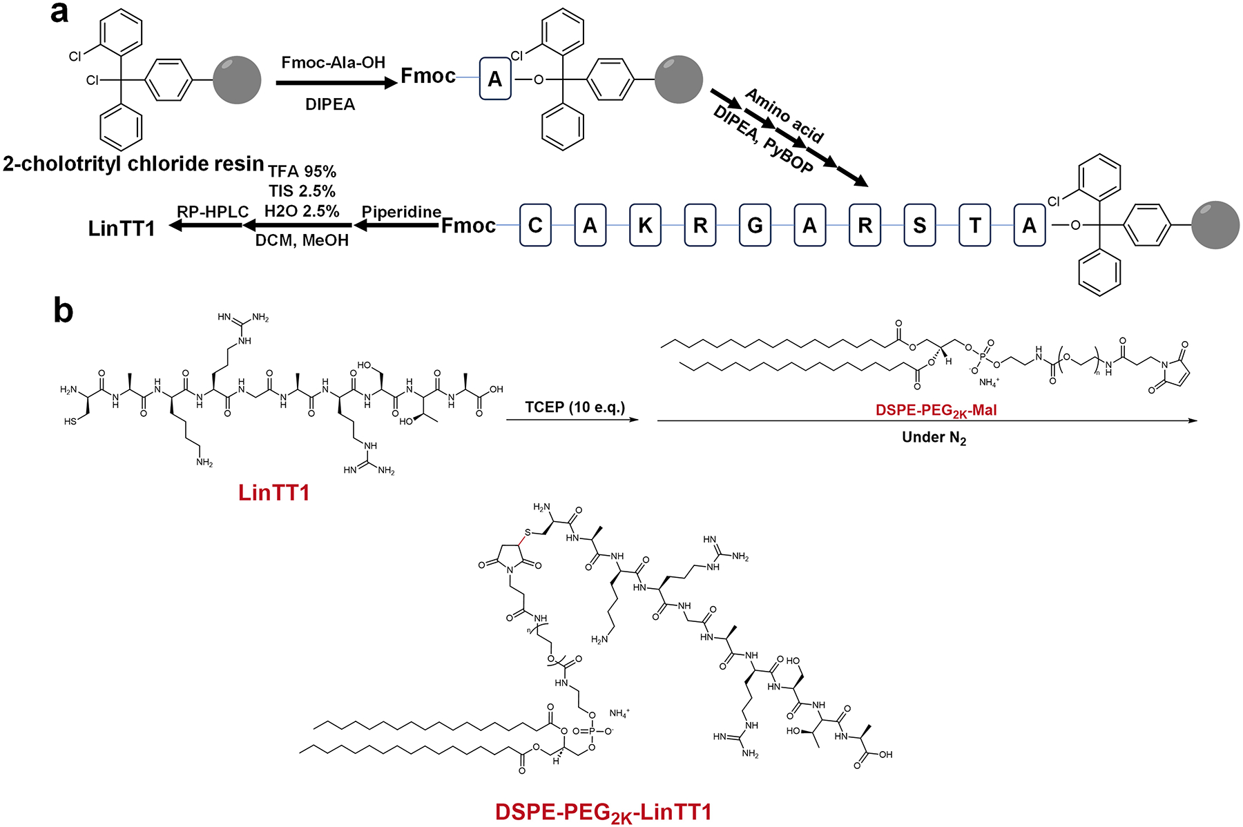
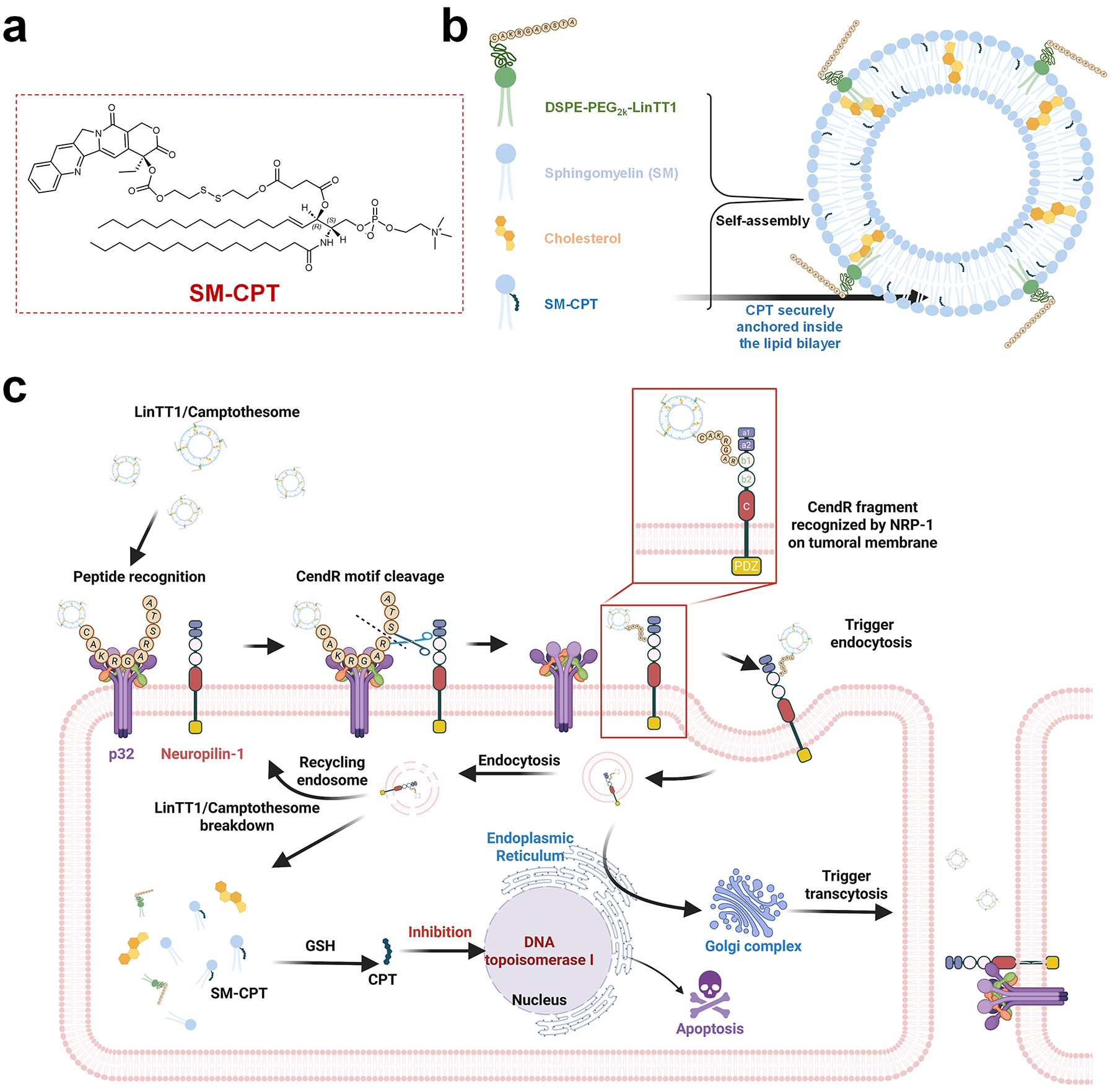
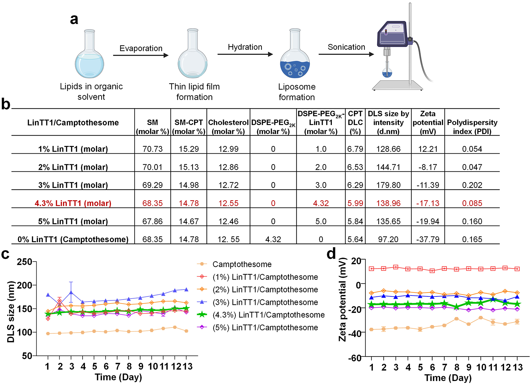
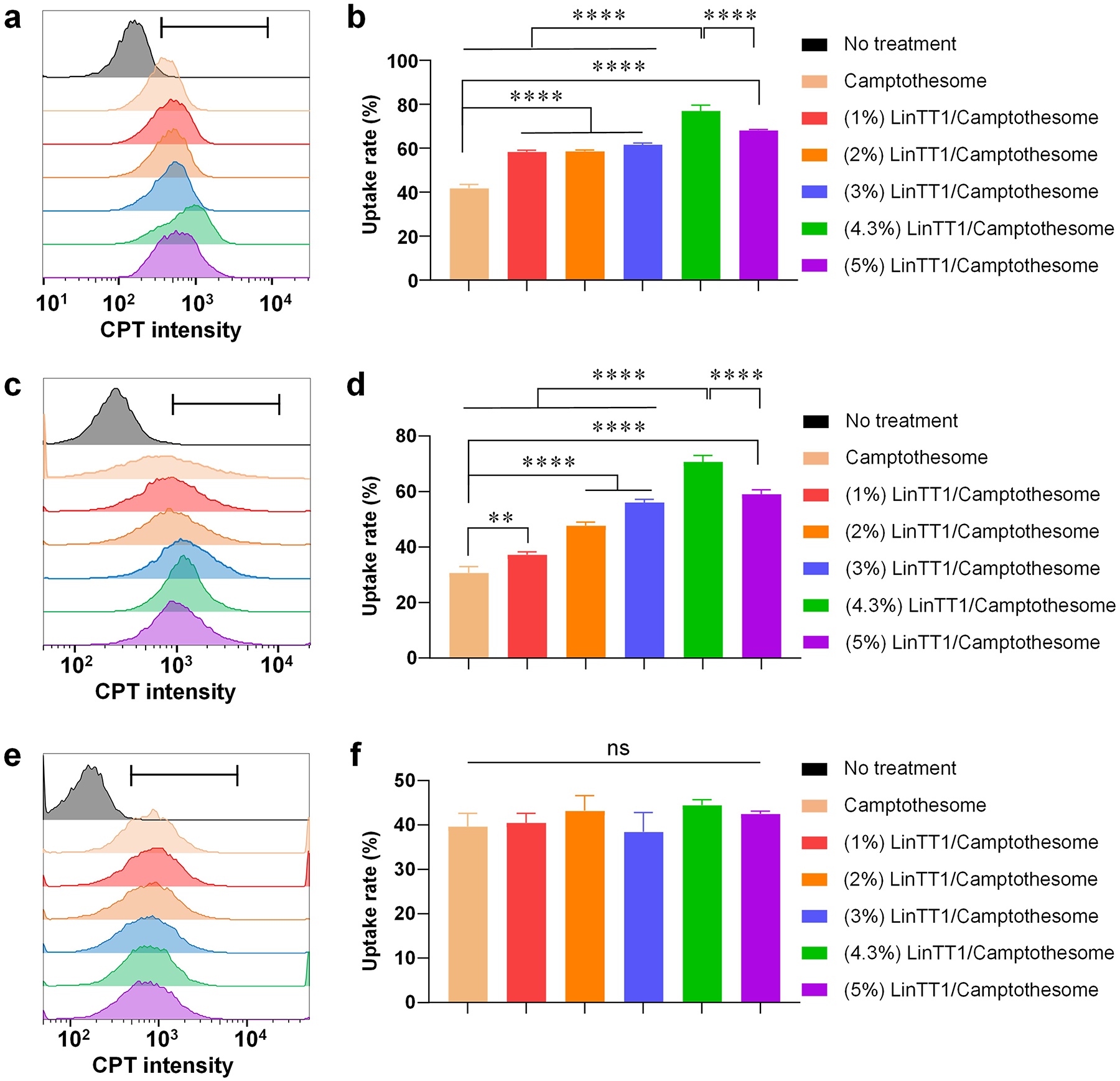
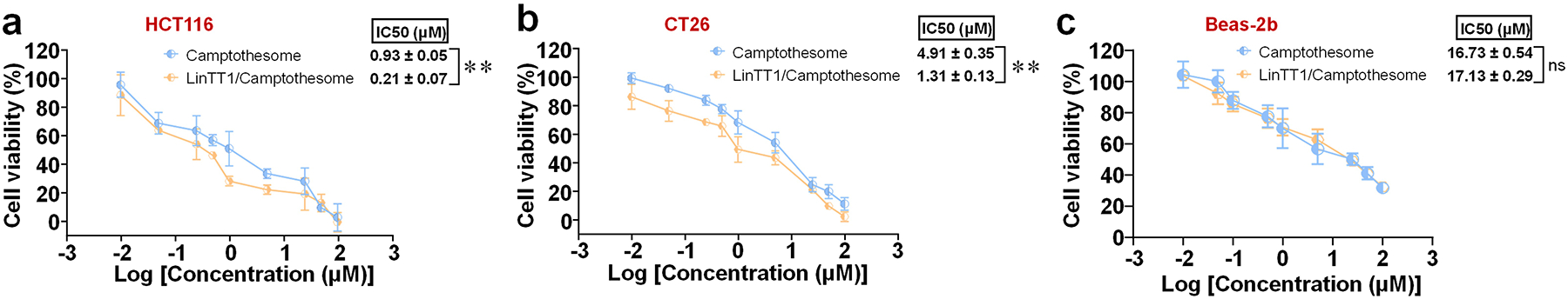
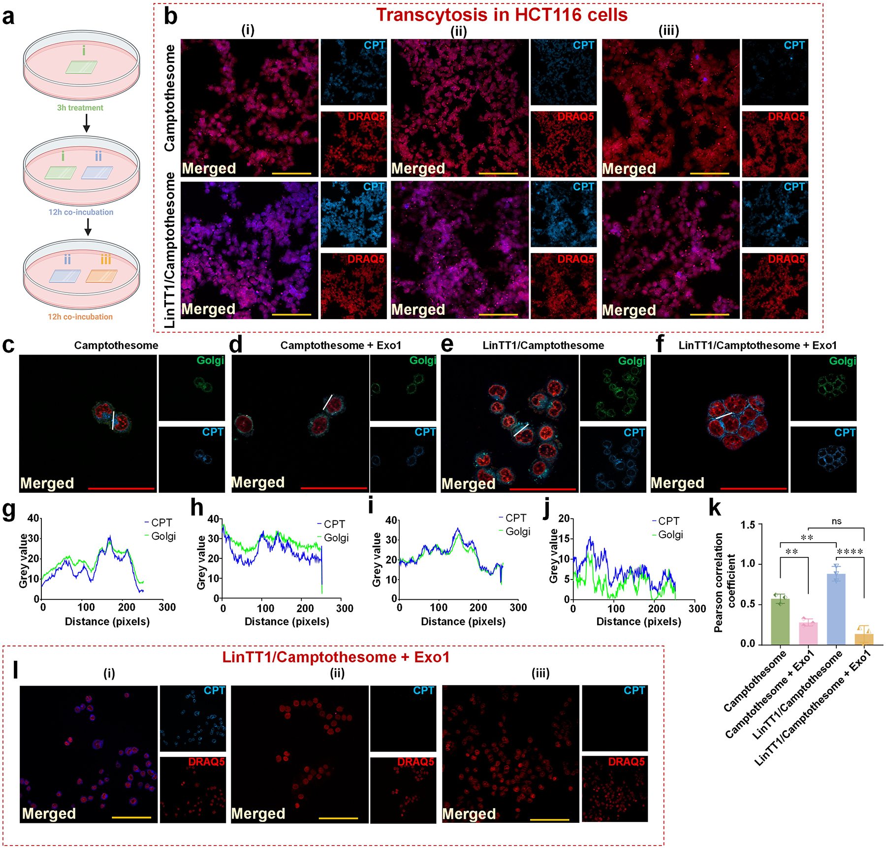
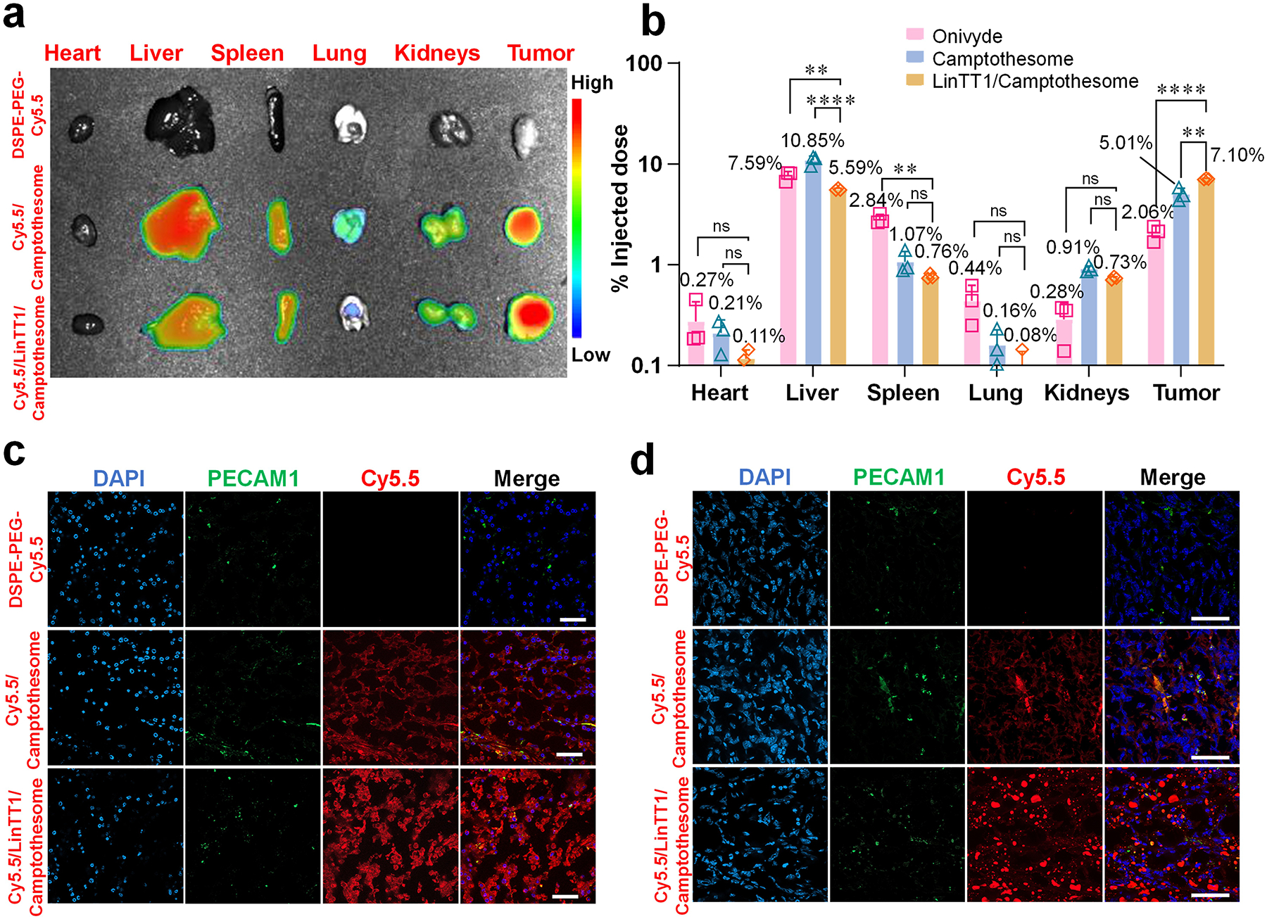
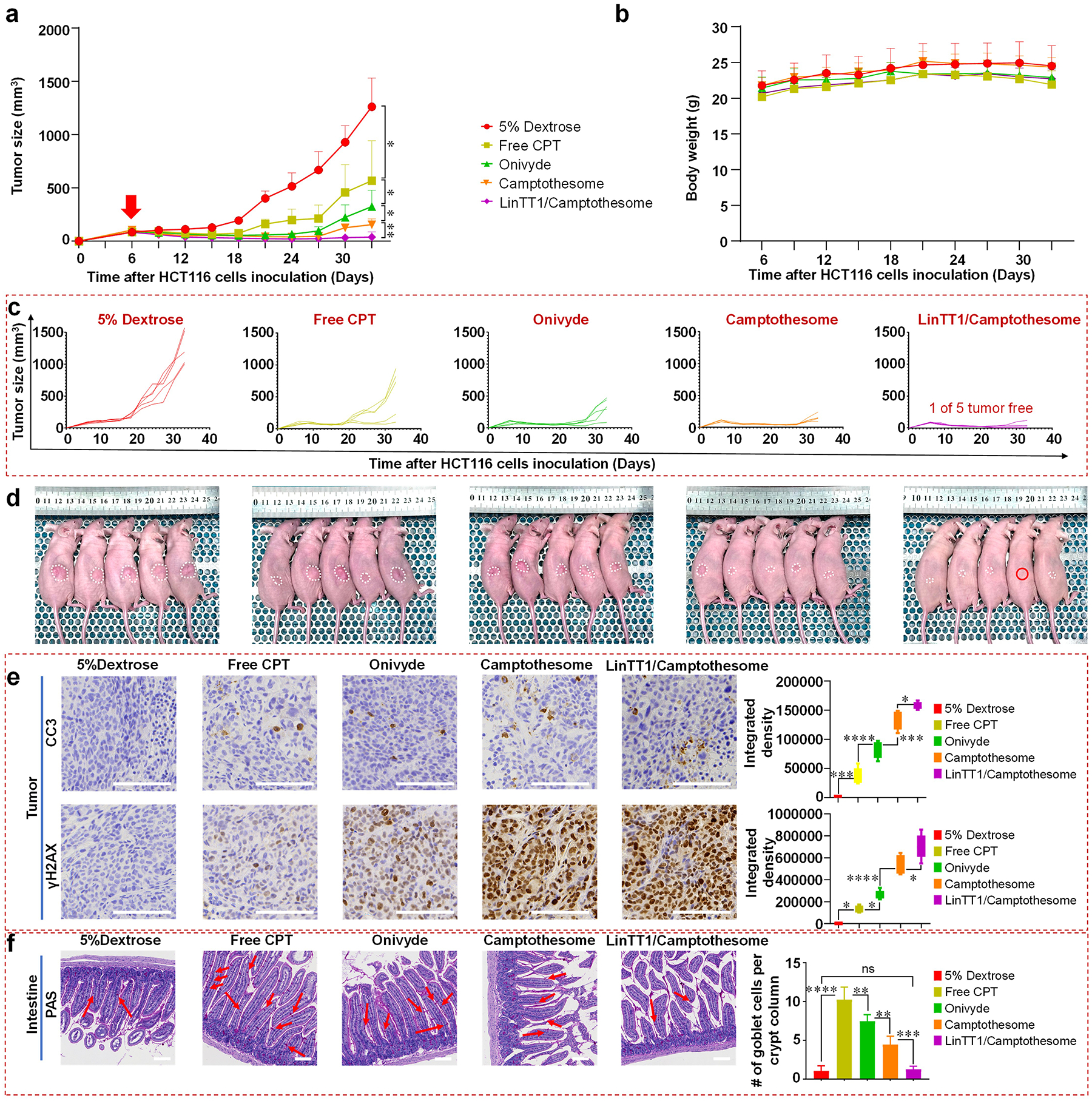
Camptothesome, a sphingomyelin (SM)-conjugated camptothecin (CPT) vesicular nanotherapeutic, addresses the poor solubility and lactone instability of CPT while enhancing drug loading, pharmacokinetics, and tumor distribution compared to CPT physically entrapped in conventional liposomes. Despite these improvements, the tumor uptake remains limited. To further enhance the tumor delivery efficiency and minimize the off-target distribution, we functionalize Camptothesome with the LinTT1 peptide, a CendR motif, which binds to overexpressed p32 proteins on tumor cell surface, initiating effective transcytosis for deep tumor penetration. Via systematic screening, the optimal peptide ratio on Camptothesome is identified. LinTT1/Camptothesome significantly increases cancer cell uptake without affecting normal cell internalization, resulting in enhanced anti-colorectal cancer cells activity. Additionally, decorating Camptothesome with the LinTT1 cell-penetrating peptide enables effective transcytosis via a Golgi-dependent intracellular trafficking mechanism, significantly improving the intratumoral delivery while reducing distribution to normal tissues. In a human HCT116 xenograft colorectal cancer (CRC) mouse model, LinTT1/Camptothesome demonstrates superior antitumor efficacy compared to both Camptothesome and Onivyde by upregulating cleaved caspase-3 and γH2AX. Our study substantiates the potential of leveraging a tumor-penetrating peptide to enhance the tumor delivery efficiency of Camptothesome, maximizing its therapeutic index for improved treatment of human CRC. STATEMENT OF SIGNIFICANCE: Despite the improved tumor delivery achieved by Camptothesome, its tumor distribution and penetration remain limited. This is because the enhanced permeability and retention effect only facilitates nanotherapeutic distribution to tumor periphery through leaky vasculature. The C-end Rule (CendR) motif-neuropilin receptor system enhances tumor-homing peptides by binding to cellular surface receptors, triggering transcytosis. Herein, LinTT1, the most potent CendR peptide that binds to the overexpressed p32 receptor on cancer cells, was effectively engineered onto Camptothesome using thiol-maleimide lipid chemistry. The LinTT1/Camptothesome significantly enhanced tumor uptake and penetration while minimizing accumulation in normal tissues, demonstrating remarkable anticancer efficacy in a human xenograft colorectal cancer model. Our findings highlight the critical role of tumor-homing peptides in unlocking the full therapeutic potential of Camptothesome.

Keywords: Camptothesome; Colorectal cancer; Nanotherapeutics; Tumor-homing LinTT1 peptide; p32.


Conflict of interest statement

Declaration of competing interest J.L. has applied for patents related to the Camptothesome technology. The other authors declare that they have no known competing financial interests or personal relationships that could have appeared to influence the work reported in this paper.

Figures

Figure 1.
Figure 1.. Synthesis mechanism of DSPE-PEG2K-LinTT1.
Synthesis schematic of LinTT1 (a) and DSPE-PEG2K-LinTT1 (b).
Figure 2.
Figure 2.. Development of peptide-decorated SM-derived CPT liposomal nanovesicle (LinTT1/Camptothesome).
a, Chemical structure of SM-CPT [3]. b, Self-assembly into LinTT1/Camptothesome. c, Schematic mechanism of peptide-mediated Camptothesome transcytosis.
Figure 3.
Figure 3.. Development and optimization of peptide-decorated Camptothesome.
a, LinTT1/Camptothesome is synthesized by thin-film hydration method, followed with probe sonication, as previously established [3, 6]. b, A table listing the physicochemical characterizations of LinTT1/Camptothesome comprised of SM/SM-CPT/Cholesterol/DSPE-PEG2K/DSPE-PEG2K-LinTT1 with different molar ratios. DLS, dynamic light scattering; d.nm, diameter value in nanometers. Data in DLS size by intensity, zeta potential, and polydispersity index are expressed as mean (n=3). Monitoring the DLS size (c) and zeta potential (d) of Camptothesome with different molar ratios of peptide in 5% Dextrose at 4°C. Data in c and d are expressed as mean ± SD (n=3).
Figure 4.
Figure 4.. LinTT1 peptide enhanced the cellular uptake of Camptothesome in CRC cancer cells but not in normal cells.
Representative flow cytometry histogram of CPT intensity in HCT116 (a), CT26 (c), and Beas-2b (e) cells treated by Camptothesome or Camptothesome with different peptide ratios at 50μg CPT/mL for 3 hours. The black gate indicates the CPT subset analyzed by FlowJo. b, d, f, Quantitative cellular uptake analyzed by GraphPad. Data in b, d, and f are expressed as mean ± SD (n=3). Statistical significance was determined by one-way ANOVA followed by Tukey’s multiple comparisons test. ns not significant, **P<0.01, ****P<0.0001.
Figure 5.
Figure 5.. Coating LinTT1 onto Camptothesome fortified the killing effects in cancer cells but not in normal cells.
Cytotoxicity study of (4.3%) LinTT1/Camptothesome in (a) HCT116, (b) CT26, and (c) Beas-2b cells at 48 hours by MTT assay. Data are expressed as mean ± SD (n=3). Statistical significance was determined by two-tailed, unpaired Student’s t-test. ns not significant, **P<0.01.
Figure 6.
Figure 6.. LinT11/Camptothesome triggers transcytosis via intracellular trafficking to Golgi.
a, Illustration to show the experimental procedure to investigate transcytosis mechanism. b, Microscopic fluorescence imaging of Camptothesome (upper panel) and LinTT1/Camptothesome (lower panel). Scale bar: 100 μm. c-f, Colocalization of Golgi and CPT signal in HCT116 cells treated with Camptothesome or LinTT1/Camptothesome for 3 hours at 50 μg CPT/mL, with or without pretreatment of the exocytosis blocker (Exo1). Scale bar: 50 μm. g-j, Fluorescence intensity profile of CPT signal and Golgi across cells along the randomly selected white line in c-f were analyzed by pixel intensity with ImageJ. k, Pearson correlation coefficient analysis of CPT and Golgi from c-f. l, Transcytosis of LinTT1/Camptothesome (3 hours, 50 μg CPT/mL) was blocked after being pretreated with Exo1 for 1 hour. The experiment procedures were the same as a. Scale bar: 100 μm. Data in k is expressed as mean ± SD (n=3). Statistical significance was determined by two-tailed, unpaired Student’s t-test. ns not significant, **P<0.01, ****P<0.0001.
Figure 7.
Figure 7.. CendR motif, LinTT1, extends circulation time and enhances tumor delivery of Camptothesome.
a, Mouse Lago optical imaging of ex-vivo imaging of different organs at 48 hours after an I.V. injection of DSPE-PEG-Cy5.5, Cy5.5/Camptothesome (20 mg CPT/kg), and Cy5.5/LinTT1/Camptothesome (20 mg CPT/kg) in subcutaneous HCT116 tumor mice (n=3, tumors: ~300 mm3). b, Tissue biodistribution at 48 hours in subcutaneous HCT116 tumor mice (n=3, tumors: ~300 mm3). Mice received a single I.V. injection for Onivyde (33.7 mg irinotecan/kg), Camptothesome and LinTT1/Camptothesome at e.q. 20 mg CPT/kg. c, Investigate the ability of LinTT1/Camptothesome to enable deep tumor penetration in the HCT116 tumor model. Scale bar: 50 μm. d, Investigate the ability of LinTT1/Camptothesome to enable deep tumor penetration in the 4T1 tumor model. Scale bar: 50 μm. Data in c is expressed as mean ± SD (n=3). Statistical significance was determined by one-way ANOVA followed by Tukey’s multiple comparisons test. ns not significant, **P<0.01, ****P<0.0001.
Figure 8.
Figure 8.. LinTT1/Camptothesome was superior to Camptothesome in anti-CRC efficacy.
a, Tumor growth curves in subcutaneous HCT116 xenograft CRC tumor model (tumors: ~100 mm3). Mice were intravenously injected with a single dose of 5% dextrose, free CPT (5 mg CPT/kg, MTD), Onivyde (33.4 mg irinotecan/kg, eq. 20 mg CPT/kg), Camptothesome and LinTT1/Camptothesome at 20 mg CPT/kg on day 6. The red arrow indicates the date that mice received the single I.V. injection. b, Mouse body weight. c, Individual tumor growth curves in subcutaneous HCT116 xenograft CRC tumor model. d, Tumor-bearing mice images on day 33. Red circle indicates tumor free mouse, white dash line circles tumor volume on each mouse. e, Representative IHC staining of tumors for CC-3 (upper panel) and γH2AX (lower panel) from d and respective quantitative analysis. f, Representative histologic sections of the intestine mucosa stained by periodic acid-Schiff (PAS) reaction and counterstained with haematoxylin from d and respective quantitative analysis. Red arrow indicates the mucin-containing goblet cells. Scale bar: 100 μm. Data in a, b, and data in (f, right panel) are presented as a mean ± SD (n=5), and data in (e, right panel) are presented as box-and-whisker plots (n=5). Statistical significance was analyzed by two-tailed, unpaired Student’s t-test. ns not significant, *P<0.05, **P<0.01, ***P<0.001, ****P<0.0001.

Similar articles

References

    1. Jiang Y, Li W, Wang Z, Lu J, Lipid-Based Nanotechnology: Liposome, Pharmaceutics 16(1) (2024) 34. - PMC - PubMed
    1. Wang Z, Li W, Jiang Y, Park J, Gonzalez KM, Wu X, Zhang QY, Lu J, Cholesterol-modified sphingomyelin chimeric lipid bilayer for improved therapeutic delivery, Nature Communications 15(1) (2024) 2073. - PMC - PubMed
    1. Wang Z, Little N, Chen J, Lambesis KT, Le KT, Han W, Scott AJ, Lu J, Immunogenic camptothesome nanovesicles comprising sphingomyelin-derived camptothecin bilayers for safe and synergistic cancer immunochemotherapy, Nature Nanotechnology 16(10) (2021) 1130–1140. - PMC - PubMed
    1. Zhang X, Zhao W, Nguyen GN, Zhang C, Zeng C, Yan J, Du S, Hou X, Li W, Jiang J, Functionalized lipid-like nanoparticles for in vivo mRNA delivery and base editing, Science advances 6(34) (2020) eabc2315. - PMC - PubMed
    1. Wang Z, Li W, Jiang Y, Tran TB, Cordova LE, Chung J, Kim M, Wondrak G, Erdrich J, Lu J, Sphingomyelin-derived nanovesicles for the delivery of the IDO1 inhibitor epacadostat enhance metastatic and post-surgical melanoma immunotherapy, Nature Communications 14(1) (2023) 7235. - PMC - PubMed