. 2025 May 16;16(1):394.
doi: 10.1038/s41419-025-07722-w.

ZSCAN21 mediates the pathogenic transcriptional induction of α-synuclein in cellular and animal models of Parkinson's disease

Affiliations

ZSCAN21 mediates the pathogenic transcriptional induction of α-synuclein in cellular and animal models of Parkinson's disease

Alina Kozoriz et al. Cell Death Dis. .

Abstract

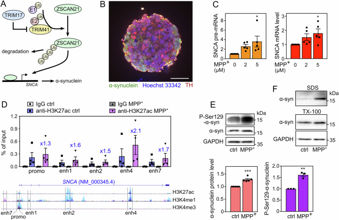
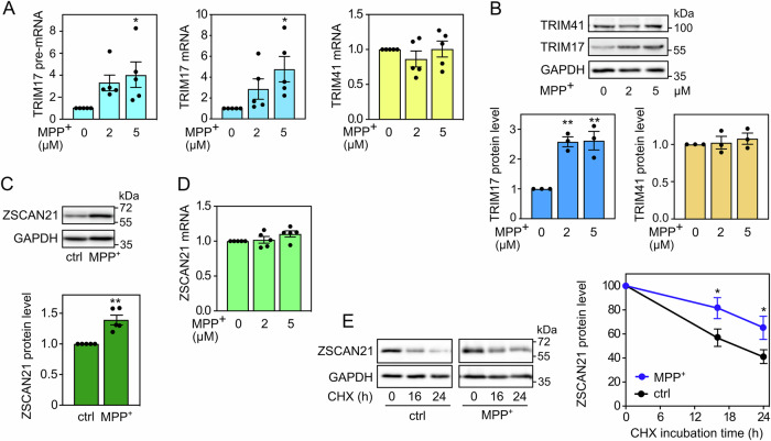
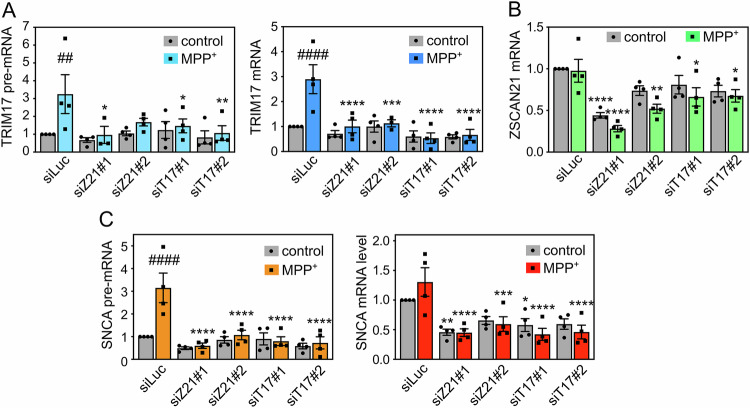
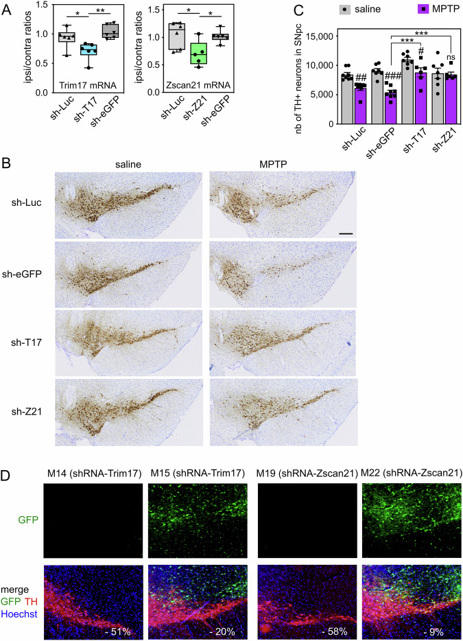
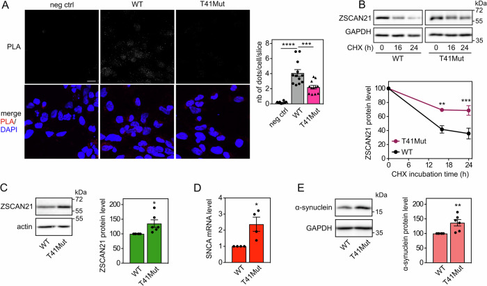
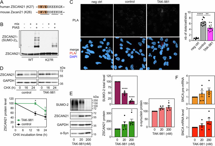
The expression level of α-synuclein is thought to play a crucial role in the pathogenesis of Parkinson's disease. However, little is known about the molecular mechanisms regulating the transcription of its gene, SNCA, particularly in the context of the disease. The transcription factor ZSCAN21 has been shown to act on SNCA, but whether ZSCAN21 is actually involved in the induction of SNCA transcription in Parkinson's disease is unknown. To address this question, we used the MPTP mouse model and LUHMES-derived dopaminergic neuronal spheroids, subjected to Parkinson's disease-related neurotoxins and mutations. We show that MPP+-treated spheroids recapitulate the main features of α-synuclein pathology and that MPP+-triggered transcriptional induction of SNCA is associated with ZSCAN21 stabilisation. Importantly, knock-down of ZSCAN21 prevents both the MPP+-triggered increase in α-synuclein mRNA and pre-mRNA levels in LUHMES-derived spheroids and the death of dopaminergic neurons in the substantia nigra of MPTP-treated mice. These effects are recapitulated by knockdown of TRIM17, a ZSCAN21 stabiliser which prevents its ubiquitination and degradation mediated by TRIM41. Moreover, reducing the interaction between ZSCAN21 and TRIM41, either by inserting Parkinson's disease-associated mutations into the TRIM41 gene or by preventing SUMOylation of ZSCAN21, results in both stabilisation of ZSCAN21 and induction of SNCA. Taken together, our data strongly suggest that ZSCAN21 is a crucial transcription factor for pathogenic α-synuclein expression and neurodegeneration in Parkinson's disease, pointing to its regulators, TRIM17 and TRIM41, as original therapeutic targets for a neuroprotective treatment of Parkinson's disease.


Conflict of interest statement

Competing interests: The authors declare no competing interests. Ethics: All the experimental and surgical procedures were conducted in accordance with the European (Directive 2010/63/UE), Spanish (Real Decreto 53/2013) and Catalan (Decret 214/97) legislation on the protection of animals used for experimental and other scientific purposes, and approved by the Vall d’Hebron Research Institute (VHIR) Ethical Experimentation Committee to ensure the use of the minimum necessary number of animals and the application of protocols that cause the least pain, suffering or distress to animals.

Figures

Fig. 1
Fig. 1. LUHMES-derived DA neuronal spheroids treated with MPP+ recapitulate α-synuclein upregulation and pathology.
A Working hypothesis. Our previous results indicate that ZSCAN21 increases the transcription of the SNCA gene, TRIM41 is an E3 ubiquitin-ligase for ZSCAN21 that induces its proteasomal degradation, and TRIM17 inhibits TRIM41-mediated ubiquitination and degradation of ZSCAN21. B A spheroid of LUHMES-derived DA neurons after 7 days of differentiation. The spheroid was cleared, stained with antibodies against synuclein (green), TH (red) and Hoechst 33342 (blue), and analysed by confocal microscopy coupled with a spinning disk (Dragonfly). The scale bar is 50 μm. C After 6 days of differentiation, LUMHES spheroids were treated or not with 2 μM or 5 μM MPP+ for 24 h. Then, total RNA was extracted, and the pre-mRNA and mRNA levels of SNCA were estimated by quantitative RT-PCR, using YWHAZ and TBP as reference genes. The graph shows mean ± SEM and individual results of five independent experiments. *Significantly different from non-treated cells (0) (one-way ANOVA followed by Dunnett’s multiple comparison test). D LUMHES spheroids were treated or not with 2 μM MPP+ for 24 h. ChIP assays were performed using an antibody against H3K27ac or rabbit IgG used as a negative control. Quantitative PCR was carried out on the immunoprecipitates and the input chromatin using primers specific for the promoter region and different enhancers. Data are expressed as the percentage of the input chromatin used for each immunoprecipitation and are the individual values and means ± SEM of four independent experiments. The ratios of enrichment in MPP+ vs control conditions with the H3K27ac antibody are indicated for each DNA sequence above the MPP+ columns. NB: the data are scattered because the enrichment rate is quite different from one ChIP experiment to another. However, in all experiments, a higher enrichment of all DNA sequences was seen in MPP+ vs control conditions, indicating a real reproducibility. Below the graph, a schematic representation of the human SNCA gene from the UCSC genome browser is shown. It depicts a single representative transcript (NCBI RefSeq NM_000345.4) and the epigenetic marks measured in ChIP-seq experiments on 7 cell lines from the ENCODE project. H3K27ac and H3K4me1 marks are often found near active regulatory elements, and H3K4me3 marks near promoters. The regulatory elements studied by ChIP are indicated. E LUMHES spheroids were treated or not with 2 μM MPP+ for 24 h. Then, total protein extracts were analysed by western blot using antibodies against α-synuclein, Ser129-phosphorylated α-synuclein and GAPDH. The intensity of the α-synuclein bands was quantified, normalised by the intensity of the GAPDH bands and expressed relative to the values obtained with non-treated cells. The graph shows mean ± SEM and individual points from three to five independent experiments. *Significantly different from non-treated cells (unpaired t-test). F LUMHES spheroids were treated or not with 2 μM MPP+ for 29 h. Then, Triton-X100-soluble and SDS-soluble protein fractions were analysed by western blot using antibodies against α-synuclein and GAPDH. These data are representative of two independent experiments.
Fig. 2
Fig. 2. MPP+-induced SNCA transcription correlates with TRIM17 induction and ZSCAN21 stabilisation.
A After 6 days of differentiation, LUMHES spheroids were treated or not with 2 or 5 μM MPP+ for 24 h. Then, total RNA was extracted, and the pre-mRNA/mRNA levels of TRIM17 and the mRNA levels of TRIM41 were estimated by quantitative RT-PCR, using YWHAZ and TBP as reference genes. The graph shows mean ± SEM and individual results of five independent experiments. *: significantly different from non-treated cells (0) (one-way ANOVA followed by Dunnett’s multiple comparison test). B LUMHES spheroids were treated as in (A), and total protein extracts were analysed by western blot using antibodies against TRIM17, TRIM41 and GAPDH. The intensity of the TRIM protein bands was quantified, normalised by the intensity of the GAPDH bands and expressed relative to the values obtained with non-treated cells. The graph shows mean ± SEM and individual points from three independent experiments. *Significantly different from non-treated cells (one-way ANOVA followed by Dunnett’s multiple comparison test). C LUMHES spheroids were treated or not with 2 μM MPP+ for 24 h. Then, total protein extracts were analysed by western blot using antibodies against ZSCAN21 and GAPDH. The results were analysed as in (B). The graph shows mean ± SEM and individual points from five independent experiments. *: significantly different from non-treated cells (unpaired t-test). D LUMHES spheroids were treated as in (A). Then, total RNA was extracted, and the mRNA level of ZSCAN21 was determined and analysed as in (A). E LUMHES spheroids were treated or not with 2 μM MPP+ for 24 h and with 40 μg/ml cycloheximide (CHX) for 0 h, 16 h or 24 h, as indicated. Total proteins were analysed by western blot using antibodies against ZSCAN21 and GAPDH. For each experiment, the amount of ZSCAN21 was normalised by the level of GAPDH in each condition and plotted against CHX incubation time. Data are the mean ± SEM of four independent experiments. *Significantly different from non-treated cells at the indicated time point (two-way ANOVA followed by Sidak’s multiple comparison test).
Fig. 3
Fig. 3. ZSCAN21 potentially binds to “enhancer 4” of SNCA.
A Schematic representation of the human SNCA gene from the UCSC genome browser depicting a single representative transcript (NCBI RefSeq NM_000345.4), two peaks of eGFP-ZSCAN21 ChIP-seq in HEK293 cells from the ENCODE project (ENCSR253CKN) and the H3K27ac marks (often found near active regulatory elements) on seven cell lines from the ENCODE project. B Consensus binding sequence for ZSCAN21 from Factorbook (https://factorbook.org) determined by the MEME software using the eGFP-ZSCAN21 ChIP-seq data (ENCSR253CKN). C Sequence of the eGFP-ZSCAN21 ChIP-seq peak in “enhancer 4” circled in red in (A), with consensus binding sites for ZSCAN21 highlighted in orange and yellow.
Fig. 4
Fig. 4. Knockdown of TRIM17 and ZSCAN21 prevents induction of SNCA transcription in LUHMES cells.
LUHMES cells were transfected with a control siRNA against Luciferase (siLuc), two different siRNAs against TRIM17 (siT17#1 and siT17#2) or two different siRNAs against ZSCAN21 (siZ21#1 and siZ21#2) at the time of plating for 3D-differentiation. After 7 days of differentiation, LUMHES spheroids were treated or not with 2 μM MPP+ for 24 h and total RNA was extracted. The pre-mRNA and mRNA levels of TRIM17 (A), the mRNA level of ZSCAN21 (B) the pre-mRNA level and mRNA levels of SNCA (C) were estimated by quantitative RT-PCR, using RSP16 and HPRT1 as reference genes. The graphs show mean ± SEM and individual results of four independent experiments. *Significantly different from siLuc in the same condition (control or MPP+, two-way ANOVA followed by Dunnett’s multiple comparison test). #: significantly different from cells treated with the same siRNA but not treated with MPP+ (two-way ANOVA followed by Sidak’s multiple comparison test).
Fig. 5
Fig. 5. Knock-down of Trim17 and Zscan21 in SN protect DA neurons from MPTP-induced neurodegeneration in mice.
A AAV vectors expressing control shRNAs (sh-Luc and sh-eGFP) or shRNAs against Trim17 (sh-T17) and Zscan21 (sh-Z21) were unilaterally injected in the midbrain of mice. Four weeks after injection, the mRNA levels of Trim17 and Zscan21 were measured both in the contralateral and ipsilateral midbrains of each mouse by RT-qPCR using β2-microglobulin and Hmbs as reference genes. Ipsilateral/contralateral mRNA ratios are presented as box plots, with minimum, maximum and median indicated. *Significant difference (one-way ANOVA followed by Sidak’s multiple comparison test). B Four weeks after injection of AAV vectors as in A, mice were subjected to or not (saline) to subacute MPTP treatment and were sacrificed three weeks after the last MPTP injection. Brain sections were stained with anti-TH antibody by immunohistochemistry, and representative images of the ipsilateral SN in the different conditions are shown. The scale bar is 200 μm. C The total number of TH-positive neurons in the SNpc of mice was counted by stereology in the different conditions. #Significantly different from control mice (saline) injected with the same AAV (two-way ANOVA followed by Sidak’s multiple comparison test); *Significantly different from sh-eGFP in the MPTP condition (two-way ANOVA followed by Dunnett’s multiple comparison test). NB: differences are also significant when compared with sh-Luc; in saline conditions, only shT17 is different from sh-Luc or sh-eGFP. D Some brain sections from the same mice analysed in B were used for a double immunofluorescence staining of TH and GFP. Representative pictures of the ipsilateral SNpc of MPTP-treated mice are shown. For each mouse, the decrease of the total number of TH-positive neurons in the SNpc, compared to the mean of control mice (saline) injected with the same shRNA, is indicated in white. NB: in mice exhibiting a strong MPTP-induced neurodegeneration, GFP is not expressed in the SNpc, whereas in mice showing a strong neuroprotection, GFP expression is strong in the SNpc.
Fig. 6
Fig. 6. Mutations of TRIM41 impair its interaction with ZSCAN21, resulting in ZSCAN21 stabilization and increased α-synuclein expression.
A 2D-differentiated LUHMES cells on a glass coverslip were fixed and subjected to in situ PLA using rabbit anti-TRIM41 and mouse anti-ZSCAN21 antibodies. Each bright spot (white or red in the merged picture) indicates that the two proteins are in close proximity. Negative control was obtained by omitting anti-ZSCAN21 antibody. Images were analysed by confocal microscopy. To better visualize the differences in PLA intensity, maximum intensity projection was applied to the z-stacks of images. Nuclear staining was performed using DAPI and visualised in merged pictures. The number of dots per cell was determined in one slice of each image using Fiji. The graph shows the mean ± SEM and individual data from 12 images per condition, including a total of more than 250 cells, from one experiment representative of two independent experiments. *Significant difference (one-way ANOVA followed by Dunnett’s multiple comparison test). The scale bar is 10 μM. NB: because the size and intensity of the dots are reduced in T41Mut cells, they are barely visible in the figure, but they could be counted. B 3D-differentiated WT or T41Mut LUHMES cells were treated with 40 μg/ml cycloheximide (CHX) for 0 h, 16 h or 24 h. Total proteins were analysed by western blot using antibodies against ZSCAN21 and GAPDH. For each experiment, the amount of ZSCAN21 was normalised by the level of GAPDH in each condition and plotted against CHX incubation time. Data are the mean ± SEM of five independent experiments. *Significantly different from WT cells at the indicated time point (two-way ANOVA followed by Sidak’s multiple comparison test). C WT or T41Mut LUHMES cells were differentiated into spheroids for 7 days. Then, total protein extracts were analysed by western blot using antibodies against ZSCAN21 and actin. The intensity of the ZSCAN21 bands was quantified, normalised by the intensity of actin and expressed relative to the values obtained with WT cells. The graph shows mean ± SEM and individual points from six independent experiments. *Significantly different from WT cells (unpaired t-test). D WT or T41Mut LUHMES cells were differentiated as in (C). Then, the mRNA levels of SNCA were estimated by quantitative RT-PCR, using GAPDH and GUSB as reference genes. The graph shows mean ± SEM and individual results of four independent experiments. *Significantly different from WT cells (unpaired t-test). E WT or T41Mut LUHMES cells were differentiated as in (C). Total protein extracts were analysed using antibodies against α-synuclein and GAPDH, and data were treated as in (C).
Fig. 7
Fig. 7. Inhibition of SUMOylation impairs the TRIM41/ZSCAN21 interaction, resulting in ZSCAN21 stabilisation and increased α-synuclein expression.
A Negatively charged amino acid-dependent SUMOylation motif (NDSM) containing K27 of human ZSCAN21 and K26 of mouse Zscan21. The core consensus sequence (ΨKxE, where Ψ is a hydrophobic residue and x any amino acid) is in orange. The acceptor Lys residue and the determining Glu residue are in bold. Amino acids conserved between human and mouse are underlined. B In vitro translated 3 × Flag-ZSCAN21 (either the WT form or the K27R mutant) was incubated with in vitro SUMOylation reaction mix (containing SUMO-2, E1 and E2 enzymes), in the presence or the absence of the E3 enzyme PIASxα, as indicated, for 1 h at 37 °C. Poly-SUMOylated forms of ZSCAN21 were detected by immunoblotting using anti-Flag antibody. C 2D-differentiated LUHMES cells were treated with 200 nM TAK-981 for 24 h. Then, neurons were fixed and subjected to in situ PLA, and images were analysed as described in Fig. 6A. The graph shows the mean ± SEM of 10 images per condition, including a total of more than 220 cells (except for negative control), from one experiment representative of three independent experiments. *Significant difference (one-way ANOVA followed by Dunnett’s multiple comparison test). D 3D-differentiated LUHMES cells were treated or not with 200 nM TAK-981 for 24 h and 40 μg/ml cycloheximide (CHX) for 0 h, 16 h or 24 h. Total protein extracts were analysed by western blot using antibodies against ZSCAN21 and GAPDH. For each experiment, the amount of ZSCAN21 was normalised by the level of GAPDH in each condition and plotted against CHX incubation time. Data are the mean ± SEM of four independent experiments. *Significantly different from non-treated cells at the indicated time point (two-way ANOVA followed by Sidak’s multiple comparison test). E 3D-differentiated LUHMES cells were treated with 0 nM, 20 nM or 200 nM of TAK-981 for 24 h. Total protein extracts were analysed by western blot using antibodies against SUMO-2, ZSCAN21, α-synuclein and GAPDH. The intensity of the bands was quantified, normalized by the intensity of the GAPDH bands and expressed relative to the values obtained with non-treated cells. The graph shows mean ± SEM and individual points from five independent experiments. *Significantly different from non-treated cells (one-way ANOVA followed by Dunnett’s multiple comparison test). F 3D-differentiated LUHMES cells were treated with 0 nM, 20 nM, or 200 nM of TAK-981 for 24 h. Then, total RNA was extracted, and the pre-mRNA and mRNA levels of SNCA were estimated by quantitative RT-PCR, using B2M and RSP16 as reference genes. The graph shows mean ± SEM and individual results of five independent experiments. The statistical analysis does not show any significant difference.

Similar articles

References

    1. Dorsey ER, Sherer T, Okun MS, Bloem BR. The emerging evidence of the Parkinson pandemic. J Parkinsons Dis. 2018;8:S3–S8. - PMC - PubMed
    1. Poewe W, Seppi K, Tanner CM, Halliday GM, Brundin P, Volkmann J, et al. Parkinson disease. Nat Rev Dis Prim. 2017;3:17013. - PubMed
    1. Blauwendraat C, Nalls MA, Singleton AB. The genetic architecture of Parkinson’s disease. Lancet Neurol. 2020;19:170–8. - PMC - PubMed
    1. Polymeropoulos MH, Lavedan C, Leroy E, Ide SE, Dehejia A, Dutra A, et al. Mutation in the alpha-synuclein gene identified in families with Parkinson’s disease. Science. 1997;276:2045–7. - PubMed
    1. Mochizuki H, Choong CJ, Masliah E. A refined concept: alpha-synuclein dysregulation disease. Neurochem Int. 2018;119:84–96. - PubMed

MeSH terms