Review
. 2025 May 16;16(1):393.
doi: 10.1038/s41419-025-07714-w.

Unraveling immunosenescence in sepsis: from cellular mechanisms to therapeutics

Affiliations
Review

Unraveling immunosenescence in sepsis: from cellular mechanisms to therapeutics

Yanghanzhao Wang et al. Cell Death Dis. .

Abstract

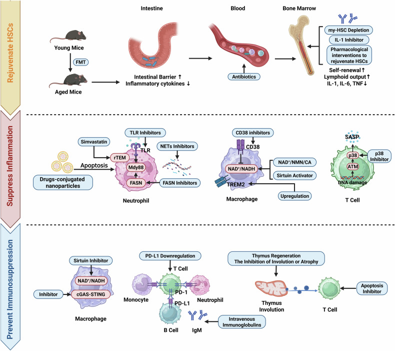
Sepsis is a life-threatening multiple organ dysfunction resulting from a dysregulated host response to infection, and patients with sepsis always exhibit a state of immune disorder characterized by both overwhelming inflammation and immunosuppression. The aging of immune system, namely "immunosenescence", has been reported to be correlated with high morbidity and mortality in elderly patients with sepsis. Initially, immunosenescence was considered as a range of age-related alterations in the immune system. However, increasing evidence has proven that persistent inflammation or even a short-term inflammatory challenge during sepsis could trigger accelerated aging of immune cells, which might further exacerbate inflammatory cytokine storm and promote the shift towards immunosuppression. Thus, premature immunosenescence is found in young sepsis individuals, which further aggravates immune disorders and induces the progression of sepsis. Furthermore, in old sepsis patients, the synergistic effects of both sepsis and aging may cause immunosenescence-associated alterations more significantly, resulting in more severe immune dysfunction and a worse prognosis. Therefore, it is necessary to explore the potential therapeutic strategies targeting immunosenescence during sepsis.


Conflict of interest statement

Competing interests: The authors declare no competing interests.

Figures

Fig. 1
Fig. 1. Persistent inflammation or even a short-term inflammatory challenge induces accelerated aging of HSCs.
Both transient and persistent inflammatory challenge could induce accelerated aging of HSCs in bone marrow, characterized by impaired self-renewal and myeloid-biased differentiation (increased myeloid progenitor cells and decreased lymphoid progenitor cells). To be specific, during the acute phase of sepsis, overproduction of cytokines from macrophages might induce miRNA-mediated downregulation of LSD1 in HSCs, which causes an acute expansion in hyperinflammatory myeloid progenitors. Additionally, 20 consecutive days of IL-1 stimulation has been demonstrated to induce senescence of HSCs. Then, myeloid progenitor cells could further differentiate into neutrophils and monocytes/macrophages. Aged neutrophils exhibit enhanced chemotaxis, phagocytic activity, and NETs release capacity, along with reduced apoptosis. Of note, the chemotaxis of aged neutrophils remains controversial, with evidence supporting both enhanced and impaired chemotactic capacity. Additionally, aged monocytes/macrophages exhibit a pro-inflammatory phenotype, and the phagocytic activity is suppressed. Decreased lymphoid progenitor cells further differentiate into NK cells, T cells, and B cells. And these cells exhibit a decline in number and/or immune function. *Created with BioRender.com.
Fig. 2
Fig. 2. Sepsis induces aberrant aging of immune cells which further exacerbates excessive inflammation.
Under physiological conditions, fresh neutrophils are released from bone marrow, and the gene Bmal1 regulates CXCR2-dependent diurnal aging. Then, senescent neutrophils with high CXCR4 and low CD62L expression return to bone marrow and are engulfed by macrophages. However, during sepsis, gut microbiota can induce accelerated aging of neutrophils via TLRs-mediated Mdy88 pathways. And aged neutrophils exhibit functional alterations accordingly. Firstly, these cells are equipped with enhance chemotactic capacity due to the higher expression levels of β integrin via p38 MAPK activation. However, neutrophils with senescent phenotype are also reported to exhibit inaccurate migration, namely rTEM, which is induced by endothelial cell-derived EVs. Therefore, the chemotactic capacity of aged neutrophils needs further investigation. Secondly, aged neutrophils exhibit a higher phagocytic potential due to the enhanced expression levels of phagocytosis receptor Mac-1. Thirdly, they show greater capacity of releasing NETs via activation of NOX. Moreover, eCIRP, DAMPs released during sepsis, can induce APANs (CXCR4+CD62LCD40+CD86+MHCII+). This subgroup of neutrophils can release high levels of IL-12 and present antigen to CD4+ T cells, which further induce Th1 polarization and the release of IFN-γ. IFN-γ released from T cells could ultimately induced NETosis (shown in the red zoomed-in section). Additionally, eCIRP upregulates SerpinB2 by combining with TLR4 on neutrophils and further suppresses caspase-3-dependent apoptosis. LPS is reported to induce premature aging of macrophages via the NAD+ imbalance. As shown in the red zoomed-in section, LPS can upregulate PRAPs and CD38 (increase NAD+ consumption), and downregulate QPRT (limit NAD+ synthesis), both of which contribute to NAD+ imbalance and induce macrophages to exhibit SASP. Moreover, LPS upregulates BRD4 via activation of NF-κB in macrophages, and enhances the release of inflammatory factors. Bacteria are reported to induce premature aging of T cells via DNA damage. As shown in the red zoomed-in section, CDT from bacteria can induce SASP in T cells via DNA damage, since DNA damage will induce cell cycle arrest and promote release of inflammatory cytokines via ATM-p38 axis. Moreover, cutaneous L. braziliensis can downregulate hTERT and impair DNA structure, which might also induce SASP in T cells. Mature CD163+CD14+ DCs increase in sepsis patients and might contribute to the excessive release of C-reactive protein (CRP) and IL-6. *Created with BioRender.com.
Fig. 3
Fig. 3. Prematurely aged immune cells during sepsis contribute to immunosuppressive state.
After a sustained hyperinflammatory response, in addition to fresh neutrophils (Ly6Ghi), many immature neutrophils (Ly6GloCD123hi) with impaired immune functions are also released into the peripheral blood or tissues. Moreover, microbial capture via phagocytic receptor SIGNR 1 on macrophages induces T cell death-dependent histone release. Cytokines induced by histones will selectively deplete mature neutrophils and increase immature neutrophils. Additionally, bacteria-induced aged neutrophils (CXCR4hiCD62Llo) inhibit TNF-α and IFN-γ secretion from T cell via upregulation of PD-L1 and the release of arginse-1. Monocytes, NK cells, more mature DC3s and mergDCs also overexpress PD-L1, and further suppress T cell proliferation. However, mregDCs might be also equipped with antigen-presenting ability due to upregulation of CD80. As shown in the blue zoomed-in section, typhoid toxin from Salmonella induces the upregulation of senescent-related gene p16INK4a. Macrophages exhibit SASP via activation of cGAS-STING signaling pathway, and macrophages-released SASP-related components can trigger T cell to exhibit SASP and inhibit T cell proliferation. Moreover, SASP-related components could further promote SASP in macrophages via phosphorylation of GCN2 and eIF2α. Additionally, inflammatory cytokine storm during sepsis, including IL-33 or S1P, could directly induce thymus involution or atrophy, which results in aberrant aging of T cells and shows adverse impacts on immune surveillance. To be specific, senescent-related genes, such as p16 and p21, are upregulated, while activation and proliferation-related genes, such as Cd27 and Cd28, are downregulated in CD4+ T cells. Bacteria can increase the expression level of p16INK4a in B cells via DNA damage. *Created with BioRender.com.
Fig. 4
Fig. 4. The distinct phenotypic and functional alterations in immune system in aged individuals with sepsis compared with young patients.
Increased permeability of intestinal-barrier in elderly patients exacerbates the release of microbial compounds and inflammatory cytokines into the circulation. IL-1, one of the cytokines, will further induce the senescence of HSCs via microbiome/IL-1/IL-1R. LPS can stimulate increased release of IL-1 and TNF-α from old plasma cells via activation of TLR-mediated pathways, which will further induce HSCs aging. Moreover, LPS could directly induce HSCs aging by upregulating Klf5 and Stat3, and downregulating Ikzf1. The old HSCs from elderly septic patients exhibit myeloid-biased differentiation more significantly. In the circulation, neutrophils from older patients exhibit enhanced NETosis and deceased phagocytic capacity compared with those from young patients. Moreover, increased percentage of MO3 (CD14+ CD16++)/monocytes is observed in old patients, and these cells show impaired phagocytosis. The percentage of NK cells decreases in old patients, and the cytotoxicity of these cells is suppressed. DCs from elderly patients exhibit diminished antigen-presenting ability and enhanced proinflammatory activity. Enhanced T cell exhaustion has been observed in elderly patients due to increased percentage of PD-1+ T cells, and the immunosuppressive effects of Tregs are also elevated. In inflamed aged lungs, MC-derived CXCL1 drive neutrophil rTEM via ACKR1-CXCL1 axis. TREM2 expression level decreases in macrophages with aging. In several organs, such as lung, spleen, and liver, the downregulation of TREM2 induced more severe inflammation via IL-23/IL-17A axis. In the heart, TREM2 deficiency macrophages show impaired phagocytosis, which causes decreased uptake of defective mitochondria from cardiomyocytes. In the liver, exosomes-containing miR-106b-5p released from TREM2 deficiency macrophages damage hepatocytic mitochondrial structure and function via Mfn2 blockade. Compared with young septic mice, increased inflammatory B1 and B2 cells are observed in adipose tissues in aged mice. And these cells induce inflammatory macrophages via NLRP3 activation, and inhibit lipolysis via Erk pathway suppression. (The red upward arrows mean increased cell numbers or functions in aged septic individuals compared with those in young septic individuals. The blue downward arrows mean decreased cell numbers or functions in aged septic individuals compared with those in young septic individuals.) *Created with BioRender.com.
Fig. 5
Fig. 5. Potential therapeutic strategies targeting immunosenescence.
The potential treatments targeting immunosenescence could be classified as three aspects. Firstly, rejuvenating HSCs is a promising therapeutic strategy. FMT from young mice could enhance intestinal barrier and further reduce inflammatory cytokines in aged mice. Antibiotic treatment can also effectively eliminate bacteria in the circulation. Moreover, my-HSC depletion, IL-1 inhibitors and pharmacological interventions, could promote HSCs self-renewal, increase lymphoid output and suppress the release of inflammatory cytokines. Secondly, during early phase of sepsis, treatments targeting immunosenescence might suppress excessive inflammation. It is recommended to use TLR inhibitors or FASN inhibitors, which suppress TLRs/Mdy88-mediated signaling pathways, in order to inhibit accelerated aging of neutrophils. Nanoparticles-containing drugs could selectively promote neutrophil apoptosis and prevent the persistent existence of aged neutrophils. High-dose simvastatin could enhance the accuracy of neutrophil migration and thereby prevent remote organ damage. Since aged neutrophils exhibit an enhanced capacity to release NETs, NETs inhibitors can be used to suppress inflammation. Moreover, restoring the balance between NAD+ and NADH by using CD38 inhibitors, NAD+/NMN/CA, and Sirtuin activators, could induce macrophages to exhibit an anti-inflammatory phenotype. The upregulation of TREM2 could also suppress the senescent pro-inflammatory phenotype of macrophages. The usage of p38 inhibitor might inhibit SASP in T cells. Thirdly, it is necessary to prevent immunosuppressive state in the late stage of sepsis. Sirtuin inhibitors or the suppression of cGAS-STING signaling pathway might restore immune functions of macrophages. Moreover, the downregulation of PD-L1 on neutrophils, monocytes or B cells might reduce their interactions with T cells, which could inhibit T cell exhaustion and restore adaptive immune functions. The usage of IgM-enriched intravenous immunoglobulins and apoptosis inhibitors targeting lymphocytes might also improve the prognosis of patients. Finally, thymus regeneration or inhibiting the involution or atrophy of thymus can be tested in septic models or patients in the future. *Created with BioRender.com.

Similar articles

Cited by

References

    1. Cajander S, Kox M, Scicluna BP, Weigand MA, Mora RA, Flohé SB, et al. Profiling the dysregulated immune response in sepsis: overcoming challenges to achieve the goal of precision medicine. Lancet Respir Med. 2024;12:305–22. - PubMed
    1. Giamarellos-Bourboulis EJ, Aschenbrenner AC, Bauer M, Bock C, Calandra T, Gat-Viks I, et al. The pathophysiology of sepsis and precision-medicine-based immunotherapy. Nat Immunol. 2024;25:19–28. - PubMed
    1. Hotchkiss RS, Monneret G, Payen D. Sepsis-induced immunosuppression: from cellular dysfunctions to immunotherapy. Nat Rev Immunol. 2013;13:862–74. - PMC - PubMed
    1. Wang N, Lu Y, Zheng J, Liu X. Of mice and men: laboratory murine models for recapitulating the immunosuppression of human sepsis. Front Immunol. 2022;13:956448. - PMC - PubMed
    1. Gentile LF, Cuenca AG, Efron PA, Ang D, Bihorac A, McKinley BA, et al. Persistent inflammation and immunosuppression: a common syndrome and new horizon for surgical intensive care. J Trauma Acute Care Surg. 2012;72:1491–501. - PMC - PubMed

LinkOut - more resources