

Genetic and imaging features of CADASIL patients with acute ischemic stroke
- PMID: 40379656
- PMCID: PMC12084288
- DOI: 10.1038/s41598-025-00220-1
Genetic and imaging features of CADASIL patients with acute ischemic stroke
Abstract
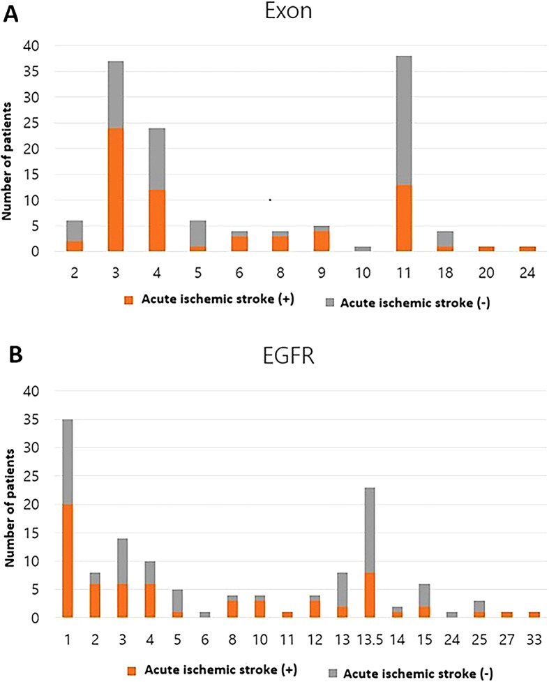
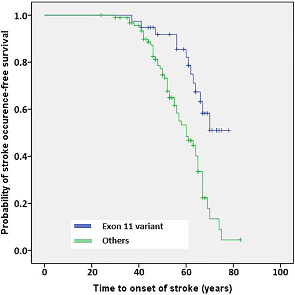
Cerebral autosomal dominant arteriopathy with subcortical infarcts and leukoencephalopathy (CADASIL), which is caused by mutations in the NOTCH3 gene, is associated with early-onset strokes. However, the specific genetic and imaging characteristics associated with acute ischemic stroke (AIS) in patients with CADASIL remain unclear. We reviewed CADASIL patients with NOTCH3 mutations, dividing them into two groups based on the presence of clinically relevant AIS lesions on diffusion-weighted imaging, observed at any time, regardless of the timing of CADASIL diagnosis. Clinical, imaging, and genetic features were compared between these groups. Genetic variations were categorized by exon location: specifically, exon 3 including Arg75Pro, and exon 11 including Arg544Cys, were examined in detail. Factors associated with AIS in CADASIL patients were analyzed. A total of 141 patients were included, of whom 70 (49.6%) were diagnosed with AIS. While there were no significant differences in vascular risk factors between the two groups, patients with AIS had a higher prevalence and greater number of lacunes (p < 0.001) and exhibited more severe white matter changes (p = 0.007). CADASIL patients with AIS had a higher rate of exon 3 variant and a lower rate of exon 11 variant compared to those without AIS. Multivariable analysis revealed that exon 11 variants were associated with a reduced risk (aOR = 0.270, 95% CI 0.099-0.733; p = 0.010). CADASIL who experienced AIS had unique genetic and imaging characteristics when compared to those who did not experience AIS.
© 2025. The Author(s).
Conflict of interest statement
Declarations. Competing interests: The authors declare no competing interests. Ethical approval: This study protocol was reviewed and approved by from the Institutional Review Board of Asan medical center, approval number (2023 − 0381).
Figures
References
-
- Lin, H. J. et al. Modifiable vascular risk factors contribute to stroke in 1080 NOTCH3 R544C carriers in Taiwan biobank. Int. J. Stroke (2024). 19,105 – 13. - PubMed
MeSH terms
Substances
Grants and funding
LinkOut - more resources
Full Text Sources
Medical
Miscellaneous
