. 2025 May 16;16(1):4579.
doi: 10.1038/s41467-025-59919-4.

Inhibiting peptidylarginine deiminases (PAD1-4) by targeting a Ca2+ dependent allosteric binding site

Affiliations

Inhibiting peptidylarginine deiminases (PAD1-4) by targeting a Ca2+ dependent allosteric binding site

Leslie A Dakin et al. Nat Commun. .

Abstract

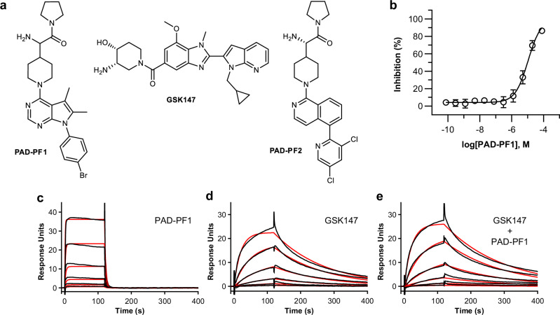
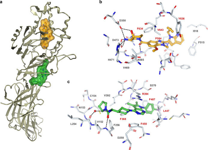
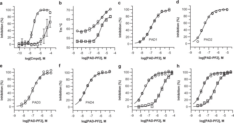
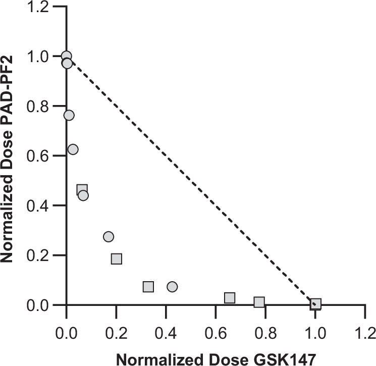
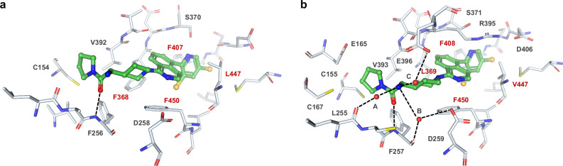
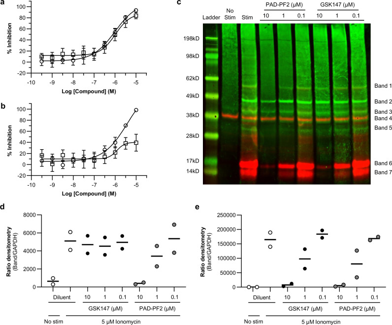
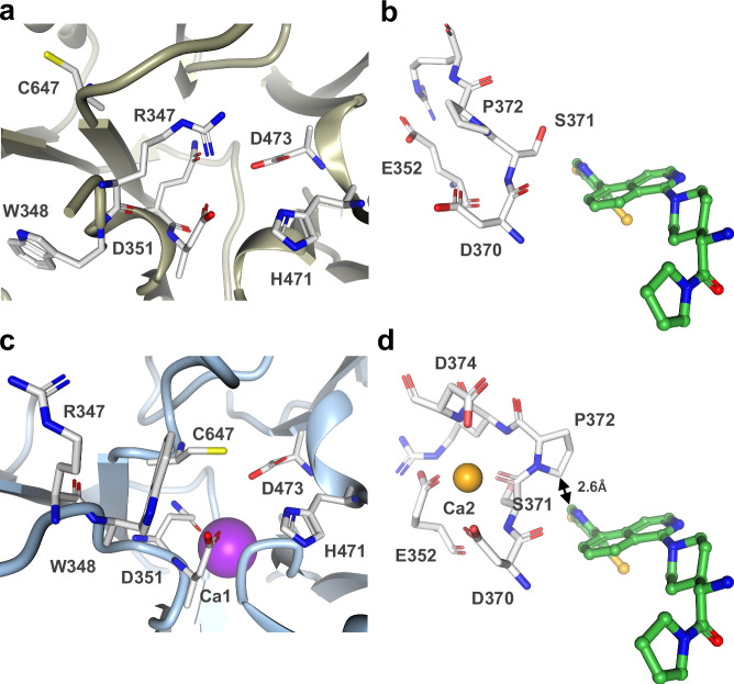
Peptidylarginine deiminases (PAD1-4) are calcium dependent enzymes responsible for protein citrullination, a post-translational modification converting arginine residues to citrulline. Elevated levels of citrullinated proteins have been associated with rheumatoid arthritis, neurodegenerative diseases, and cancers. Though highly selective PAD4 inhibitors have been described, inhibitors to the broader family currently are limited to covalent substrate analogs. Herein, we describe an allosteric binding pocket common to PAD1-4 suitable for the identification of potent, non-covalent enzyme inhibitors. A ligand-based virtual screen is utilized to identify a PAD4 inhibitor for which surface plasmon resonance confirms target binding but non-competitively with a known PAD4 ligand. We further show through co-crystal structure analysis that the ligand binds PAD4 at an allosteric pocket resulting in stabilization of a catalytically inactive, calcium-deficient enzyme conformation. A ligand designed based on this site potently inhibits all four PAD isozymes and prevents protein citrullination in neutrophils with a broader protein repertoire than observed with a PAD4-selective inhibitor.


Conflict of interest statement

Competing interests: All authors were employees of Pfizer Inc. at the time this work was performed.

Figures

Fig. 1
Fig. 1. Ligand-based virtual screen identified PAD-PF1 as a PAD4 inhibitor and non-competitive binder with GSK147.
a Chemical structures of PAD-PF1, GSK147, and PAD-PF2. b PAD-PF1 inhibits PAD4-mediated citrullination of peptide substrate Atto655-(Ser-Arg-Gly-Ala)3 in a fluorescence quenching (FQ) assay in the presence of 0.4 mM Ca2+. Data points and error bars represent the mean and SD of quadruplicate determinations of a representative experiment. IC50 = 15.9 µM, pIC50 = 4.80 (0.26), mean (SD), n = 29 biologically independent experiments. ce Representative surface plasmon resonance (SPR) traces characterizing binding of respective ligands to Bap-tagged PAD4 captured on streptavidine sensor. The red line represents the fitted curve. c PAD-PF1. KD = 2.82 µM, pKD = 5.55 (0.12), mean (SD), n = 4 technical replicates. The highest concentration of PAD-PF1 tested was 10 µM, with five more 3-fold dilutions. d, GSK147. KD = 0.469 µM, pKD = 6.33 (0.016), mean (SD), n = 4 technical replicates. The highest concentration of GSK147 tested was 3.33 µM with five more 3-fold dilutions. e Same concentration series of GSK147 in the presence of 10 µM PAD-PF1. KD = 0.380 µM, pKD = 6.42 (0.071), mean (SD), n = 4 technical replicates. Source data are provided as a Source Data file.
Fig. 2
Fig. 2. PAD-PF1 binds to an allosteric binding pocket distinct from GSK147.
a Crystal structure of human full-length recombinant PAD4 simultaneously bound to GSK147 (orange carbon sticks and van der Waals surface) and PAD-PF1 (green carbon sticks and van der Waals surface). Protein backbone displayed as a ribbon diagram. b PAD4 binding pocket of GSK147 (orange carbon sticks). c PAD4 binding pocket of PAD-PF1 (green carbon sticks). In (b, c), side chains interacting with the small molecules are shown as white carbon sticks, hydrogen bonds are dotted lines, and water molecules are represented as red spheres. Key protein residues with hydrophobic interactions with the ligand are labeled in red.
Fig. 3
Fig. 3. PAD-PF2 potently inhibits PAD1-4 isoforms in a Ca2+-dependent manner.
a PAD-PF2 (open circles) inhibits PAD4-mediated citrullination of peptide substrate Atto655-(Ser-Arg-Gly-Ala)3 employing a fluorescence quenching (FQ) assay in the presence of 0.4 mM Ca2+. Data points and error bars represent the mean and SD of three independent experiments in duplicate. IC50 = 42.7 nM, pIC50 = 7.37 (0.03), mean (SD), n = 3 independent experiments. The enantiomer of PAD-PF2 (ent-PAD-PF2, open squares) was less potent. IC50 = 26.0 µM, pIC50 = 4.59 (0.45), mean (SD), n = 3 independent experiments. b PAD-PF2 induces a thermal stabilization of PAD2 (open circles, EC50 = 2.04 µM) and PAD4 (open squares, EC50 = 1.95 µM) protein. Individual data points shown for a duplicate determination. cf Inhibition of the respective PAD isoform by PAD-PF2 determined using the GDH-enzyme coupled assay in the presence of 0.25 mM Ca2+. Individual data points shown for a duplicate determination along with the fitted IC50 curves (line). c PAD1, IC50 = 109 nM. d PAD2, IC50 = 28.5 nM. e PAD3, IC50 = 106 nM. f PAD4, IC50 = 24.0 nM. g, h Calcium dependence of PAD2 or PAD4 inhibition by PAD-PF2. Inhibition was determined using the GDH enzyme-coupled assay. Data points from duplicate experiments performed at high (open squares) and low (open circles) Ca2+ concentrations are plotted together, and the fitted IC50 curves are shown (line). g The determined IC50 values for PAD2 inhibition were 27.9 nM and 3.09 µM (0.25 and 1.5 mM Ca2+, respectively). h The determined IC50 values for PAD4 inhibition were 20.1 nM and 694 nM (0.25 and 1.5 mM Ca2+, respectively). Source data are provided as a Source Data file.
Fig. 4
Fig. 4. PAD-PF2 and GSK147 synergistically inhibit PAD4.
The method of Loewe was used to assess synergy between PAD-PF2 and GSK147 towards inhibition of PAD4. Serial dilutions of the inhibitors (0–3.6 μM) were tested in all combinations (two-by-two matrix) under the PAD4 GDH-coupled assay condition with 1.5 mM CaCl2. Plotted are the normalized doses of the two compounds that were observed to produce half-maximal activity. For circles, x = [GSK147]/GSK147 IC50 in the absence of PAD-PF2, y = PAD-PF2 IC50/PAD-PF2 IC50 in the absence of GSK147. For squares, x = GSK147 IC50/GSK147 IC50 in the absence of PAD-PF2, y = [PAD-PF2]/PAD-PF2 IC50 in the absence of GSK147. Isobole represented by the dotted line. Source data are provided as a Source Data file.
Fig. 5
Fig. 5. PAD-PF2 binds similarly to both PAD4 and PAD2.
a Crystal structure of human full-length recombinant PAD4 bound to PAD-PF2 (green carbon sticks). The disordered side chains of Asp369 and Ser370 were constructed using the program ADDS. b Crystal structure of human full-length recombinant PAD2 bound to PAD-PF2 (green carbon sticks). In a, b, side chains interacting with the small molecules are shown as white carbon sticks, hydrogen bonds are dotted lines, and water molecules are represented as red spheres. Key protein residues with hydrophobic interactions with the ligand are labeled in red.
Fig. 6
Fig. 6. PAD-PF2 inhibits ionomycin induced protein citrullination in human isolated neutrophils.
a, b Dose-dependent inhibition of ionomycin-induced protein citrullination was observed for both PAD-PF2 (open circles) and GSK147 (open squares) in human isolated neutrophils. Data points represent mean ± S.D. from four separate experiments. Determination of citrullinated proteins performed by flow cytometry employing detection by either (a) anti-CitH3 antibody: PAD-PF2: IC50 = 1.0 µM, pIC50 = 5.98 (0.20); GSK147: IC50 = 1.1 µM, pIC50 = 5.97 (0.11) (mean (SD), n = 4 biologically independent experiments) or (b), F95 antibody: PAD-PF2: IC50 = 1.5 µM, pIC50 = 5.83 (0.15); GSK147: IC50 = 1.1 µM, pIC50 = 5.98 (0.14) (mean (SD), n = 4 biologically independent experiments). c Western blot analysis of human isolated neutrophil lysates, either unstimulated or stimulated with 5 µM ionomycin in the absence or presence of three concentrations of PAD-PF2 or GSK147. Citrullinated proteins were detected with either F95 (green) or CitH3 (red) antibodies. Control proteins include CitH3 (Bands 6 and 7) and GAPDH (Band 4). d, e Western blot densitometry of (d) Band 1 and (e) Band 6 representing citrullinated histone H3 as a ratio of GAPDH protein loading control. Bars represent the mean from two biologically independent experiments. Source data are provided as a Source Data file.
Fig. 7
Fig. 7. PAD-PF2 binding prevents PAD2 from forming active protein conformation by interfering with Ca2+ binding.
a, b Crystal structure of human full-length recombinant PAD2 bound to PAD-PF2 (PDB 9DOL). a Active site pocket with residues in catalytically inactive orientations. b Putative calcium ion (Ca2) binding domain with residue D374 disordered. PAD-PF2 (green carbon sticks). c, d, Crystal structure of holoenzyme PAD2 F221/222 A mutant (PDB 4N2C). c Active site pocket with calcium ion (Ca1, purple). d Calcium ion (Ca2, yellow) binding domain overlayed with position of PAD-PF2 (green carbon sticks) from PAD2:PAD-PF2 structure demonstrating potential clash with Pro372.

Similar articles

Cited by

References

    1. Vossenaar, E. R. et al. PAD, a growing family of citrullinating enzymes: genes, features and involvement in disease. BioEssays25, 1106–1118 (2003). - PubMed
    1. Taki, H. et al. Purification of enzymatically inactive peptidylarginine deiminase type 6 from mouse ovary that reveals hexameric structure different from other dimeric isoforms. Adv. Biosci. Biotechnol.2, 304–310 (2011).
    1. Ranaivoson, F. M. et al. Crystal structure of human peptidylarginine deiminase type VI (PAD6) provides insights into its inactivity. IUCrJ11, 395–404 (2024). - PMC - PubMed
    1. Saijo, S. et al. Monomeric form of peptidylarginine deiminase type I revealed by x-ray crystallography and small-angle x-ray scattering. J. Mol. Biol.428, 3058–3073 (2016). - PubMed
    1. Funabashi, K. et al. Structures of human peptidylarginine deiminase type III provide insights into substrate recognition and inhibitor design. Arch. Biochem. Biophys.708, 108911 (2021). - PubMed

MeSH terms

LinkOut - more resources