. 2025 May 16;16(1):4569.
doi: 10.1038/s41467-025-59790-3.

Engineering tripartite gene editing machinery for highly efficient non-viral targeted genome integration

Affiliations

Engineering tripartite gene editing machinery for highly efficient non-viral targeted genome integration

Hangu Nam et al. Nat Commun. .

Abstract

Non-viral DNA donor templates are commonly used for targeted genomic integration via homologous recombination (HR), with efficiency improved by CRISPR/Cas9 technology. Circular single-stranded DNA (cssDNA) has been used as a genome engineering catalyst (GATALYST) for efficient and safe gene knock-in. Here, we introduce enGager, an enhanced GATALYST associated genome editor system that increases transgene integration efficiency by tethering cssDNA donors to nuclear-localized Cas9 fused with single-stranded DNA binding peptide motifs. This approach further improves targeted integration and expression of reporter genes at multiple genomic loci in various cell types, showing up to 6-fold higher efficiency compared to unfused Cas9, especially for large transgenes in primary cells. Notably, enGager enables efficient integration of a chimeric antigen receptor (CAR) transgene in 33% of primary human T cells, enhancing anti-tumor functionality. This 'tripartite editor with ssDNA optimized genome engineering (TESOGENASE) offers a safer, more efficient alternative to viral vectors for therapeutic gene modification.


Conflict of interest statement

Competing interests: I.M., J.W., J.S., D.L., K.X., R.S. and H.W. are paid employees from Full Circles Therapeutics, R.S. is founder and shareholder of Quintara Bioscience, Inc. Provisional patent “DNA EDITING CONSTRUCTS AND METHODS USING THE SAME” related to this study have been filed by Hao Wu and Richard Shan with Patent Application No. 63/427,370, on November 22, 2022. The remaining authors declare no competing interests.

Figures

Fig. 1
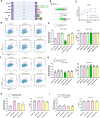
Fig. 1. Fusion of Cas9 and homologous recombination proteins enhance the ssDNA mediated knock-in.
A Schematic diagram of various Cas9-homologous recombination protein fusion constructs (enGagers) in all-in-one (AIO) plasmid format modified from Addgene plasmid #42230. Two nuclear localization signals were added to the N’ and C’-termini of the Cas9 protein. RecA is the bacteria homologous DNA repair protein and Rad51(AE)/Rad51(SEAD) are two mutant variants from eukaryotes. Brex is a 36 amino acid peptide reported to recruit Rad51 in mammalian cells. B Schematic diagram of Knock in strategy of a 2Kb (left) and 4Kb (right) cssDNA donor template for RAB11A locus. C Representative FACS profiles with gating strategy showing % of GFP transgene cassette Knock in on RAB11 locus at day 3 post electroporation for various enGagers listed in (A). D Quantification of 2Kb GFP transgene cassette Knock in fold change of various enGagers as compared to Cas9 WT at day 3 (left), 8 (middle) and 14 (right) post electroporation. E representative FACS profiles with gating strategy showing % of 4Kb GFP transgene cassette Knock in on RAB11 locus at day 3 post electroporation for various enGagers listed in (A). F Quantification of 4Kb GFP transgene cassette Knock in fold change of various enGagers as compared to Cas9 WT at day 3 (left), 7 (right) post electroporation. Note that Brex enGager does not enhance knock in efficiency. Rad51 mutants and RecA enGagers increase both small and large transgene cassette knock in by 1.57–3.04-fold. RecA enGager outperforms among the enGagers tested. Bars represent mean ± SD from 3 biological replicates.
Fig. 2
Fig. 2. Identification of mini enGagers with Cas9 fused ssDNA binding motifs in K562 cells.
A Schematic diagram of various Cas9-ssDNA binding motifs fusion constructs (enGagers) in all-in-one (AIO) plasmid format modified from Addgene plasmid #42230. Two nuclear localization signals were added to the N’ and C’-termini of the Cas9 protein. Cas9-RecA fusion construct was used as a positive control. FECO, WECO and YECO are 20 amino acid sequences previously identified as ssDNA binding motifs in various bacteria species of RecA(Voloshin et al., 1996), FECO3X, WECO3X and YECO3X are 3 tandem copies of the 20 aa peptides separated by multi-GS peptide linkers. B Schematic diagram of Knock in strategy of a 2Kb cssDNA donor template for RAB11A locus. C Representative FACS profiles with gating strategy showing % of GFP transgene cassette Knock in on RAB11 locus at day 7 post electroporation for various small enGagers listed in (A). D Quantification of 2Kb GFP transgene cassette Knock in fold change of various enGagers as compared to Cas9 WT at day 7 post electroporation. Note that Cas9-FECO fusion performs similarly with Cas9-RecA fusion in cssDNA mediated transgene integration (1.59- vs 1.58-fold). EnGagers with 3X tandem ssDNA binding peptides do not further enhance knock in efficiency does not enhance knock in efficiency. Bars represent mean ± SD from 3 biological replicates. Illustrations for panel C is generated with BioRender.
Fig. 3
Fig. 3. Identification of additional enGagers from Cas9-ssDNA binding module chimeras in K562 cells.
A Schematic diagram of various Cas9-ssDNA binding protein and peptide fusion constructs (enGagers) in all-in-one (AIO) plasmid format modified from Addgene plasmid #42230. Two nuclear localization signals were added to the N’ and C’-termini of the Cas9 protein. Cas9-RecA and Cas9-FECO fusion constructs were used as positive controls. The fusion protein or peptides include DrRecA, 20 aa motif identified from DrRecA, SSAP, Lambda Red, RecT, RadA and RadB from Archaea. B Schematic diagram of Knock in strategy of a 2Kb cssDNA donor template for RAB11A locus. C Quantification of 2Kb GFP transgene cassette Knock in fold change of various enGagers as compared to Cas9 WT at day 7 post electroporation. Note that Cas9-DrRecA and Cas9-DrRecA20AA fusion has the highest performance in knock in with 2.17- and 2.43-fold as compared to Cas9 WT, respectively. **p < 0.01, paired t-test compared to AIO Cas9 group. D Quantification of cell viability day 7 post electroporation. Bars represents mean ± SD from 3 biological replicates for (C, D). E Amino acid sequence alignment of 20AA of multiple E.coli RecA mutant variants and RecA from archaea and mammalian organism. Dr Deinococcus radiodurans, Ec Escherichia coli, Sc Saccharomyces cerevisiae, Hs Homo sapiens, Pf P. furiosus, Sso S. solfatarcus.
Fig. 4
Fig. 4. enGager mediated genome integration enhancement is ssDNA dependent.
A Schematic diagram of various Cas9-ssDNA binding protein and peptide fusion constructs (enGagers) in mRNA form. Two nuclear localization signals were added to the N’ and C’-termini of the Cas9 protein. GS is a shortened poly-GS peptide linker. B Schematic diagram of Knock in strategy of a 2Kb cssDNA donor template for RAB11A locus. C Dose titration of cssDNA from 0.3, 1, 1.5, 2 and 3ug for 2Kb GFP transgene knock-in at day 3 post electroporation. Cas9-RecA mRNA enhance around 25–30% knock in efficiency than Cas9 WT mRNA at all the cssDNA dose tested in k562 cells. D Representative FACS profiles with gating strategy showing % of 2Kb GFP transgene cassette Knock-in on RAB11 locus by cssDNA donor at day 5 post electroporation for various enGagers listed in (A) in K562 cells. E Quantification of 2Kb GFP transgene cassette Knock in fold change (left) and cell viability (right) of various enGagers as compared to Cas9 WT at day 5 post electroporation from (D). F Representative FACS profiles with gating strategy showing % of 2Kb GFP transgene cassette Knock-in on RAB11 locus by dsDNA donor at day 5 post electroporation for various enGagers listed in A in K562 cells. G Quantification of 2Kb GFP transgene cassette Knock in fold change (left) and cell viability (right) of various enGagers as compared to Cas9 WT at day 5 post electroporation from (F). H Quantification of 2Kb GFP transgene cassette Knock in fold change (left) and cell viability (right) of various enGagers mRNA as compared to Cas9 WT mRNA with cssDNA donor at 5 days post-delivery in HEK293 cells by lipofectamine 3000 transfection. I Quantification of 2Kb GFP transgene cassette Knock in fold change (left) and cell viability (right) of various enGagers mRNA as compared to Cas9 WT mRNA with dsDNA donor at 5 days post-delivery in HEK293 cells by lipofectamine 3000 transfection. Bars represents mean ± SD from 3 biological replicates. Illustrations for panels (E, GI) are generated with BioRender.
Fig. 5
Fig. 5. enGagers in mRNA form enhance genome integration on various locus and large payload.
A Schematic diagram of Knock in strategy of 2Kb (css76), 4Kb (css116) and 8Kb (css167) cssDNA donor templates for RAB11A locus. B Quantification of % of 2Kb (left), 4Kb (middle) and 8Kb (right) GFP transgene cassette Knock-in for various mRNA enGagers RAB11A day 13 post electroporation in K562 cells. C Quantification of cell viability for 2Kb (left), 4Kb (middle) and 8Kb (right) GFP transgene cassette Knock-in for various mRNA enGagers on RAB11A locus day 13 post electroporation in K562 cells. D Schematic diagram of Knock in strategy of 2Kb (css27), 4Kb (css88) cssDNA donor templates for B2M locus. E Quantification of % of 2Kb (left) and 4Kb (right) GFP transgene cassette Knock-in for various mRNA enGagers on B2M locus day 13 post electroporation in K562 cells. F Quantification of cell viability for 2Kb (left) and 4Kb (right) GFP transgene cassette Knock-in for various mRNA enGagers on B2M locus day 13 post electroporation in K562 cells. Bars represents mean ± SD from 3 biological replicates for (B, C, E, F). ***p < 0.001; ns non-significant, paired t-test compared to mRNA-Cas9 groups for C&E. G Schematic diagram showing enGager mRNA/sgRNA/cssDNA delivery into cells using LNP formulation. Once the editor components were delivered into the cytoplasm, the enGager mRNA is translated into endonuclease protein which forms a complex with sgRNA and cssDNA donor template. The assembled tripartite editing machinery complex then can be effectively shuttled into the nucleoplasm and tether onto the target genomic locus for transgene integration. Quantification of GFP transgene knock-in on RAB11A locus in HEK293 cells (H) and HepG2 cells (I) by LNP delivery at day 2, day 6 and day 9 post-delivery. Data were compared to mRNA-WT Cas9. Bars represents mean ± SD from 2 biological replicates. Illustrations for panel (G) is generated with BioRender.
Fig. 6
Fig. 6. CAR-T engineering by enGager with superior efficiency than WT Cas9.
A Schematic diagram of Knock in strategy of 3Kb CD19/CD22 dual CAR (css62) cssDNA donor templates for TRAC locus in primary T cells. B Quantification of % of CD19/CD22 CAR Knock-in using cssDNA donor analyzed by CD19 binder for various doses of Cas9 WT and Cas9-GS-FECO enGager mRNA at 1 ug, 2 ug and 4 ug. Data were collected at day 7 and day 11 post electroporation of primary T cells. Cas9-GS-FECO enGager achieves ~ 4- to 6-fold higher CAR-T engineering efficiency than Cas9 WT. Bars represents mean ± SD from 2 biological replicates. C Characterization of the engineered CAR-T cells or mock treated T cells for total cell count, cell proliferation fold change and cell viability over time. D NALM6 leukemia lymphocyte killing curve of unengineered T cells, CD19-CD22 dual CAR-T cells engineered with 2 ug of WT Cas9 mRNA and 2 ug of GS-FECO enGager mRNA over the course of 96 h. Effect (T cells): Target (NALM6 cells) are at 2.25:1 for left panel, 4.5:1 for middle panel and 9:1 for right panel. E NALM6 cell killing function of CAR-T cells at 24 h for E:T ratio at 2.25:1, 4.5:1 and 9:1. Bars represents mean ± SD from 2 biological replicates. F Schematic diagram of engineered enGagers with single stranded DNA binding protein (SSBP) can recruit cssDNA donor template and form a tripartite editing machinery for efficient translocation of the entire editing complex from cytoplasm to nucleus. As a result, the donor DNA has higher effective local concentration in the nucleus for more efficient homologous directed genome integration. This process works more prominently with cssDNA. Illustrations for panel (F) is generated with BioRender.

Update of

Similar articles

Cited by

References

    1. Cong, L. et al. Multiplex genome engineering using CRISPR/Cas systems. Science339, 819–823 (2013). - PMC - PubMed
    1. Doudna, J. A. & Charpentier, E. The new frontier of genome engineering with CRISPR-Cas9. Science346, 1258096 (2014). - PubMed
    1. Hsu, P. D., Lander, E. S. & Zhang, F. Development and applications of CRISPR-Cas9 for genome engineering. Cell157, 1262–1278 (2014). - PMC - PubMed
    1. Gilbert, L. A. et al. CRISPR-mediated modular RNA-guided regulation of transcription in eukaryotes. Cell154, 442–451 (2013). - PMC - PubMed
    1. Mali, P. et al. RNA-guided human genome engineering via Cas9. Science339, 823–826 (2013). - PMC - PubMed

MeSH terms

LinkOut - more resources