. 2025 May 16;15(1):17040.
doi: 10.1038/s41598-025-02071-2.

Experimental study of cerebral edema and expression of VEGF and AQP4 in the penumbra area of rat brain trauma

Affiliations

Experimental study of cerebral edema and expression of VEGF and AQP4 in the penumbra area of rat brain trauma

Li Ai et al. Sci Rep. .

Abstract

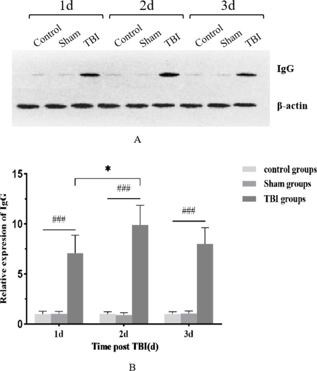
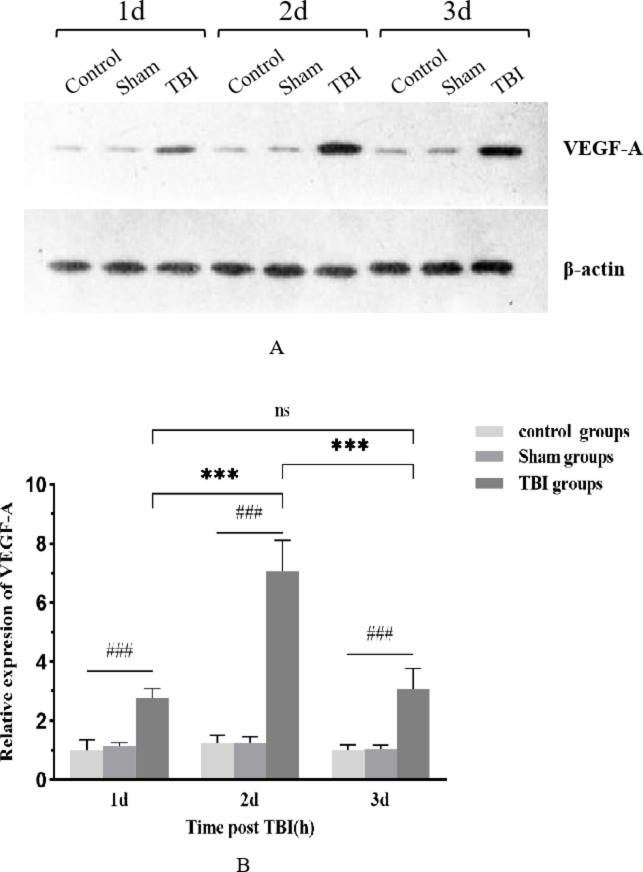
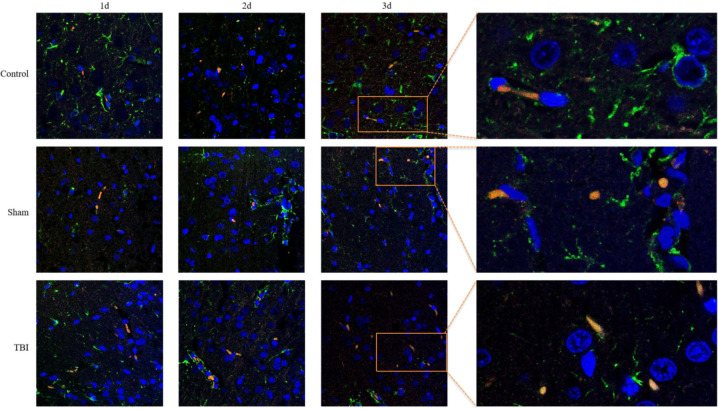
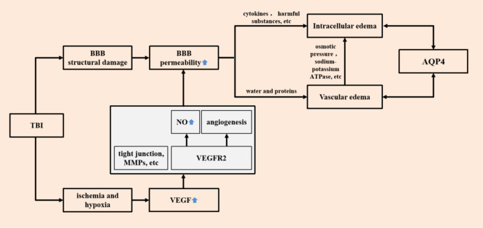
Traumatic brain injury (TBI) has a high disability rate and a high fatality rate. Traumatic penumbra (TP) is a potentially reversible area around the core area of brain trauma with cerebral edema as the main pathological change, which is a breakthrough to improve the prognosis of patients with TBI and reduce the mortality and disability rate of TBI. Unfortunately, the pathophysiological mechanism of TP is still not fully understood. In this study, we established a moderate traumatic brain injury model in rats and detected pathological molecular markers in TP. Protein content of IgG, VEGF, and AQP4 were detect respectively by HE, Immunofluorescence, and western blot. To investigate the time-varying characteristics of TP, to provide a reference for research and development and screening of TBI targeted drugs. Our experiment showed mainly intracellular edema and vascular edema in TP, first intracellular then vascular edema was dominant. IgG, VEGF, and AQP4 in TP increased significantly. On the second day, AQP4 decreased, and third day AQP4 increased again. We found that in the early stage of TBI cerebral edema developed and it is related to the increase of BBB permeability, upregulation of VEGF and AQP4. Suggesting potential targets for treatment of TP.

Keywords: AQP4; BBB; Cerebral edema; Traumatic penumbra; VEGF.


Conflict of interest statement

Declarations. Competing interests: The authors declare no competing interests. Institutional review board statement: All animal experiments were reviewed and approved by the Institutional Animal Care and Committee of The Seventh People’s Hospital of Chongqing (approval number 479/ March 19,2020). All the experiments were carried out in the army special occupational disease prevention and Control Laboratory of Field Surgery Research Department, Army Characteristic Medical Center, Army Medical University. All experimental animals were treated in accordance with GB/T 35892 − 2018 Guidelines for ethical review of the welfare of experimental animals and guidelines for the management and use of experimental animals (8th edition).

Figures

Fig. 1
Fig. 1
Pathological features of brain tissue in the TP region across different observation time points in each group. Cell morphology and structure appeared normal in both the control and Sham groups. In the TBI group, glial cells exhibited swelling, increased cell volume, transparent and lightly stained cytoplasm, and characteristic vacuolar changes at each time point observed (indicated by black dovetail arrows). Additionally, spaces surrounding vascular endothelial cells were notably widened (indicated by black arrows).
Fig. 2
Fig. 2
Histogram illustrating the IgG protein content in the TP region at various observation times across groups, accompanied by representative cropped blots from different gels (A,B) (N = 5). The IgG protein content in the TBI group significantly increased at each observed time point, reaching its peak at 2 days post-injury. ### indicates a significant difference between the TBI group and both the control and Sham groups (p < 0.001). * indicates a significant difference between the 2-day and 1-day post-injury groups within the TBI group (p < 0.05).
Fig. 3
Fig. 3
Histogram showing VEGF protein content in the TP region at different observation time points across groups, accompanied by representative cropped blots from different gels (A,B) (N = 5). VEGF protein content in the TBI group significantly increased at each observed time point, peaking at 2 days post-injury. ### indicates a significant difference between the TBI group and both the control and Sham groups (p < 0.001). *** indicates a significant difference between the 2-day and 1-day post-injury groups within the TBI group (p < 0.001). ‘ns’ indicates no significant difference between the 1-day and 3-day post-injury groups within the TBI group (p > 0.05).
Fig. 4
Fig. 4
The distribution of AQP4 in each group at different time points was observed by immunofluorescence double labeling. Cell nuclei are marked in blue, GFAP in green, and AQP4 in red.
Fig. 5
Fig. 5
Histogram illustrating the AQP4 protein content in the TP region at different observation time points for each group, accompanied by representative cropped blots from different gels (A,B) (N = 5). The AQP4 protein content in the TP region of the TBI group significantly increased at each observed time point. ### indicates a significant difference between the TBI group and both the control and Sham groups (p < 0.001). *** indicates a significant difference between the TBI 3-day group and both the 1-day and 2-day post-injury groups within the TBI group (p < 0.001). ‘ns’ indicates no significant difference between the 1-day and 2-day post-injury groups within the TBI group (p > 0.05).
Fig. 6
Fig. 6
Molecular mechanism derivation diagram for the formation and development of brain edema in the TP region in the early stage of TBI.

Similar articles

References

    1. Wang, H. et al. TMT-based proteomics analysis to screen potential biomarkers of acute-phase TBI in rats. Life Sci.264, 118631 (2021). - PubMed
    1. van Gils, A. et al. Management of mild traumatic brain injury. Pract. Neurol.20 (3), 213–221 (2020). - PubMed
    1. Pitkänen, A. et al. Posttraumatic epilepsy - disease or comorbidity? Epilepsy Behav.38, 19–24 (2014). - PubMed
    1. Dewan, M. C. et al. Estimating the global incidence of traumatic brain injury. J. Neurosurg.130 (4), 1080–1097 (2018). - PubMed
    1. Lu, C. et al. Advances in diagnosis, treatments, and molecular mechanistic studies of traumatic brain injury. Biosci. Trends. 9 (3), 138–148 (2015). - PubMed

Substances

LinkOut - more resources