. 2025 May 16;15(1):17102.
doi: 10.1038/s41598-025-00517-1.

Evaluation of tumor targets selected from public genomic databases for imaging of pancreatic ductal adenocarcinoma

Affiliations

Evaluation of tumor targets selected from public genomic databases for imaging of pancreatic ductal adenocarcinoma

Nada Badr et al. Sci Rep. .

Abstract

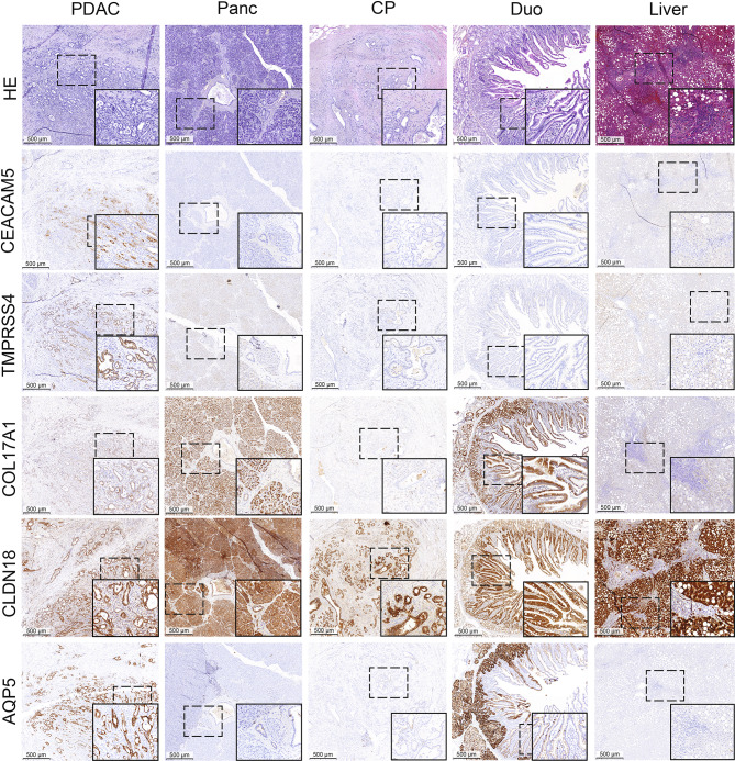
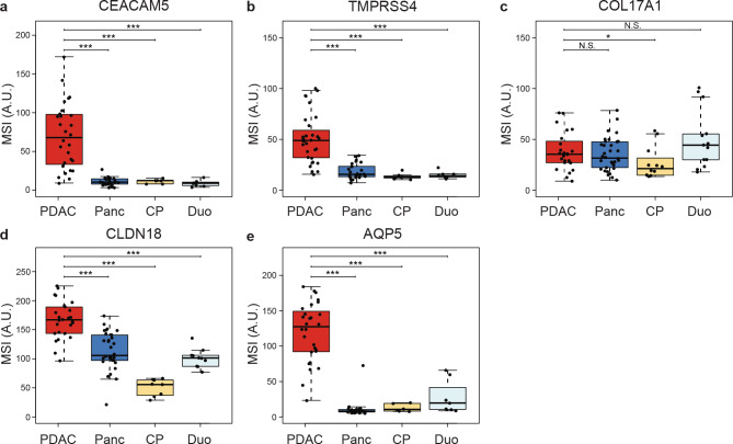
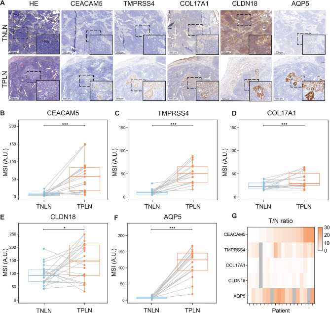
Pancreatic ductal adenocarcinoma (PDAC) is an aggressive malignancy with a 5-year survival rate of approximately 5-7%, and complete surgical resection remains the only curative treatment but is often unfeasible. Fluorescence-guided surgery (FGS) using tumor-targeted probes may improve tumor visualization and facilitate complete resection. This study aimed to identify and validate tumor targets for FGS during PDAC resection procedures. RNA expression data from over 4000 cell surface genes, obtained from public genomic databases, were analyzed to identify genes encoding PDAC-associated proteins. Eleven potential tumor targets were identified, including CEACAM5, TMPRSS4, COL17A1, CLDN18, and AQP5. Protein expression was evaluated by immunohistochemistry (IHC) in tissues from 44 PDAC and 7 chronic pancreatitis (CP) patients. All targets, except COL17A1, showed significantly higher expression in PDAC tissue compared to healthy pancreatic, CP, and duodenal tissue (p < 0.001), as well as in tumor-positive versus tumor-negative lymph nodes. Especially CEACAM5, TMPRSS4, and AQP5 were identified as the most promising targets for distinguishing PDAC from healthy tissues and detecting lymph node metastasis during FGS. The development of probes targeting multiple markers, such as AQP5 with CEACAM5 and/or TMPRSS4, may help overcome interpatient variability and enhance detection across patients.

Keywords: Data-driven approach; Fluorescence-guided surgery; Molecular imaging; Pancreatic cancer; Tumor-target selection.


Conflict of interest statement

Declarations. Competing interests: The authors declare no competing interests. Ethics approval and consent to participate: All procedures involving human participants complied with the ethical standards of the institutional and/or national research committee and followed the principles outlined in the 1964 Helsinki Declaration, including its amendments or equivalent ethical standards. The Ethics Committee of the Leiden University Medical Center reviewed and approved this study (protocol code B20.052, approval date: 17 December 2020). The requirement for informed consent was waived by the Ethics Committee.

Figures

Fig. 1
Fig. 1
Representative immunohistochemical staining images of tissue sections stained with anti-CEACAM5, anti-TMPRSS4, anti-COL17A1, anti-CLDN18, and anti-AQP5. Scale bars represent 500 μm. HE: hematoxylin and eosin staining. Tissue types: PDAC: pancreatic ductal adenocarcinoma, Panc: pancreas, CP: chronic pancreatitis, Duo: duodenal tissue, Liver: liver tissue.
Fig. 2
Fig. 2
Mean staining intensity (MSI) in tissue sections immunohistochemically stained with anti-CEACAM5, anti-TMPRSS4, anti-COL17A1, anti-CLDN18, and AQP5. (a) MSI for CEACAM5 expression. (b) MSI for TMPRSS4 expression. (c) MSI for COL17A1 expression. (d) MSI for CLDN18 expression. (e) MSI for AQP5 expression. Thick lines indicate medians, boxes represent the interquartile ranges (IQRs), whiskers indicate the 95% confidence intervals (CI), and circles data from individual patients. Horizontal bars indicate significance levels (Mann-Whitney U-test: ***, p < 0.001; **, p < 0.01; *, p < 0.05; N.S.; not significant). Tissue types: PDAC: pancreatic ductal adenocarcinoma, Panc: pancreas, CP: chronic pancreatitis, DUO: duodenal tissue.
Fig. 3
Fig. 3
Paired analysis of the mean staining intensities (MSIs) of targets in tissue sections from patients with both pancreatic ductal adenocarcinoma (PDAC) and pancreas (Panc) tissue available. (a) MSI for CEACAM5 expression. (b) MSI for TMPRSS4 expression. (c) MSI for COL17A1 expression. (d) MSI for CLDN18 expression. (e) MSI for AQP5 expression. Dots and lines represent data from individual patients. Thick lines indicate medians and boxes represent interquartile ranges (IQRs). Horizontal bars indicate significance levels (Wilcoxon-signed rank test: ***, p < 0.001; **, p < 0.01; *, p < 0.05; N.S.; not significant). (f) Heat map showing the T/N ratios (MSI PDAC/MSI Panc) for patients with available PDAC and Panc data. Gray regions indicate missing data.
Fig. 4
Fig. 4
(a) Representative immunohistochemical (IHC) staining of tissue sections stained with anti-CEACAM5, anti-TMPRSS4, anti-COL17A1, anti-CLDN18, and anti-AQP5. Scale bars represent 500 µm. HE: hematoxylin and eosin staining. Tissue types: TNLN: Tumor-negative lymph node, TPLN: Tumor-positive lymph node. (bf) Paired analysis of the mean staining intensities (MSIs) of targets in tissue sections from patients with both TNLN and TPLN tissues available. (b) MSI for CEACAM5 expression. (c) MSI for TMPRSS4 expression. (d) MSI for COL17A1 expression. (e) MSI for CLDN18 expression. (f) MSI for AQP5 expression. Dots and lines represent data of individual patients. Thick lines indicate medians and boxes represent interquartile ranges (IQRs). Horizontal bars indicate significance levels (Wilcoxon-signed rank test: ***, p < 0.001; **, p < 0.01; *, p < 0.05; N.S. = not significant). (g) Heat map showing the T/N ratios (MSI TPLN / MSI TNLN) for patients in whom TNLN and TPLN data were available. Gray regions indicate missing data.

References

    1. Adamska, A., Domenichini, A. & Falasca, M. Pancreatic ductal adenocarcinoma: current and evolving therapies. Int. J. Mol. Sci.1810.3390/ijms18071338 (2017). - PMC - PubMed
    1. Tummers, W. S. et al. Impact of resection margin status on recurrence and survival in pancreatic cancer surgery. Br. J. Surg.106, 1055–1065. 10.1002/bjs.11115 (2019). - PMC - PubMed
    1. Tummers, W. S. et al. Selection of optimal molecular targets for tumor-specific imaging in pancreatic ductal adenocarcinoma. Oncotarget8, 56816–56828. 10.18632/oncotarget.18232 (2017). - PMC - PubMed
    1. Vuijk, F. A. et al. Molecular targets for diagnostic and intraoperative imaging of pancreatic ductal adenocarcinoma after neoadjuvant FOLFIRINOX treatment. Sci. Rep.10, 16211. 10.1038/s41598-020-73242-6 (2020). - PMC - PubMed
    1. Gerritsen, A. et al. Preoperative characteristics of patients with presumed pancreatic cancer but ultimately benign disease: a multicenter series of 344 pancreatoduodenectomies. Ann. Surg. Oncol.21, 3999–4006. 10.1245/s10434-014-3810-7 (2014). - PubMed

MeSH terms