Review
. 2025 Jun;32(6):608-621.
doi: 10.1038/s41417-025-00907-7. Epub 2025 May 16.

Structural regulation of PLK1 activity: implications for cell cycle function and drug discovery

Affiliations
Review

Structural regulation of PLK1 activity: implications for cell cycle function and drug discovery

Danda Chapagai et al. Cancer Gene Ther. 2025 Jun.

Abstract

Polo Like Kinase 1 (PLK1), a key regulator of mitosis whose overexpression is often associated with poor survival rates in cancer, continues to be widely investigated as an oncology drug target with clinical trials evaluating second and third generation inhibitors. In addition to the conserved N-terminal kinase domain (KD), a unique characteristic of the Polo-Like kinase family is the C-terminal polo-box domain (PBD). The PBD contains a phosphopeptide binding site that recognizes substrates primed by other kinases and furthermore is responsible for subcellular localization of PLK1 to specific sites in the nucleus including centrosomes and kinetochores. Another role of the PBD is its regulatory ability through domain-domain interactions with the KD to maintain an autoinhibited state of PLK1. Insights into post translational modifications and the PBD - KD domain-domain association have been obtained and show that key events in PLK1 regulation include phosphosubstrate binding, T210 phosphorylation and engagement with the Bora protein. These can induce an open and active conformation where the domain-domain inhibitory interactions no longer dominate. Further regulatory events recently described include the interchange between monomeric and dimeric forms, which can also serve to inhibit or activate PLK1 during the cell cycle. Different oligomeric forms of PLK1, existing as homodimers and heterodimers with PLK2, have been identified and likely play context dependent roles. This review provides an overview of recent information describing structural and mechanistic insights into inhibition of PLK1 and the temporal and spatial requirements of its activation and regulation. It also covers recent insights into the conformational regulation of other members of the Polo-Like kinase family. The implications of the conformational regulation of PLK1 with respect to cell cycle function and drug discovery are significant and are therefore discussed in detail.


Conflict of interest statement

Competing interests: In addition to his primary affiliation at the University of South Carolina, CM is President and CSO of PPI Pharmaceuticals, LLC, developing inhibitors as PLK1 as next generation cancer therapeutics. No other authors declare competing interests.

Figures

Fig. 1
Fig. 1. Domain architecture of PLK1 showing KD, IDL region and PBD.
The polo-cap is abbreviated as PC and the two polo-boxes are PB1 and PB2. The D-Box and K492 are two sites shown to be involved in PLK1 degradation. Key amino acid residues identified in the kinase domain include the catalytic Lys82 and two sites of regulatory phosphorylation (Ser137 and Thr210). In the PBD, Trp414, Leu490, Lys492, His538 and Lys540 are critical residues for phosphorecognition.
Fig. 2
Fig. 2. Overlay of PBD-targeted ligands, highlighting the convergence of interactions with the cryptic pocket of PLK1.
Five CP engaging ligands (see also Fig. 3A) position an aromatic ring similarly as shown left. Yellow: 4X9R (imidazolium-containing phosphopeptide macrocycle 3B); cyan: 5NEI (polotyrin); green: 5NMM (Alpha-Bromo-3-Iodotoluene); magenta: 5NN2 (Z228588490); grey: 4LKM (PL-74); Blue: 3P7 FDPPLHSpTA, PBIP1 residues 71–79); orange: 5J19 AAFSS[pT]PK, Pon residues 58–65).
Fig. 3
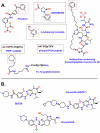
Fig. 3. Chemical Structures of PLK1 Inhibitors.
A PBD-targeted ligands, that engage the cryptic pocket of PLK1. Diverse ligands including peptides, peptidomimetics and small molecules that bind to the PBD are shown. The convergent ring engaging the CP (Fig. 2) is highlighted in the red boxes and letters for peptides. B ATP competitive inhibitors of the PLK1 kinase domain.
Fig. 4
Fig. 4. Overlay of ATP competitive inhibitors of PLK1 obtained to date and highlighting the distinct binding modes observed.
The binding mode of an ATP analogue with carbon atoms highlighted in yellow is shown for reference. Magenta represents BI2536, volasertib and similar ligands; cyan represents onvansertib and similar ligands; green (2OWB) is a 1,5-dihydropyrrolo[3,4-c]pyrazole derivative.
Fig. 5
Fig. 5. Monomer-dimer regulation of PLK1 activity [77].
PLK1 assumes an inactive closed and autoinhibited structure in G1 and S phases of the cell cycle. To fully sequester in an inactive state, Bora binding stabilizes dimeric PLK1 which is maintained until late G2 when Bora degradation and Aur-A phosphorylation results in a monomeric and open conformation of PLK1 which is fully activated both in terms of its catalytic activity and ability to bind to and recruit substrates in specific subcellular locations. Monomeric PLK1 has an exposed nuclear localization signal which then facilities its translocation into the nucleus. Figure was created using Biorender.
Fig. 6
Fig. 6. Autoinhibited structure of PLK1 generated using AlphaFold [93].
The structure generated was found to be in the autoinhibited, closed conformation state, and shows the IDL (purple) bridging the KD and PBD, a key detail absent in the Danio structure [69]. The kinase domain represents the overlay of the co-crystal structure of the isolated KD structure bound to BI2536. The two polo boxes of the PBD are shown in ribbon form. When the KD inhibitor BI2536 (lavender surface) was modeled into the active site, a severe steric clash occurs with Pro403 of the PB1 of the PBD. Experimental data strongly suggests that this results in an open but catalytically inactive conformation of PLK1.
Fig. 7
Fig. 7. Comparison of AlphaFold models generated for PLKs 1–3.
Individual PLK1s shown at the top of the figure are superimposed just using the KD residues to highlight the differences in the positions of the PBD in each case and this is further highlighted by the overlay figure. The compact autoinhibited PLK1 structure is not observed in PLK2 or PLK3. A significant finding from the PLK2 structure is the helical content of the IDL, its engagement with the CP and the 7th strand of the β6α motif.
Fig. 8
Fig. 8. Model of the conformational regulation of PLK1.
PLK1 is in an inactive state (red KD) when autoinhibited by a KD–PBD interaction or in a dimer promoted by PBD interactions. Binding of Bora relieves either form, but still retains PLK1 in an inactive state until recruitment of Aur-A which activates PLK1 through T210 phosphorylation (green KD). Phosphosubstrate binding has also been shown to lead to an increase in activity of PLK1. Monomeric PLK1 will be susceptible to degradation through exposure of the destruction box and Lys492 in the PBD (Fig. 1), Abbapolin PBD inhibitors have been shown to induce monomeric PLK1 and lead to their proteasomal degradation (left) [95]. ATP binding site inhibitors such as BI2536 induce a catalytically inactive open conformation of PLK1 [88, 89] (right).

Similar articles

References

    1. Eckerdt F, Yuan J, Strebhardt K. Polo-like kinases and oncogenesis. Oncogene. 2005;24:267–76. - PubMed
    1. Zitouni S, Nabais C, Jana SC, Guerrero A, Bettencourt-Dias M. Polo-like kinases: structural variations lead to multiple functions. Nature Rev Mol cell Biol. 2014;15:433–52. - PubMed
    1. Strebhardt K. Multifaceted polo-like kinases: drug targets and antitargets for cancer therapy. Nature Rev. 2010;9:643–60. - PubMed
    1. Lee HS, Min S, Jung YE, Chae S, Heo J, Lee JH, et al. Spatiotemporal coordination of the RSF1-PLK1-Aurora B cascade establishes mitotic signaling platforms. Nat Commun. 2021;12:5931. - PMC - PubMed
    1. Singh P, Pesenti ME, Maffini S, Carmignani S, Hedtfeld M, Petrovic A, et al. BUB1 and CENP-U, Primed by CDK1, Are the Main PLK1 Kinetochore Receptors in Mitosis. Mol Cell. 2021;81:67–87.e9. - PMC - PubMed

MeSH terms

LinkOut - more resources