Review
. 2025 May 16;23(1):349.
doi: 10.1186/s12951-025-03435-6.

The biological activity and potential of probiotics-derived extracellular vesicles as postbiotics in modulating microbiota-host communication

Affiliations
Review

The biological activity and potential of probiotics-derived extracellular vesicles as postbiotics in modulating microbiota-host communication

Xiaoming Zhang et al. J Nanobiotechnology. .

Abstract

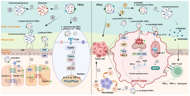
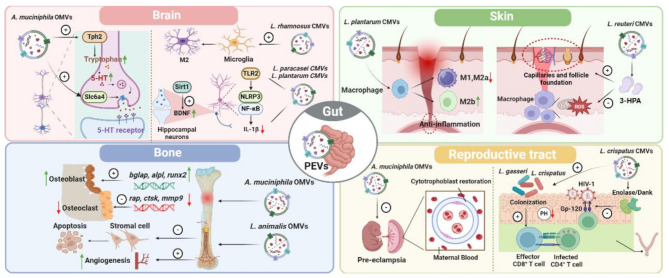
Probiotics such as Lactobacillus and Bifidobacterium spp. have been shown to be critical for maintaining host homeostasis. In recent years, key compounds of postbiotics derived from probiotic metabolism and cellular secretion have been identified for their role in maintaining organ immunity and regulating intestinal inflammation. In particular, probiotic-derived extracellular vesicles (PEVs) can act as postbiotics, maintaining almost the same functional activity as probiotics. They also have strong biocompatibility and loading capacity to carry exogenous or parental active molecules to reach distal organs to play their roles. This provides a new direction for understanding the intrinsic microbiota-host communication mechanism. However, most current studies on PEVs are limited to their functional effects/benefits, and their specific physicochemical properties, composition, intrinsic mechanisms for maintaining host homeostasis, and possible threats remain to be explored. Here, we review and summarize the unique physicochemical properties of PEVs and their bioactivities and mechanisms in mediating microbiota-host communication, and elucidate the limitations of the current research on PEVs and their potential application as postbiotics.

Keywords: Extracellular vesicles (EVs); Host homeostasis; Microbiota-host communication; Postbiotics; Probiotic-derived extracellular vesicles (PEVs).


Conflict of interest statement

Declarations. Ethics approval and consent to participate: Not applicable. Consent for publication: Not applicable. Competing interests: The authors declare no competing interests.

Figures

Fig. 1
Fig. 1
Schematic representation of probiotics-derived Extracellular Vesicles (PEVs). Figure 1 depicts the cell wall structure of Gram-negative and Gram-positive probiotics and the process of extrusion of Outer membrane vesicle (OMV) and Cytoplasmic membrane vesicle (CMV) from the cell wall, and also includes the morphology of the bilayer lipid membrane structure of OMV and CMV as well as the internal structure of the membrane proteins, cytoplasmic It also includes the bilayer lipid membrane structure of OMV and CMV, as well as their internal structures including membrane proteins, cytoplasmic proteins, nucleic acids and bacterial cell wall components, which visualize the differences in the production, secretion and composition of OMV and CMV
Fig. 2
Fig. 2
Schematic representation of the role of PEVs in the maintenance of host intestinal homeostasis. Figure 2 firstly demonstrates the functions of various PEVs in maintaining intestinal homeostasis by down-regulating inflammatory signaling pathways to alleviate inflammation; regulating the levels of intestinal epithelial cell tight junction proteins to maintain the integrity of the intestinal barrier and improving the intestinal microbiota. In addition, PEVs enhanced the tumor-killing effect of CD8+ T cells by regulating intestinal microbial metabolites; induced tumor cells apoptosis by regulating the expression of mitochondrial and endoplasmic reticulum stress and apoptosis proteins; and induced the polarization of macrophages toward M1-type killing phenotypes, which emphasized the anticolonic tumor effect of PEVs
Fig. 3
Fig. 3
Schematic representation of PEVs in regulating the health of host distal organs. Figure 3 is a schematic illustration of PEVs regulating host distal organ health, mainly demonstrating the biological activities of PEVs in the brain, bone, skin and reproductive tract. In the brain, PEVs ameliorate neuroinflammation by inhibiting microglia activation, reverse neuronal damage and improve learning memory by promoting brain-derived neurotrophic factor (BDNF) expression and serotonin signaling. In bone, PEVs promote bone healing by inhibiting osteoclasts, promoting osteoblast formation, and facilitating revascularization. In the skin, PEVs induced macrophage polarization to reduce skin inflammation and promoted wound healing through local revascularization and hair follicle establishment. In the reproductive tract, PEVs resisted HIV-1 infection by both interfering with HIV-1 binding to CD4+ T cells and promoting colonization by Lactobacillus; they also rescued pregnancy outcome by ameliorating placental trophoblast cell stress and fetal injury in disease states

References

    1. Yilmaz B, Macpherson AJ. Delving the depths of ‘terra incognita’ in the human intestine - the small intestinal microbiota. Nat Rev Gastroenterol Hepatol. 2024. - PubMed
    1. de Vos WM, et al. Gut Microbiome and health: mechanistic insights. Gut. 2022;71(5):1020–32. - PMC - PubMed
    1. Kumar R, et al. Recent advancements in the development of modern probiotics for restoring human gut Microbiome dysbiosis. Indian J Microbiol. 2020;60(1):12–25. - PMC - PubMed
    1. Kechagia M, et al. Health benefits of probiotics: a review. ISRN Nutr. 2013;2013:481651. - PMC - PubMed
    1. Aja E et al. Health effects and therapeutic potential of the gut microbe Akkermansia muciniphila. Nutrients. 2025;17(3). - PMC - PubMed

LinkOut - more resources