. 2025 May 17;25(1):103.
doi: 10.1007/s10142-025-01611-3.

3'UTR RNA editing driven by ADAR1 modulates MDM2 expression in breast cancer cells

Affiliations

3'UTR RNA editing driven by ADAR1 modulates MDM2 expression in breast cancer cells

Elanur Almeric et al. Funct Integr Genomics. .

Abstract

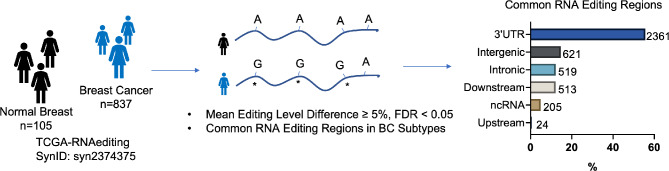
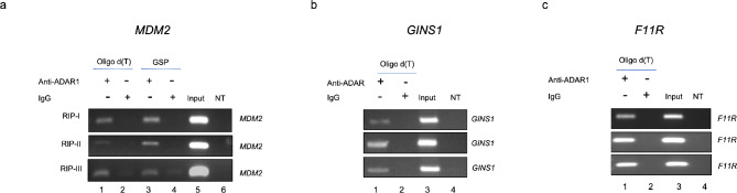
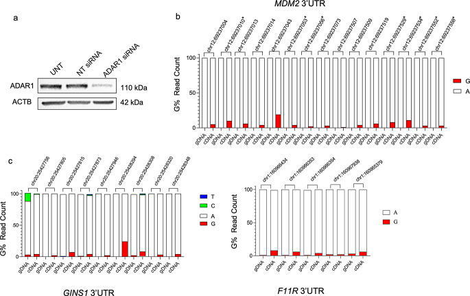
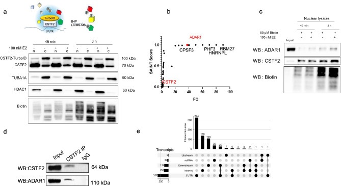
Epitranscriptomic changes in the transcripts of cancer related genes could modulate protein levels. RNA editing, particularly A-to-I(G) editing catalyzed by ADAR1, has been implicated in cancer progression. RNA editing events in the 3' untranslated region (3'UTR) can regulate mRNA stability, localization, and translation, underscoring the importance of exploring their impact in cancer. Here, we performed an in silico analysis to detect breast cancer enriched RNA editing sites using the TCGA breast cancer RNA-seq dataset. Notably, the majority of differential editing events mapped to 3' untranslated regions (3'UTRs). We confirmed A-to-I(G) editing in the 3'UTRs of MDM2 (Mouse Double Minute 2 homolog), GINS1 (GINS Complex Subunit 1), and F11R (Junctional Adhesion Molecule A) in breast cancer cells. RNA immunoprecipitation with ADAR1 antibody confirmed the interaction between ADAR1 and MDM2, GINS1, and F11R 3'UTRs. ADAR1 knockdown revealed decreased editing levels, establishing ADAR1 as the editing enzyme. A reporter assay for MDM2, an oncogene overexpressed mostly in luminal breast cancers, demonstrated that RNA editing enhances protein expression, in agreement with reduced MDM2 protein levels in ADAR1 knockdown cells. Further exploration into the mechanisms of 3'UTR editing events revealed an interaction between ADAR1 and CSTF2, a core component of the polyadenylation machinery, as identified through biotin-based proximity labeling mass spectroscopy, and co-immunoprecipitation experiments. Furthermore, CSTF2 knockdown reduced both ADAR1 and MDM2 protein levels. Our findings highlight implications for MDM2 regulation by ADAR1-dependent 3'UTR RNA editing and present an interplay between RNA editing on 3'UTRs and the mRNA polyadenylation machinery. These results improve our understanding of ADAR1's role in cancer-associated 3' UTR RNA editing and its potential as a therapeutic target.

Keywords: 3’UTR; ADAR1; CSTF2; F11R; GINS1; MDM2; Proximity biotinylation; RNA editing.


Conflict of interest statement

Declarations. Ethics approval and consent to participate: Not applicable. Consent for publication: Not applicable. Competing interests: The authors declare no competing interests.

Figures

Fig. 1
Fig. 1
TCGA breast cancer (n = 837) and normal breast tissue (n = 105) RNA-seq data was screened for A-to-I(G) editing events. Informative RNA editing sites were identified in at least five tumor and non-tumor sample pairs. Differential editing was assessed using the Wilcoxon test, with significant sites defined by FDR < 0.05 and a mean editing level difference ≥ 5% between tumor and non-tumor samples. Majority of significant editing sites map to 3’UTRs. The upstream and downstream regions were defined as 1 kilobase (kb) sequences flanking the annotated transcription start and end sites of the gene, respectively. ncRNA: Noncoding RNA
Fig. 2
Fig. 2
A-to-I(G) editing events in the 3’UTRs of MDM2 (a), GINS1 (b), and F11R (c) in MCF7 cells. gDNA (genomic DNA) or cDNA templated PCR amplicons were sequenced and G% values were calculated based on NGS read counts. The NGS confirmed A-to-I(G) editing events that were not initially predicted are indicated by *
Fig. 3
Fig. 3
RIP-PCR for MDM2 (a), GINS1 (b) and F11R (c). For RNA-IP, ADAR1 (Abcam, ab168809), and rabbit IgG (Cell Signaling, 2729) antibodies were used. cDNAs were synthesized from RNA samples isolated after RIP with oligo d(T) primers (lanes 1, 2), or gene-specific primers (GSP) for MDM2 (lanes 3,4). Three independent biological replicates are shown for each RIP experiment and PCR. Lane 5 for MDM2, and lanes 3 for GINS1 and F11R RIP-PCRs had MCF7 cDNA (Input) as positive control for PCR. NT lane was no template/negative control
Fig. 4
Fig. 4
siRNA-mediated ADAR1 knockdown and RNA editing in MCF7 cells, a. Western blot shows ADAR1 protein levels in ADAR1 siRNA or NT (non-targeting) siRNA transfected cells. UNT: Untransfected cells. The same blots were hybridized with ACTB antibody to test sample loading, b. A-to-I(G) editing ratios of MDM2, c. A-to-I(G) editing ratios of GINS1, and A-to-I(G) editing ratios of F11R in ADAR1 knockdown MCF7 cells
Fig. 5
Fig. 5
a. MDM2 mRNA expression levels (log2-transformed RSEM TPM values (TPM + 0.001) in GTEx breast normal tissue (n = 179), TCGA breast cancer normal tissue (n = 113), and TCGA breast cancer tumor subtypes (luminal A (lumA,0 n = 562), luminal B (lumB, n = 214), basal (n = 190), HER2-enriched (n = 81), and normal-like (n = 39)). Asterisks above the violin plots indicate statistical significance compared to TCGA normal breast tissue data, while asterisks below the violin plots indicate statistical significance compared to GTEx normal breast tissue. One-way ANOVA with Tukey correction for multiple comparisons was performed (**** p < 0.0001, *** p < 0.001, ** p < 0.01, and “ns” for non-significant), b. MDM2 expression in lumA and lumB breast tumors compared with matched normal tissue (paired t-test, **** p < 0.0001), c. Kaplan-Meier analysis of relapse-free survival (RFS) in TCGA BRCA lumA and lumB patients based on MDM2 transcript levels. Patients were grouped into ‘high’ (red) and ‘low’ (black) expression categories based on “Auto select best cutoff” option using data upload. For lumA, Hazard ratio (HR) is 1.98 (95% CI: 1.06–3.71) with a log-rank p-value of 0.021. For lumB, log-rank p-value is 0.048 (HR:3.37). The number of patients at low vs high risk at different time points is indicated below the graphs
Fig. 6
Fig. 6
Effect of RNA editing on MDM2 protein levels, (a) Dual luciferase reporter assay with MDM2 3’UTR edit rich region. Non-edited (A) and edited (G) oligos were cloned downstream of the luciferase gene in pMIR. MCF7 cells were transiently transfected, and Firefly/Renilla luciferase read-outs from the constructs were normalized to that of empty pMIR (EV) (*p < 0.05, ***p < 0.0005, n = 3 independent transfections, one-way ANOVA, Tukey’s HSD), (b) ADAR1 and MDM2 protein levels in ADAR1 siRNA transfected (24 h, 48 h, 72 h) MCF7 cells. The same blots were hybridized with ACTB antibody to test sample loading. The image is representative of 3 independent experiments. Graphs show densitometric quantification of MDM2 and ADAR1 bands normalized to NT siRNA transfected cells (**p < 0.005, ***p < 0.0005, ****p < 0.0001, one-way ANOVA, Tukey’s HSD). NT: non-targeting siRNA transfected cells, UNT: Untransfected cells, (c) NGS and G% reads for the fifteen RNA editing positions for the 3’UTR of MDM2 in normal breast tissue. Fold changes in RNA editing percentage values between MCF7 cells and normal breast cDNA are shown on the bars
Fig. 7
Fig. 7
ADAR1 interacts with CSTF2, a. Schematic shows the proximity biotinylation approach to detect protein interactions at 3’UTRs using CSTF2_TurboID_HA fusion protein expression and biotinylation in MCF7 cells. Biotinylated target proteins were immunoprecipitated (B-IP) and analyzed by LC-MS/MS. Western blot analysis of CSTF2-TurboID transfected MCF7 cells treated with 100 nM E2 or ethanol for 45 min–3 h, and 50 µM biotin. Nuclear and cytoplasmic lysates were subjected to western blot analysis using anti-CSTF2 antibody to detect CSTF2_TurboID_HA fusion and endogenous CSTF2. HDAC1 and TUBA1A antibodies were used to validate the nuclear and cytoplasmic fractions. Anti-Biotin antibody was used for biotinylation assessment, b. Biotinylated nuclear proteins were identified through LC-MS/MS analysis. ADAR1 was found to be significantly enriched among the biotinylated nuclear proteins (SAINT score 1), c. ADAR1 was confirmed to be biotinylated by the CSTF2-TurboID fusion. MCF7 cells were transfected with CSTF2-TurboID. After 24 h, cells were incubated with 50 µM Biotin in the presence or absence of E2 for 45 min and 3 h. Isolated nuclear and cytoplasmic lysates were used for affinity capture with streptavidin beads. Eluted proteins were used for western blot analysis with anti-ADAR1, anti-CSTF2 and anti-Biotin antibodies, d. Nuclear extracts (500 µg) of MCF7 cells were subjected to Co-IP with CSTF2 or isotype-matched IgG. Input was 25 µg nuclear lysate. Immunoprecipitated proteins were then subjected to immunoblotting using CSTF2 or ADAR1 antibodies. WB: western blotting, e. Upset plot showing intersection of RNA editing events on mRNAs. Majority of RNA editing events were only detected on 3’UTRs. Each row represents a genomic feature, and the filled dots indicate the intersection of editing regions in individual mRNAs. The vertical bars indicate the number of genes in each category
Fig. 8
Fig. 8
Effect of CSTF2 knockdown on ADAR1 and MDM2 protein levels. Western blot analysis of CSTF2 siRNA transfected MCF7 cells (24, 48, and 72 h) showed decreased ADAR1 and MDM2 protein levels. The same blots were hybridized with ACTB antibody to test sample loading. The image is representative of 3 independent experiments. Graphs show densitometric quantification of bands normalized to NT bands (ns: not significant, *p < 0.05, **p < 0.01, ****p < 0.0001; n = 3 biological replicates, one-way ANOVA, Tukey’s HSD). NT: non-targeting siRNA transfected cells

Similar articles

References

    1. Aran D, Camarda R, Odegaard J, Paik H, Oskotsky B, Krings G, Goga A, Sirota M, Butte AJ (2017) Comprehensive analysis of normal adjacent to tumor transcriptomes. Nat Commun 8(1):1077. 10.1038/s41467-017-01027-zPMID: 29057876; PMCID: PMC5651823 - PMC - PubMed
    1. Ayaz G, Yasar P, Olgun CE, Karakaya B, Kars G, Razizadeh N, Yavuz K, Turan G, Muyan M (2019) Dynamic transcriptional events mediated by estrogen receptor alpha. Front Biosci (Landmark Ed).;24(2):245–276. 10.2741/4716. PMID: 30468654 - PubMed
    1. Bahn JH, Lee JH, Li G, Greer C, Peng G, Xiao X (2012) Accurate identification of A-to-I RNA editing in human by transcriptome sequencing. Genome Res 22(1):142–150. 10.1101/gr.124107.111Epub 2011 Sep 29. PMID: 21960545; PMCID: PMC3246201 - PMC - PubMed
    1. Bahn JH, Ahn J, Lin X, Zhang Q, Lee JH, Civelek M, Xiao X (2015) Genomic analysis of ADAR1 binding and its involvement in multiple RNA processing pathways. Nat Commun 6:6355. 10.1038/ncomms7355PMID: 25751603; PMCID: PMC4355961 - PMC - PubMed
    1. Bass BL (2002) RNA editing by adenosine deaminases that act on RNA. Annu Rev Biochem 71:817–846. 10.1146/annurev.biochem.71.110601.135501Epub 2001 Nov 9. PMID: 12045112; PMCID: PMC1823043 - PMC - PubMed

MeSH terms