Quantitative electroencephalogram and machine learning to predict expired sevoflurane concentration in infants
- PMID: 40381151
- PMCID: PMC12474631
- DOI: 10.1007/s10877-025-01301-2
Quantitative electroencephalogram and machine learning to predict expired sevoflurane concentration in infants
Abstract
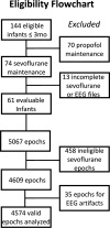
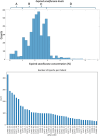
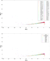
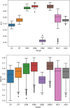
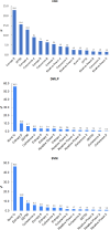
Processed electroencephalography (EEG) indices used to guide anesthetic dosing in adults are not validated in young infants. Raw EEG can be processed mathematically, yielding quantitative EEG parameters (qEEG). We hypothesized that machine learning combined with qEEG can accurately classify expired sevoflurane concentrations in young infants. Knowledge from this may contribute to development of future infant-specific EEG algorithms. Frontal EEG collected from infants ≤ 3 months were time-matched as one-minute epochs to expired sevoflurane (eSevo). Fifteen qEEG parameters were extracted from each epoch and eight machine learning models combined the qEEG to classify each epoch into one of four eSevo levels (%): 0.1-1.0, 1.0-2.1, 2.1-2.9, and > 2.9. 64 epochs formed the post hoc SHAP dataset to determine the qEEG that contributed most to the model. The remaining epochs were randomly split 50 times into 80/20 training/testing sets. Accuracy and F1-score determined model performance. 42 infants provided 4574 epochs. The top classifiers K-nearest neighbors, default multi-layer perceptron, and support vector machine achieved 67.5-68.7% accuracy. Burst suppression ratio and entropy β were the top contributors to the models. Post hoc analysis performed without burst suppression ratio yielded similar prediction performance. In young infants, machine learning applied to qEEG predicted eSevo levels with moderate success. Burst suppression ratio, the most important contributor, represented an efficient EEG feature that encapsulated underlying EEG changes seen on other qEEG features. These results provided insight into EEG parameter selection and optimal machine learning models used for future development of infant-specific EEG algorithms.
Keywords: Infant electroencephalogram EEG; Machine learning; Pediatric EEG anesthesia; Quantitative EEG anesthesia; SHAP EEG analysis.
© 2025. The Author(s).
Conflict of interest statement
Declarations. Conflict of interest: Drs. Yuan and Kurth received consultant fees from Masimo Inc. that are unrelated to this study. Dr Yuan received a research grant from Masimo Inc. that is unrelated to this study. The remaining authors declare that they have no conflicts of interest.
Figures

References
-
- Katoh T, Ikeda K. The minimum alveolar concentration (MAC) of sevoflurane in humans. Anesthesiology. 1987;66(3):301–3. - PubMed
-
- Chan MTV, Hedrick TL, Egan TD, et al. American Society for enhanced recovery and perioperative quality initiative joint consensus statement on the role of neuromonitoring in perioperative outcomes: electroencephalography. Anesth Analg. 2020;130(5):1278–91. - PubMed
-
- Klein AA, Meek T, Allcock E, et al. Recommendations for standards of monitoring during anaesthesia and recovery 2021: Guideline from the Association of Anaesthetists. Anaesthesia. 2021;76(9):1212–23. - PubMed
-
- Fleischmann A, Georgii M-T, Schuessler J, Schneider G, Pilge S, Kreuzer M. Always assess the raw electroencephalogram: why automated burst suppression detection may not detect all episodes. Anesth Analg. 2023;136(2):346–54. - PubMed
MeSH terms
Substances
Grants and funding
LinkOut - more resources
Full Text Sources

