. 2025 Jun 20;6(2):103819.
doi: 10.1016/j.xpro.2025.103819. Epub 2025 May 15.

Protocol to chemically deplete phagocytic hemocytes in Anopheles gambiae using clodronate liposomes

Affiliations

Protocol to chemically deplete phagocytic hemocytes in Anopheles gambiae using clodronate liposomes

Hyeogsun Kwon et al. STAR Protoc. .

Abstract

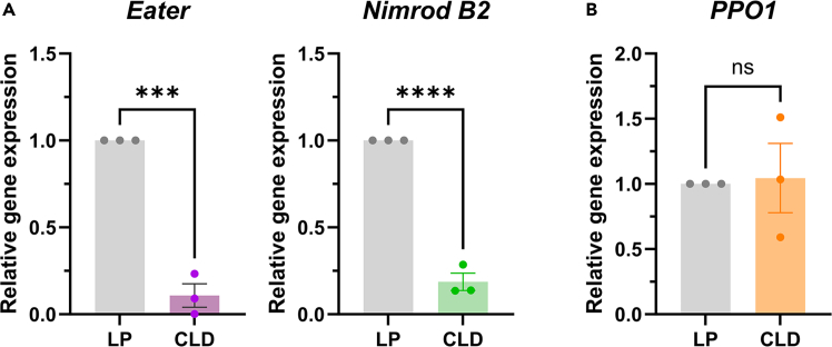
Understanding the roles of phagocytic hemocytes in mosquito innate immunity has been significantly limited due to the lack of genetic tools. Here, we present a protocol for depleting phagocytic hemocytes in Anopheles gambiae mosquitoes using clodronate liposomes. We describe steps for mosquito injection, as well as validation by microscopy, quantitative real-time PCR (real-time qPCR), and flow cytometry analysis. This protocol allows for the delineation of phagocytic hemocyte function in mosquito immunity, which can be more broadly applied to other arthropod systems. For complete details on the use and execution of this protocol, please refer to Kwon et al.1.

Keywords: Cell separation/fractionation; Genetics; Immunology; Model Organisms; Molecular Biology; Protocols in Entomology; Special Issue.


Conflict of interest statement

Declaration of interests The authors declare no competing interests.

Figures

None
Graphical abstract
Figure 1
Figure 1
Validation of granulocyte depletion using a hemocytometer Examples of hemocyte subtypes when visualized on a hemocytometer using light microscopy (A). Granulocytes display distinct morphological characteristics based on their adherent properties and larger size. Both prohemocytes and oenocytoids are round, with oenocytoids distinguished by their relatively larger size and cupped-like appearance. Cell debris, such as fat body cells, can occasionally be observed and distinguished as clusters of large cells that are distinct from the morphology of the mosquito hemocyte subtypes. The percentage of granulocytes from control liposome (LP) or clodronate liposome (CLD) treatments can be assessed to confirm the depletion of granulocytes at 24 h post-injection (B). Error bars represent the mean ± SEM of three independent replicates. Data were tested for normality and analyzed using an unpaired t-test (∗∗∗P < 0.001) using GraphPad Prism 10. Scale bar, 10 μm.
Figure 2
Figure 2
Confirmation of phagocyte depletion by IFA after clodronate liposome treatment Immunofluorescence assays (IFAs) were performed to examine perfused hemocytes from control liposome (LP) or clodronate liposome (CLD) treatments. Attached hemocytes are visualized using FITC-WGA (green), CM-DiI (red) and DAPI (blue), to clearly demonstrate the reduced number of attached granulocytes in CLD-treated mosquitoes. Scale bar, 20 μm.
Figure 3
Figure 3
Confirmation of phagocytic hemocyte depletion using real-time qPCR analysis The depletion of phagocytic hemocytes was confirmed by assessing the expression of the phagocytic cell markers, Eater and Nimrod B2, in control liposome (LP)- or with clodronate liposome (CLD)-treated mosquitoes (A). In contrast, PPO1, a marker for non-phagocytic hemocytes, displayed no difference in expression between LP- and CLD-treated groups (B). Bars represent the mean ± SEM of three independent biological replicates (dots). Statistical analysis was performed using an unpaired t-test in GraphPad Prism 10.0. Asterisks indicate statistical significance (∗∗∗, P < 0.001; ∗∗∗∗, P < 0.0001). ns, not significant.
Figure 4
Figure 4
Assessment of phagocytic hemocyte proportions using flow cytometry The proportion of phagocytic hemocytes was evaluated between control liposome (LP) and clodronate liposome (CLD) treatments using flow cytometry. Double-positive hemocytes were identified by gating based on fluorescent signal thresholds: red for bead uptake and green for WGA staining, located in the upper right quadrant (denoted by red box). The impact of CLD on phagocytic hemocyte depletion was assessed by comparing the percentage of double-positive (phagocytic) hemocytes between the LP and CLD samples (29.4 vs. 15.5). This example displays an approximate 47% reduction (29.4–15.5 / 29.4) in the number of phagocytic cells following CLD treatment under naïve conditions.

Similar articles

References

    1. Kwon H., Smith R.C. Chemical depletion of phagocytic immune cells in Anopheles gambiae reveals dual roles of mosquito hemocytes in anti- Plasmodium immunity. Proc. Natl. Acad. Sci. 2019;116:14119–14128. doi: 10.1073/pnas.1900147116. - DOI - PMC - PubMed
    1. Ramesh Kumar J., Smith J.P., Kwon H., Smith R.C. Use of clodronate liposomes to deplete phagocytic immune cells in Drosophila melanogaster and Aedes aegypti. Front. Cell Dev. Biol. 2021;9 doi: 10.3389/fcell.2021.627976. - DOI - PMC - PubMed
    1. Adegoke A., Ribeiro J.M.C., Brown S., Smith R.C., Karim S. Rickettsia parkeri hijacks tick hemocytes to manipulate cellular and humoral transcriptional responses. Front. Immunol. 2023;14 doi: 10.3389/fimmu.2023.1094326. - DOI - PMC - PubMed
    1. Adegoke A., Hanson J., Smith R.C., Karim S. Ehrlichia chaffeensis co-opts phagocytic hemocytes for systemic dissemination in the Lone Star tick, Amblyomma americanum. J. Innate Immun. 2024;16:66–79. doi: 10.1159/000535986. - DOI - PMC - PubMed
    1. Rolandelli A., Laukaitis-Yousey H.J., Bogale H.N., Singh N., Samaddar S., O’Neal A.J., Ferraz C.R., Butnaru M., Mameli E., Xia B., et al. Tick hemocytes have a pleiotropic role in microbial infection and arthropod fitness. Nat. Commun. 2024;15 doi: 10.1038/s41467-024-46494-3. - DOI - PMC - PubMed

LinkOut - more resources