. 2025 Jan 2;19(1):wraf097.
doi: 10.1093/ismejo/wraf097.

Independently evolved extracellular electron transfer pathways in ecologically diverse Desulfobacterota

Affiliations

Independently evolved extracellular electron transfer pathways in ecologically diverse Desulfobacterota

Dario R Shaw et al. ISME J. .

Abstract

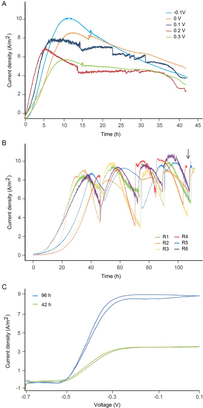
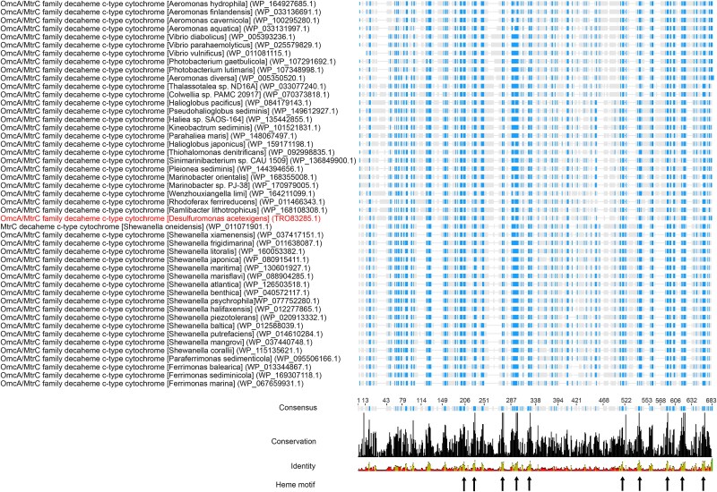
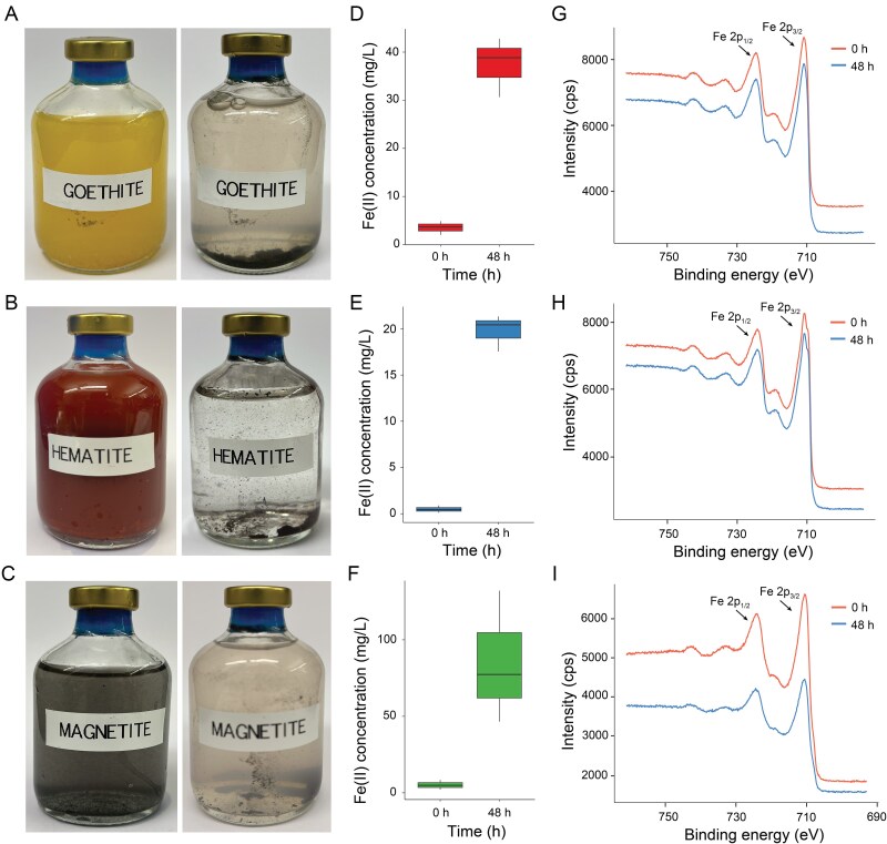
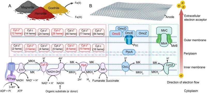
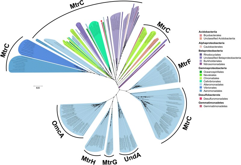
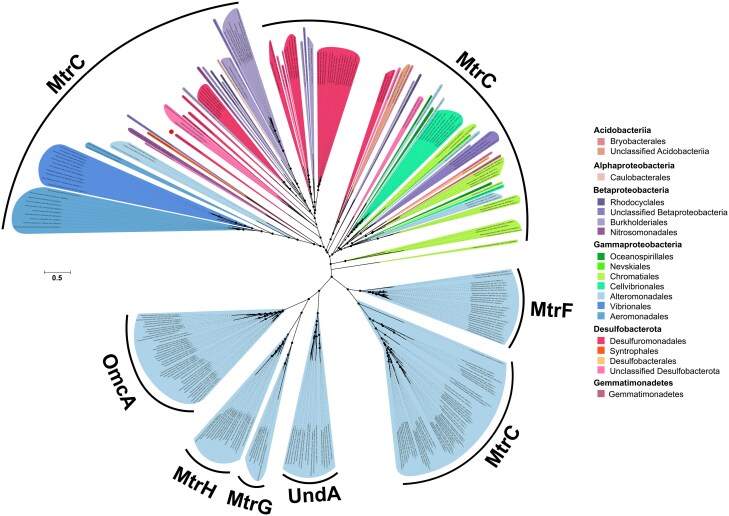
Extracellular electron transfer plays a role in the biogeochemical cycling of carbon, metals, sulfur, and nitrogen, and has wide-ranging biotechnological applications. The metal-reducing (Mtr), outer-membrane cytochrome (Omc), and porin-cytochrome (Pcc) pathways facilitate electron transfer to insoluble electron acceptors via trans-outer membrane cytochrome complexes. Although these pathways perform a similar function, they are phylogenetically unrelated, indicating independent evolutionary origins. Here, we report an extracellular electron transfer mechanism in which the high-current producing bacterium Desulfuromonas acetexigens differentially co-expresses, at transcript and protein levels, the porin-cytochrome, outer-membrane cytochrome, and metal-reducing pathways, along with high-molecular-weight cytochromes containing a large number of hemes (up to 86 heme-binding motifs), under extracellular electron transfer growth conditions (i.e. electrode under set potential or naturally occurring iron oxide minerals as the electron acceptor). Additionally, we identified over 40 Desulfobacterota species from diverse ecological environments that encode the outer-membrane cytochrome and metal-reducing pathways, with the majority also expressing the porin-cytochrome pathway. The newly identified metal-reducing proteins in Desulfobacterota form a major lineage, greatly expanding the known diversity of these proteins. To our knowledge, mtrCAB genes have not been reported in the Desulfobacterota phylum (formerly classified as Deltaproteobacteria), nor has any electroactive organism been shown to express these phylogenetically distant pathways simultaneously. These findings have ecological implications, challenging the belief that certain extracellular electron transfer pathways are exclusive to specific taxa, and suggesting that these pathways are more widespread than previously thought. Additionally, this reveals a previously unrecognized versatility in microbial electron transfer mechanisms that can be exploited in biotechnological applications.

Keywords: Desulfobacterota; Desulfuromonas; electroactive bacteria; electron transport; extracellular electron transfer; iron-reducing bacteria; multiheme cytochromes.


Conflict of interest statement

The authors declare that are no competing interests regarding the publication of this article.

Figures

Figure 1
Figure 1
Electroactivity of D. Acetexigens. (A) Chronoamperometry of a single-chamber multiple working electrode MEC inoculated with D. Acetexigens and operated under different set potentials. (B) Chronoamperometry of single-chamber MECs inoculated with D. Acetexigens and operated in fed-batch mode at a set potential of −0.1 V vs Ag/AgCl. The black arrow indicates the sampling point for differential expression analyses. (C) cyclic voltammetry (CV) response (1 mV s−1 scan rate) of D. Acetexigens at different time intervals of batch cycle operation with acetate as the electron donor and a set potential of −0.1 V vs Ag/AgCl.
Figure 2
Figure 2
Sequence alignment of outer membrane OmcA/MtrC family decaheme c-type cytochromes from various taxonomic groups. Representative sequences from a multiple sequence alignment of MtrC family decaheme proteins are shown. The name of each species appears in brackets, followed by the accession number for each protein in parentheses. MtrC from D. Acetexigens appears highlighted. Residues with conservation >60% in the sequences appear in color. Arrows indicate the heme binding sites, which correspond to conservation levels >90%. The complete alignment of the Omc/MtrC family decaheme proteins is available in Supplementary Fig. 8.
Figure 3
Figure 3
Incubations of D. acetexigens with iron oxides as the sole electron acceptor for growth. (A–C) Photographs of vials inoculated with D. acetexigens and using acetate as the electron donor at 0 h (left) and 48 h (right) after incubation with goethite (A), hematite (B), and magnetite (C). (D–F) iron (II) concentration in the medium at 0 h and 48 h after incubation of D. acetexigens with goethite (D), hematite (E), and magnetite (F). G–I XPS spectrum of the iron oxides before and after the incubation with goethite (G), hematite (H), and magnetite (I). The arrows indicate the Fe 2p1/2 and Fe 2p3/2 peaks characteristic of the iron oxides [90].
Fig<b>ure</b> 4
Figure 4
Molecular model of EET in D. acetexigens. The EET metabolic pathways utilized by D. acetexigens to deliver electrons to external insoluble electron acceptors were constructed with proteins that showed a significant response (P < 0.05) to the iron oxides and electrode as the electron acceptor. The green-dashed box (box on the right) indicates the homolog metal-reducing (Mtr) pathway with Shewanella spp. The blue-dashed box (box in the center) indicates the homolog EET pathway with Geobacter spp. The cytochromes that are exclusively found in Desulfuromonas spp. are shown within the red-dashed box (box on the left). The black arrows indicate the direction of the reactions and proton (H+) transport across membranes. The red arrow indicates the direction of the electron flow to the external electron acceptor. Protein names in red indicate proteins found in multiple copies. Protein names marked with an asterisk (*) have undefined intracellular location, which could be either periplasmic or in the outer membrane. Identifiers of the proteins used in the model are listed in Supplementary Table 16.
Figure 5
Figure 5
Phylogenetic relationship among known OmcA/MtrC decaheme c-type cytochromes from diverse bacteria and the newly identified D. acetexigens MtrC detected in this study. Maximum-likelihood phylogenetic tree constructed from a concatenated alignment of the known diversity of OmcA/MtrC decaheme c-type cytochromes and the D. acetexigens MtrC detected in this study. Color codes were assigned by taxonomic order. Bootstrap support values ≥90% from 1000 resamples are indicated with filled black circles. The scale bar represents average changes per amino acid position. MtrC, OmcA, MtrH, MtrG, UndA, and MtrF indicate members of the MtrC family of proteins. MtrC of D. acetexigens is indicated with a red dot. A full resolution image of the tree is available in Supplementary Fig. 11.
Figure 6
Figure 6
Phylogenomic relationship and environmental distribution of bacteria that encode phylogenetically distant Mtr, Omc and Pcc pathways. (A) Maximum likelihood phylogenetic tree constructed from 71 single-copy core genes of the Desulfobacterota genomes identified in this study encoding Mtr genes. Bootstrap support values ≥90% from 1000 resamples are indicated with filled black circles. The accession number for each genome is provided in parentheses. The tree was rooted using Shewanella oneidensis MR-1 genome as the outgroup. (B) Genetic occurrence of Mtr, Omc, and Pcc genes in the genomes identified in this study and their environmental distribution. Filled colored dots indicate the presence of a gene or set of genes in a genome or the presence of the organism in a given environment. “EET” indicates that the EET capability has been observed using graphene oxide (GO), Fe(III) oxides or electrode as the electron acceptor. Unfilled dots in “EET” indicate that the bacteria have not been tested for EET capability. “Wastewater” in environmental distribution indicates that the species has been observed in wastewater or wastewater treatment systems. “Bioelectrochemical system” in environmental distribution indicates the detection of the organism in electrode biofilms from bioelectrochemical reactors. Detailed characteristics and full description of the genomes, genes, and environments are found in Supplementary Table 17.
Figure 7
Figure 7
Phylogenetic relationship among known OmcA/MtrC decaheme c-type cytochromes from diverse bacteria and the Desulfobacterota MtrC detected in this study. Maximum-likelihood phylogenetic tree constructed from a concatenated alignment of the known diversity of OmcA/MtrC decaheme c-type cytochromes and the Desulfobacterota MtrC detected in this study. Color codes were assigned by taxonomic order. Bootstrap support values ≥90% from 1000 resamples are indicated with filled black dots. The scale bar represents the average changes per amino acid position. MtrC, OmcA, MtrH, MtrG, UndA, and MtrF indicate members of the MtrC family of proteins. MtrC of D. acetexigens is indicated with a red dot. Full resolution image of the tree is available in Supplementary Fig. 14.

References

    1. Methé BA, Nelson KE, Eisen JA. et al. Genome of Geobacter sulfurreducens: metal reduction in subsurface environments. Science 2003;302:1967–9. 10.1126/science.1088727 - DOI - PubMed
    1. Ishii S, Suzuki S, Norden-Krichmar TM. et al. A novel metatranscriptomic approach to identify gene expression dynamics during extracellular electron transfer. Nat Commun 2013;4:1601. 10.1038/ncomms2615 - DOI - PubMed
    1. Shi L, Dong H, Reguera G. et al. Extracellular electron transfer mechanisms between microorganisms and minerals. Nat Rev Microbiol 2016;14:651–62. 10.1038/nrmicro.2016.93 - DOI - PubMed
    1. Shaw DR, Ali M, Katuri KP. et al. Extracellular electron transfer-dependent anaerobic oxidation of ammonium by anammox bacteria. Nat Commun 2020;11:2058. 10.1038/s41467-020-16016-y - DOI - PMC - PubMed
    1. Kumar A, Hsu LHH, Kavanagh P. et al. The ins and outs of microorganism–electrode electron transfer reactions. Nat Rev Chem 2017;1:0024. 10.1038/s41570-017-0024 - DOI

LinkOut - more resources