. 2025 May 17;16(1):4595.
doi: 10.1038/s41467-025-59721-2.

Conformational trajectory of the HIV-1 fusion peptide during CD4-induced envelope opening

Affiliations

Conformational trajectory of the HIV-1 fusion peptide during CD4-induced envelope opening

Bhishem Thakur et al. Nat Commun. .

Abstract

The hydrophobic fusion peptide (FP), a critical component of the HIV-1 entry machinery, is located at the N terminus of the envelope (Env) gp41 subunit. The receptor-binding gp120 subunit of Env forms a heterodimer with gp41. The gp120/gp41 heterodimer assembles into a homotrimer, in which FP is accessible for antibody binding. Env conformational changes or "opening" that follow receptor binding result in FP relocating to a newly formed interprotomer pocket at the gp41-gp120 interface where it is sterically inaccessible to antibodies. The mechanistic steps connecting the entry-related transition of antibody accessible-to-inaccessible FP configurations remain unresolved. Here, using SOSIP-stabilized Env ectodomains, we visualize that the FP remains accessible for antibody binding despite substantial receptor-induced Env opening. We delineate stepwise Env opening from its closed state to a functional CD4-bound symmetrically open Env in which we show that FP was accessible for antibody binding. We define downstream re-organizations that lead to the formation of a gp120/gp41 cavity into which the FP buries to become inaccessible for antibody binding. These findings improve our understanding of HIV-1 entry and delineate the entry-related conformational trajectory of a key site of HIV vulnerability to neutralizing antibody.


Conflict of interest statement

Competing interests: The authors declare the following competing interests: B.T. and P.A. have applied for patents on HIV-1 Envelope modifications related to this work. The other authors declare no competing interest.

Figures

Fig. 1
Fig. 1. Time-dependent conformational changes in HIV-1 BG505 SOSIP Env upon incubation with CD4.
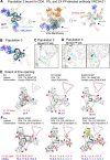
A Structure of pre-fusion, pre-receptor, closed HIV-1 Env (PDB: 5I8H) bound to broadly neutralizing, fusion peptide-directed antibody VRC34.01. The Env is shown in surface representation with the gp120 subunits colored light gray, and within the gp120 subunits, the V1V2 loop colored wheat, V3 loop olive and the residues contributing to the bridging sheet in the open Env colored red. The gp41 subunits are colored black with the fusion peptide (FP) colored cyan. The antibody VRC34.01 is shown in ribbon representation bound to its FP-centered epitope. B Structure of pre-fusion, CD4-bound open HIV-1 Env bound to CD4-induced antibody 17b. The Env is colored similarly as in panel A. CD4 is shown as a yellow ribbon and 17b Fab is shown as an orange ribbon. C Surface plasmon based binding (SPR) analysis monitoring FP burial. Env was incubated at 25 °C with either sCD4 alone or with CD4 and the coreceptor mimicking antibody 17b. At different time-points after incubation, binding was measured to the fusion peptide targeting antibody VRC34.01. Some elements of the SPR schematic were created in BioRender. Acharya, P. (2025) https://BioRender.com/m74zcsz. D Simultaneous Env opening and fusion peptide burial were measured by incubating Env with CD4 and at different time-points injecting over a VRC34.01 IgG or a 17b IgG surface. Source data for panels (C, D) are provided as a Source Data file. Data shown are representative of at least two independent experiments. E Cryo-EM reconstructions of three distinct populations of CD4/17b-bound, partially open Env bound to VRC34.01. Population 1 is bound to VRC34.01 at all three sites. The blue arrows indicate sites unoccupied by VRC34.01 in Populations 2 and 3. Source Data.
Fig. 2
Fig. 2. A partially open intermediate on the HIV-1 entry pathway retains FP accessibility to antibody binding.
A Three views of Population 1 structure shown in cartoon representation with gp41 colored black, gp120 gray, CD4 yellow, VRC34.01 blue, 17b orange. Glycans are shown as sticks. FP within the gp41 subunit is colored cyan. Within gp120, bridging sheet is colored red and α0 helix green. B Population 1 coordinates including Env (gp120 in gray, gp41 in black) and CD4 (yellow) fitted into the in situ cryo-ET reconstruction of a partially open CD4-bound Env (EMD-29294). C (Left to right) Vectors describing position of gp120 relative to gp41. The gp120 structure (blue), gp120 V1/V2 region (green), and gp41 (orange) in the closed state overlayed with the centroid locations depicting the dihedral, angles, and distances describing the position of gp120 relative to gp41. Dihedral, angle, and distance values for closed, intermediate, and open state structures. Data shown as scatter dot plots with horizontal lines indicating the mean and standard deviation. List of structures used for the calculations is provided in Supplementary Table 3. Source data are provided as a Source Data file. D (Left) Population 1 protomer shown in surface representation zoomed-in at the location of the FP. FP is shown in cartoon representation. The gp120 subunit is colored light gray, gp41 black, FP cyan and FPPR pale green. (Middle) One protomer of the partially open Env bound to CD4, 17b Fab and 8ANC195 Fab (PDB ID: 6CM3) shown in surface representation zoomed-in at the location of the FP (shown in cartoon representation). The gp120 subunit is colored gray, gp41 black, FP dark teal and FPPR light pink. (Right) Overlay of a Population 1 protomer with a protomer of a partially open CD4,17b,8ANC195-bound Env (PDB ID: 6CM3). The gp120 subunits were used for the superposition. Inset zooms in on the FP and FPPR. Zoomed-in panel is slightly rotated compared to zoomed-out view for better visualization. The solid lines (pale green for Population 1 and light pink for 6CM3) show the distance between FPPR residues Gln 540 and gp120 residue Phe 223. Source Data.
Fig. 3
Fig. 3. Burial of FP upon gp120 opening.
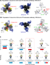
A Three views of the Population 2 structure shown in cartoon representation with gp41 colored black, gp120 light gray, CD4 yellow, VRC34.01 Fab blue, 17b Fab orange. Glycans are shown in stick representation. The FP within the gp41 subunit is colored cyan. Within gp120, the bridging sheet is colored red and the α0 helix green. Blue circle-headed arrows indicate the gp41 positions that are not bound to VRC4.01 Fab. B View of Population 3 coordinates from the viral membrane shown in cartoon representation with gp41 colored black, gp120 light gray, CD4 yellow, VRC34.01 Fab blue, 17b Fab orange. Glycans are shown in stick representation. The FP within the gp41 subunit is colored cyan. C Population 2 structure zoomed-in view of gp41 subunit that was not bound to VRC34.01 showing the buried FP in cyan and the FPPR in light green. The EM map (EMD-46671) contoured at a level of 0.105 in ChimeraX is shown as a transparent surface with fitted coordinates shown in cartoon representation. D Population 3 structure zoomed-in view of its two gp41 subunits that were not bound to VRC34.01, showing the buried FP in cyan and the FPPR in light green. The EM map (EMD-46672) contoured at a level of 0.123 in ChimeraX is shown as a transparent surface with fitted coordinates shown in cartoon representation. E, F Extent of Env openness measured as the distance between residue 368 (blue spheres) and residue 124 (red spheres) in (E) previously published Env conformational states and (F). Env conformational states defined in this study.
Fig. 4
Fig. 4. FP trajectory upon CD4-induced Env opening.
A HIV-1 Env structures organized by extent of opening. Left to right: closed, VRC34.01-bound Env (PDB: 5I8H) with gp41 colored olive, FPPR orange and FP cyan; partially open, CD4,17b,VRC34.01-bound Env (PDB:9D90; this study) with gp41 colored black, FPPR light green and FP cyan; partially open, CD4,17b,8ANC195-bound Env (PDB:6CM3) with gp41 colored magenta, FPPR light pink and FP teal; partially open CD4,17b,VRC34.01-bound Env (EMD-46671; this study) with gp41 colored black, FPPR light green and FP cyan; fully open, CD4,17b-bound Env (PDB:5VN3) with gp41 colored blue, FPPR light blue and FP cyan. The gp120 subunit is colored gray. Inset: Zoomed-in view of region around α0 helix (green). B 180° rotated views A with brown squares around the FP (colored cyan except in partially open CD,17b,8ANC195-bound BG505 structure, where it is colored teal). C Zoomed-in views of region around FP. Red arrows indicate direction of CD4-induced Env opening from pre-CD4, closed Env to CD4-induced fully open Env. D Comparisons of gp41 organization between fully open Env with sequestered and inaccessible FP (PDB: 5VN3), and (left) P1 (transiently exposed FP), (middle) partially open Env with transiently buried FP (PDB: 6CM3), and (right) P2 (inaccessible FP). Red arrows (left and middle panels) indicate HR2 movement that allows the FPPR to re-orient creating space for FP burial. EG Locations in Env structure (PDB 4ZMJ fitted into EMDB-21412) where two fluorophores (Cy3 and Cy5 derivatives) are attached for smFRET imaging (E), three-dimensional presentation (F) and quantification (G) of conformational distribution-indicated FRET histograms observed from the gp120-gp41 perspective. The probability of each state (G), presented as mean ± s.e.m. (uncertainty), was derived from histograms (F), each compiled from Nm > 200 traces (specifically, 241, 213, 236, and 242), as detailed in Fig. S12F. The determining parameters are listed in Table S4. Virus EnvBG505 samples three primary conformational states (PT Pre-triggered, PC Prefusion Closed, and CO: CD4-bound open). PT predominates in the ligand-free condition, while VRC34 shifts the conformational landscape differently from that of the CD4-bound opening. Source data are provided as a Source Data file. Source Data.
Fig. 5
Fig. 5. Partially open early intermediates and a stepwise mechanism for CD4-induced Env opening.
A CD4,VRC34.01-bound BG505 SOSIP Env with rotation, bridging sheet (red) and α0 helix (green) formation observed in a single gp120. B CD4,VRC34.01-bound BG505 SOSIP Env with rotation, bridging sheet (red) and α0 helix (green) formation observed in two gp120 subunits. C A structure-guided mechanism for stepwise Env opening along the HIV-1 entry pathway. Top panel structures were determined previously, bottom panel structures were determined in this study. Stepwise transitions are marked with numbers within a circle on top of each structure starting from (1) the binding of a single CD4 to a closed Env trimer (PDB: 5U1F, 8FYI). This is followed by (2) opening of the Env trimer (EMD-29292) that allows additional CD4 molecules to bind. (5) A partially open Env conformation was described bound to CD4, a coreceptor (Co-R) mimicking antibody, and the gp120/gp41 interface targeting antibody 8ANC195 (PDB: 6CM3, 6EDU) where the FP was buried within a gp41 cavity. The CD4-induced opening of the HIV-1 Env culminates in the complete rotation of all the gp120 subunits that are accompanied by gp41 conformational changes and resulting in the burial of FP. This state is numbered (8) in this schematic. This study showed that the geometry of the functional entry intermediate (5) that was also visualized on membrane-associated Env (EMD-29294), was compatible with the FP being either buried or exposed, and thus, in this conformation the FP was accessible to antibodies. Further, this study filled in mechanistic gaps between (2) and (5) by showing stepwise gp120 rotations to reach this functional entry intermediate. Finally, this study visualized a stepwise mechanism for how the functional entry intermediate (5) may transition to the fully open Env (8), yet again by stepwise opening of the each gp120 subunit from its partially rotated to the fully rotated conformation, which was accompanied by burial of the FP in the corresponding protomer. Schematics of membranes and the host receptors were created in BioRender. Acharya, P. (2025) https://BioRender.com/7tpbllk.

Update of

Similar articles

References

    1. Harrison, S. C. Viral membrane fusion. Virology479, 498–507 (2015). - PMC - PubMed
    1. Pancera, M. et al. Structure and immune recognition of trimeric pre-fusion HIV-1 Env. Nature514, 455–461 (2014). - PMC - PubMed
    1. Lyumkis, D. et al. Cryo-EM structure of a fully glycosylated soluble cleaved HIV-1 envelope trimer. Science342, 1484–1490 (2013). - PMC - PubMed
    1. Kwong, P. D. et al. Structure of an HIV gp120 envelope glycoprotein in complex with the CD4 receptor and a neutralizing human antibody. Nature393, 648–659 (1998). - PMC - PubMed
    1. Zhao, M. et al. Insertion and anchoring of the HIV-1 fusion peptide into a complex membrane mimicking the human T-cell. J. Phys. Chem. B128, 12710–12727 (2024). - PubMed

MeSH terms

LinkOut - more resources