. 2025 May 17;25(1):180.
doi: 10.1186/s12935-025-03808-9.

FBXL18 promotes endometrial carcinoma progression via destabilizing DUSP16 and thus activating JNK signaling pathway

Affiliations

FBXL18 promotes endometrial carcinoma progression via destabilizing DUSP16 and thus activating JNK signaling pathway

Jie Pi et al. Cancer Cell Int. .

Abstract

Objective: The therapeutic options for patients with advanced endometrial carcinoma (EC) were still limited and the prognosis remained unfavorable. F-box and leucine-rich repeat protein 18 (FBXL18), belonging to the F-box protein family, was frequently altered in human cancer, while its functional role and underlying mechanisms in EC were largely unexplored.

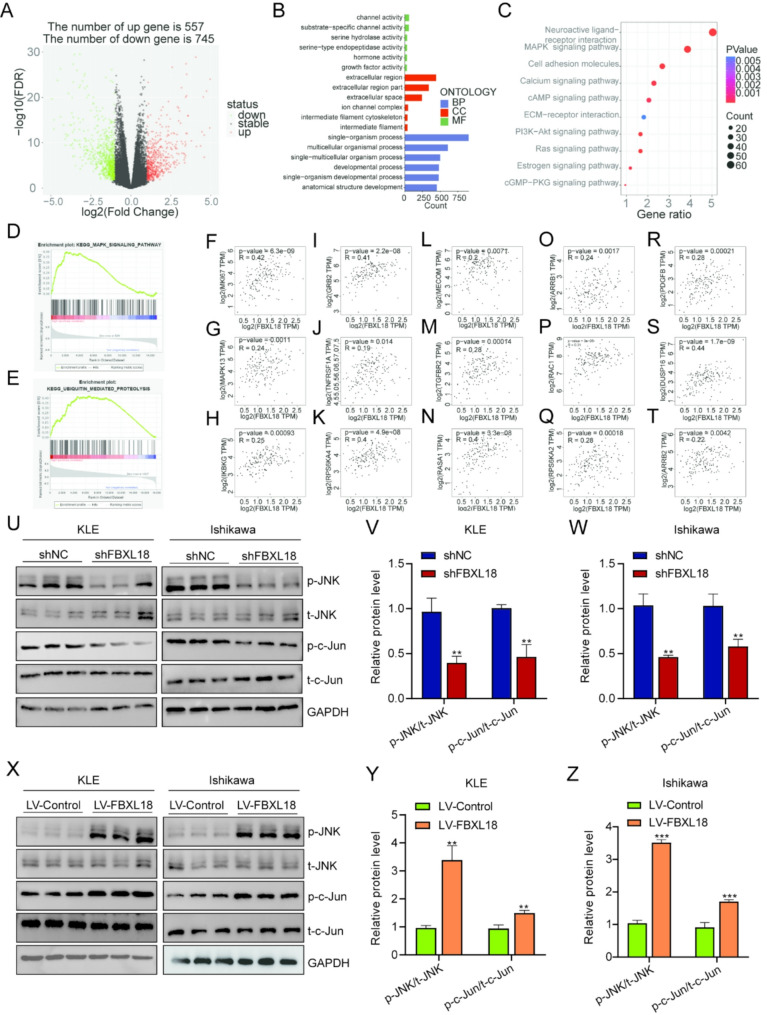
Methods: The expression of FBXL18 in EC tissues and cells were explored using data mining strategies and further experiments. Multiple in vitro assays, including CCK-8, colony formation, wound healing, and Transwell invasion assays, were performed to assess the function of FBXL18 on cell proliferation, migration, and invasion. Bioinformatic analyses, western blot, qRT-PCR, Co-immunoprecipitation and ubiquitination assays were employed to identify the downstream pathway and direct substrate of FBXL18.

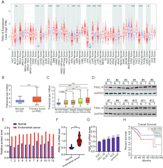
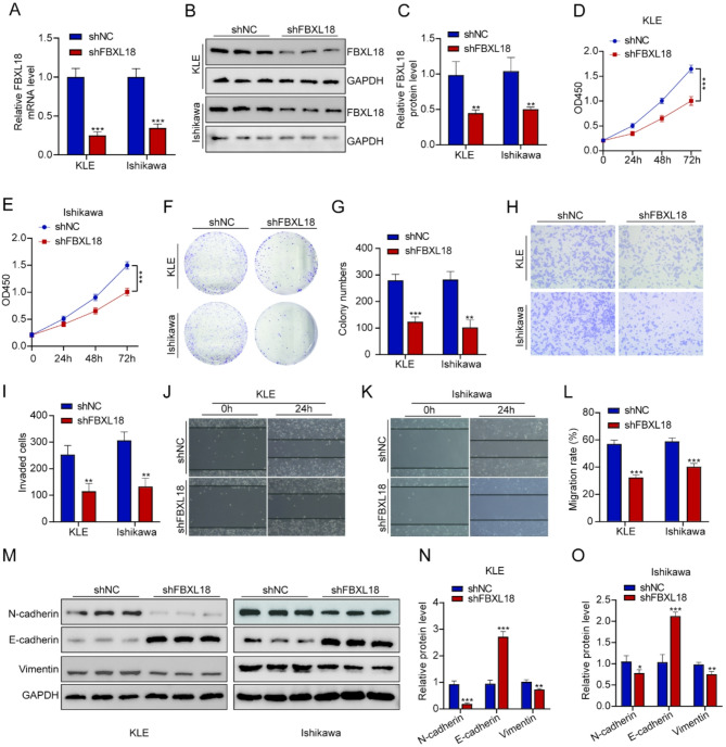
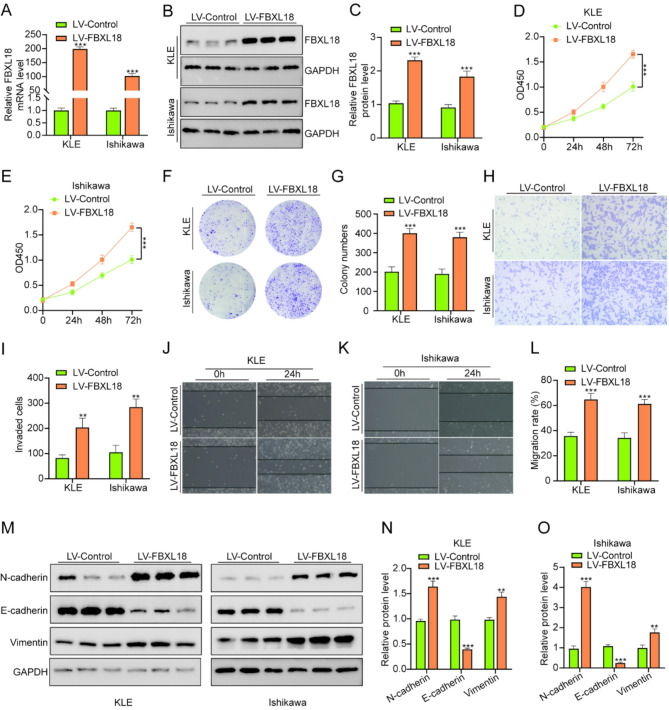
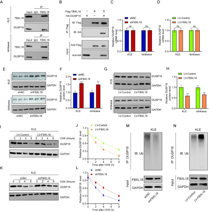
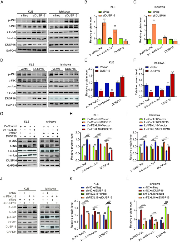
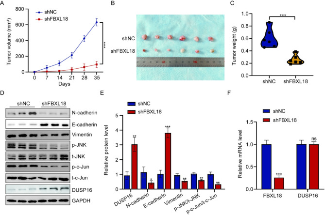
Results: FBXL18 was highly expressed in EC tissues and cell lines, and EC patients with high FBXL18 expression had poor clinical outcome. Loss- and gain-of-function assays showed that silencing FBXL18 suppressed EC cell proliferation, migration, and invasion, while overexpressing FBXL18 caused the opposite effects. Mechanistically, FBXL18 could physically interacted with DUSP16, a dual specificity phosphatase, leading to its ubiquitination and degradation, and thus activating JNK signaling pathway. Upregulation of DUSP16 in EC cells alleviated FBXL18 overexpression-induced activation of JNK signaling pathway, and reversed FBXL18 overexpression-mediated enhanced cell capacities of proliferation, migration, and invasion.

Conclusion: In summary, our study had showcased the elevated expression, prognostic prediction performance, and the malignant tumor-promoting role of FBXL18 in EC. The novel mechanisms underlying this phenotype are that FBXL18 promotes the ubiquitination and degradation of DUSP16, and thus activates JNK/c-JUN signaling to facilitate EC progression.

Keywords: DUSP16; Endometrial carcinoma; FBXL18; JNK signaling; Ubiquitination.


Conflict of interest statement

Declarations. Ethics approval and consent to participate: The experiment with patient tissue specimens was authorized by the Ethics Committee of Renmin Hospital of Wuhan University. All animal experiments were approved by the Animal Care and Use Committee of Wuhan University Renmin Hospital. Consent for publication: All authors have approved the publication of this study. Competing interests: The authors declare no competing interests.

Figures

Fig. 1
Fig. 1
FBXL18 was highly expressed in EC tissues and cells, and augmented expression predicted poor prognosis. (A) The expression of FBXL18 in various tumors from the TIMER database. (B) The mRNA expression pattern of FBXL18 in EC tissues and corresponding normal tissues was analyzed using the UALCAN database. (C) The comparison of FBXL18 mRNA level in normal tissues and EC tissues of different tumor stage. (D-E) Western blot analysis of FBXL18 protein levels in twelve pairs of EC tissues and adjacent normal tissues, and quantitative analysis. (F) qRT-PCR analysis of FBXL18 mRNA levels in twelve paired EC tissues and adjacent normal tissues. (G) qRT-PCR analyses of FBXL18 mRNA levels in normal endometrial stromal cells (ESC) and a variety of EC cells (KLE, Ishikawa, HEC-1B, RL95-2). (H) The association of FBXL18 expression and patients’ overall survival was determined. *P < 0.05; **P < 0.01; ***P < 0.001
Fig. 2
Fig. 2
Silence of FBXL18 impeded cell proliferation, migration, and invasion, and suppressed EMT in EC cells. (A) The mRNA levels of FBXL18 in EC cells of shNC and shFBXL18 groups were detected by qRT-PCR. (B-C) Western blot analysis of FBXL18 protein levels in EC cells of shNC and shFBXL18 groups, and quantitative analysis. (D-G) CCK-8 and colony formation assays were conducted to assess cell proliferation abilities in stably infected EC cells, and quantitative analyses. (H-I) Transwell invasion assay was used to evaluate invasiveness in EC cells after silencing FBXL18, and quantitative analysis. (J-L) Wound healing assay was employed to determine cell migration ability after silencing FBXL18 in KLE and Ishikawa cells, and quantitative analysis. (M-O) Western blot analysis of EMT related markers (N-cadherin, Vimentin, and E-cadherin) in EC cells of shNC and shFBXL18 groups, and quantitative analysis. *P < 0.05; **P < 0.01; ***P < 0.001
Fig. 3
Fig. 3
Enforced expression of FBXL18 accelerated cell proliferation, migration, and invasion, and promoted EMT in EC cells. (A) The mRNA levels of FBXL18 in EC cells of LV-Control and LV-FBXL18 groups were detected by qRT-PCR. (B-C) Western blot analysis of FBXL18 protein levels in EC cells of LV-Control and LV-FBXL18 groups, and quantitative analysis. (D-G) CCK-8 and colony formation assays were conducted to assess cell proliferation abilities in stably infected EC cells, and quantitative analyses. (H-I) Transwell invasion assay was used to evaluate invasiveness in EC cells after overexpressing FBXL18, and quantitative analysis. (J-L) Wound healing assay was employed to determine cell migration ability after FBXL18 overexpression in KLE and Ishikawa cells, and quantitative analysis. (M-O) Western blot analysis of EMT related markers (N-cadherin, Vimentin, and E-cadherin) in EC cells of LV-Control and LV-FBXL18 groups, and quantitative analysis. *P < 0.05; **P < 0.01; ***P < 0.001
Fig. 4
Fig. 4
FBXL18 positively regulated JNK signaling pathway in EC. (A) Volcano plot of FBXL18-related differentially expressed genes based on the RNA sequencing data of EC samples with high- and low-FBXL18 expression. (B-C) GO and KEGG enrichment analyses of FBXL18-related differentially expressed genes. (D-E) The results of GSEA revealed that MAPK and ubiquitin-mediated proteolysis were markedly enriched in FBXL18High group. (F-T) The Pearson’s correlation analyses of FBXL18 and genes of the MAPK signaling pathway. (U-W) The protein levels of JNK, p-JNK, c-JUN, and p-c-JUN in EC cells after silencing FBXL18 were determined by western blot assay, and quantitative analysis. (X-Z) The protein levels of JNK, p-JNK, c-JUN, and p-c-JUN in EC cells after overexpressing FBXL18 were measured by western blot assay, and quantitative analysis. *P < 0.05; **P < 0.01; ***P < 0.001
Fig. 5
Fig. 5
FBXL18 targeted DUSP16 as a ubiquitination substrate. (A-B) The results of Co-IP and western blotting assays revealed the endogenous and exogenous interaction of FBXL18 and DUSP16. (C-D) qRT-PCR analysis of DUSP16 mRNA levels after silencing or overexpressing FBXL18 in KLE and Ishikawa cells. (E-F) Western blot analysis of DUSP16 protein level in EC cells of shNC and shFBXL18 groups, and quantitative analysis. (G-H) Western blot analysis of DUSP16 protein level in EC cells after overexpressing FBXL18, and quantitative analysis. (I-J) DUSP16 protein half-life in EC cells of LV-Control and LV-FBXL18 groups were evaluated by CHX chase assay. (K-L) Comparison of DUSP16 protein half-life in EC cells stably knockdown of FBXL18 and control cells. (M-N) The ubiquitination status of endogenous DUSP16 after silencing or overexpressing FBXL18 in EC cells were determined by immunoprecipitation and western blotting. *P < 0.05; **P < 0.01; ***P < 0.001
Fig. 6
Fig. 6
FBXL18 promoted EC cell malignancy via DUSP16-mediated activation of JNK signaling. (A-C) Western blot analysis of DUSP16, JNK, p-JNK, c-JUN, and p-c-JUN levels in EC cells of siNeg and siDUSP16 groups, and quantitative analysis. (D-F) Western blot analysis of DUSP16, JNK, p-JNK, c-JUN, and p-c-JUN levels in EC cells after overexpressing DUSP16, and quantitative analysis. (G-I) Western blot analysis of DUSP16, JNK, p-JNK, c-JUN, and p-c-JUN levels in EC cells transfected with LV-Ctrl + Vector, LV-Ctrl + DUSP16, LV-FBXL18 + Vector, LV-FBXL18 + DUSP16, and quantitative analysis. (J-L) Western blot analysis of DUSP16, JNK, p-JNK, c-JUN, and p-c-JUN levels in EC cells transfected with shNC + siNeg, shNC + siDUSP16, shFBXL18 + siNeg, shFBXL18 + siDUSP16, and quantitative analysis. *P < 0.05; **P < 0.01; ***P < 0.001
Fig. 7
Fig. 7
Silence of FBXL18 inhibited EC growth in vivo. (A) Tumor volumes of shNC and shFBXL18 groups were determined at different defined time points. (B) Representative images of xenograft tumors in shNC and shFBXL18 groups. (C) Tumor weights of shNC and shFBXL18 groups were measured at the end point. (E) Western blot analysis of FBXL18, DUSP16, JNK, p-JNK, c-JUN, p-c-JUN, and EMT-related markers in tumor samples of shNC and shFBXL18 groups, and quantitative analysis. (F) qRT-PCR analysis of FBXL18 and DUSP16 in tumor samples of shNC and shFBXL18 groups. *P < 0.05; **P < 0.01; ***P < 0.001

Similar articles

References

    1. Murali R, Soslow RA, Weigelt B. Classification of endometrial carcinoma: more than two types. Lancet Oncol. 2014;15(7):e268–278. - PubMed
    1. Huvila J, Pors J, Thompson EF, Gilks CB. Endometrial carcinoma: molecular subtypes, precursors and the role of pathology in early diagnosis. J Pathol. 2021;253(4):355–65. - PubMed
    1. Léon-Castillo A. Update in the molecular classification of endometrial carcinoma. Int J Gynecol Cancer. 2023;33(3):333–42. - PubMed
    1. Sung H, Ferlay J, Siegel RL, Laversanne M, Soerjomataram I, Jemal A, Bray F. Global Cancer statistics 2020: GLOBOCAN estimates of incidence and mortality worldwide for 36 cancers in 185 countries. CA Cancer J Clin. 2021;71(3):209–49. - PubMed
    1. Li J, Yang H, Zhang L, Zhang S, Dai Y. Metabolic reprogramming and interventions in endometrial carcinoma. Biomed Pharmacother. 2023;161:114526. - PubMed

LinkOut - more resources