. 2025 May 17;23(1):229.
doi: 10.1186/s12964-025-02150-x.

AlkTango reveals a role for Jeb/Alk signaling in the Drosophila heart

Affiliations

AlkTango reveals a role for Jeb/Alk signaling in the Drosophila heart

G Wolfstetter et al. Cell Commun Signal. .

Abstract

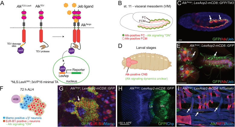
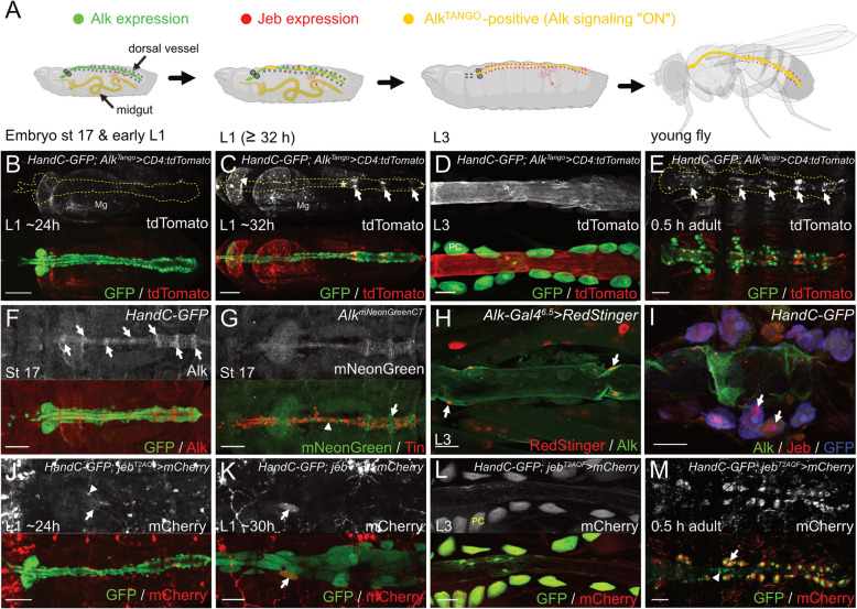
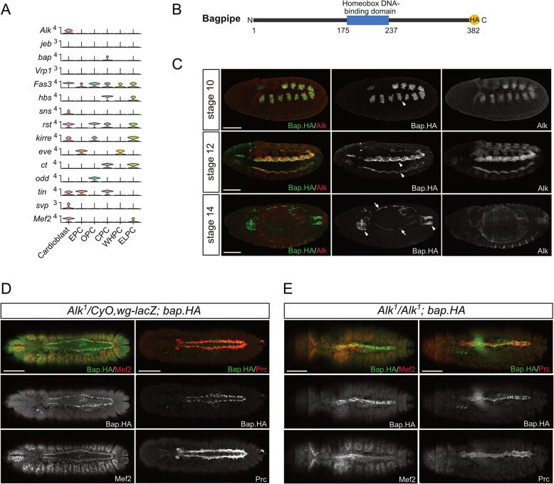
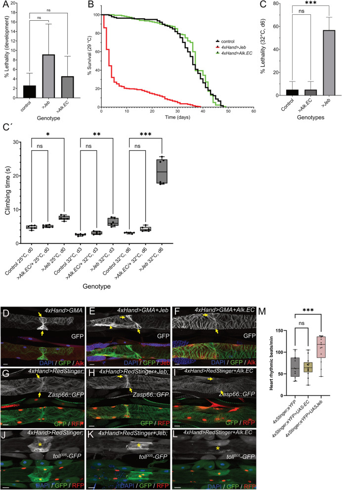
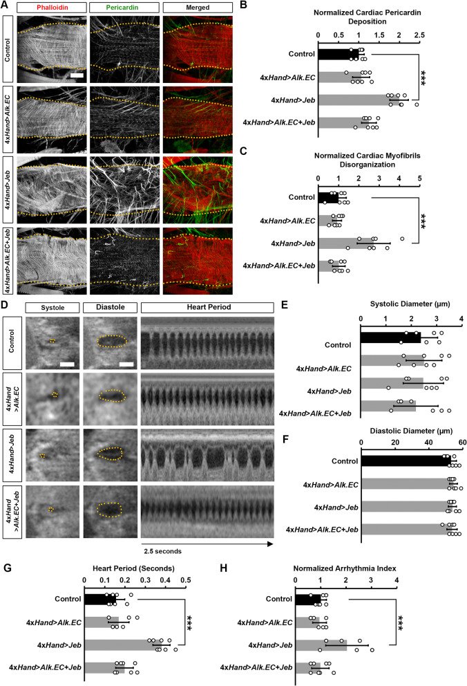
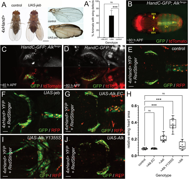
Anaplastic lymphoma kinase (Alk) signaling is important in a variety of biological contexts such as cell type specification, regulation of metabolic and endocrine programs, behavior, and cancer. In this work, we generated a Tango GPCR assay-based, dimerization-sensitive Alk activity reporter (AlkTango) and followed receptor activation throughout Drosophila development. AlkTango reports Alk activation in embryonic and larval tissues previously linked to Alk signaling. Remarkably, AlkTango was active in the heart of Drosophila larvae and adult flies. We show that cardiomyocytes express Alk from late embryonic stages to adulthood, while jeb expression in pericardial cells coincided with AlkTango activity. Perturbation of cardiac Alk signaling leads to decreased adult survival as well as lower fitness and increased lethality in response to heat stress. In keeping with a role for Alk, heart measurements reveal arrythmia and irregular muscle contraction upon ligand stimulation. Finally, activation of cardiac Alk signaling induces hyperplasia in the accessory wing hearts of adult flies.

Keywords: CRISPR/Cas9; Cardiac arrythmia; Cardiomyocyte; Jelly belly; Tango RTK activity assay; Wing hearts.


Conflict of interest statement

Declarations. Competing interests: No competing interests.

Figures

Fig. 1
Fig. 1
AlkTango reports Alk activation. A Schematic outline of the AlkTango system, comprising the AlkTCS::LexA and AlkTEV CRISPR modified Alk alleles, as well as the LexAop2-reporter. In brief: AlkTCS::LexA and AlkTEV dimerize in the presence of Alk ligand Jeb, resulting in proteolytic cleavage of an NLS.LexADBD::3xVP16 minimal TA chimera (LexA*) which initiates reporter gene expression. B Schematic outline of AlkTango activity (depicted in green), indicating Alk activation in visceral mesoderm founder cells (arrows) of AlkTango; LexAop2-mCD8::GFP embryos. Alk protein expression depicted in red. C AlkTango activity (GFP, green), indicating Alk activation in visceral mesoderm founder cells (arrows) of AlkTango; LexAop2-mCD8::GFP embryos. Anti-Alk appears in red, anti-Jeb in blue. D Schematic outline of Alk expression in the larval CNS, where Alk signaling dynamics are unclear. Alk protein expression depicted in red. E Antibody staining revealing AlkTango activity (GFP, green) in the larval CNS of AlkTango; LexAop2-mCD8::GFP animals. Anti-Alk appears in red, anti-Jeb in blue. F Schematic outline of AlkTango activation (depicted in green) in EcR-B1-positive MB γ-neurons of a third instar larva. EcR-B1 in red, mamo labels MB α’β’-neurons in blue. G AlkTango activation (GFP, green) in EcR-B1-positive MB γ-neurons of a third instar larva. EcR-B1 in red, mamo labels MB α’β’-neurons in blue. H AlkTango activity (GFP, green) in lamina and medulla neurons of a pupal optic lobe ~50 h APF. I AlkTango activation (RFP, red) in somatic muscles and motoneuron axons (arrows) of a third instar AlkTango; LexAop2-mCD4::tdTomato larva. Anti-Alk appears in green, muscles stained with phalloidin (blue). Scale bars are: 20 µm in C, 50 µm in E, H, and 100 µm in I. Schematics in B, D and F created with BioRender.com (2023)
Fig. 2
Fig. 2
Alk and Jeb are expressed in adjacent cell types in the dorsal vessel. A Schematic created with BioRender.com (2023) summarizing cardiac Alk expression, jebT2A reporter expression in surrounding tissues as well as cardiac AlkTango reporter activity in different developmental stages. Green indicates Alk expression with no or low AlkTango reporter activity, Jeb expressing tissues are marked in red, AlkTango reporter activity is shown in yellow, HandC-GFP positive lymph gland and PCs are denoted in dark grey. B AlkTango reporter activity (white, red in merge) in a freshly hatched L1 larva. The HandC-GFP reporter (green) was used to label the dorsal vessel (encircled area in b/w images), Tango associated tdTomato expression is only visible in the larval midgut (Mg). C Six hours after hatching, AlkTango reporter activity (white, red in merge) is now visible in aorta (arrowhead), ostia (arrows), and the valve region (asterisk) of the HandC-GFP labeled (green) dorsal vessel. D Antibody staining against HandC-GFP (green) and tdTomato (anti RFP antibody) reveals persistent AlkTango reporter activity (white, red in merge) in cardiomyocytes of a wandering third instar larva. PC = pericardial cell. E AlkTango reporter activity (white, red in merge) in the adult heart (encircled area in b/w image) ~30 min after emergence. Heart cells are labeled by the HandC-GFP reporter, the highest AlkTango signals appear in ostia (arrows). F Anti-Alk antibody staining (white, red in merge) in cardiomyocytes of the HandC-GFP-positive (green) dorsal vessel of a late-stage Drosophila embryo. Arrows label presumptive ostia. G Anti-Tin (red) and mNeonGreen (white, green in merge) antibody staining reveals AlkmNeonGreenCT expression in Tin-positive cardiomyocytes (arrowhead) and Tin-negative ostia (arrow) of a late-stage Drosophila embryo. H Antibody staining of the dorsal vessel of a wandering L3 larva expressing UAS-RedStinger (red) under control of AlkP6.5-Gal4. Anti Alk antibody staining appears in green; arrows indicate cardiomyocyte nuclei. I Alk expression in the adult heart of a HandC-GFP fly stained with anti-Alk (green), anti-Jeb (red), and anti-GFP (blue). Arrows indicate Jeb antibody staining in PCs. J JebT2A-QF driven QUAS-mCherry expression (white, red in merge) in a freshly hatched L1 larva. An arrow marks dendritic branches of a JebT2A-positive ddaC neuron, arrowhead labels an unspecified JebT2A-positive cell close to the heart. K Six hours afterhatching, JebT2A-QF driven QUAS-mCherry expression (white, red in merge)can be detected in single pericardial cells (arrow) of the dorsal vessel (labeled green by HandC-GFP). L Antibody staining against mCherry (anti-RFP antibody) and HandC-driven GFP reveals Jeb reporter activity in pericardial cells (PC) of a third instar larva. M Jeb reporter activity appears in pericardial cells (arrow) but not cardiomyocytes (arrowhead) of the adult heart ~30 min after emergence. Scale bars are 10 µm in J; 20 µm in E and F; 50 µm in B, C, G, H, I, and K; 100 µm in D, and L
Fig. 3
Fig. 3
Alk mutants do not display obvious defects in specification of the embryonic heart. A Stacked violin plot illustrating the expression levels of genes associated with VM fusion downstream of Alk. The genes shown are Alk, jeb, bap, Vrp1, Fas3, hbs, sns, rst, kirre, eve, ct, odd, tin, svp and Mef2. Each column represents a cell cluster identified within the cardiogenic progenitor population, and the stacked violins depict the distribution of expression for each gene across clusters. Data was derived from the single-cell RNA sequencing dataset (GSE168774) and the plot was generated using the scCustomize R package. B Schematic representation of the CRISPR/Cas9-mediated C-terminal HA tag knock-in at the endogenous bap gene locus, creating the bapHA allele. C bapHA embryos stained for HA (green) and Alk (red). BapHA is expressed in the VM at stages 10-12 where it exhibits strong nuclear localization in founder cells at stage 12 (arrowheads). Expression is observed in both the foregut and hindgut at stage 14 (arrowheads). BapHA is also present in early cardiac precursor cells at stage 14 (arrows). D-E Stage 15–16 embryos stained for BapHA (green) and the heart markers Mef2 and Prc (red). In Alk1 heterozygote controls, Bap is expressed in pericardial cells (D). In Alk1 mutants, the midgut development is affected, but the heart structure and the expression of heart markers, including Bap, Mef2, and Prc, remain unaffected (E)
Fig. 4
Fig. 4
Jeb overexpression affects later heart function. Column graph depicting developmental lethality (in %) of 4xHand-Gal4 control (mean = 2.612 +/- 2.594 SD), 4xHand>UAS-jeb (mean = 9.166 +/- 6.396 SD), and 4xHand>UAS-Alk.EC (mean = 4.569 +/- 4.201 SD). One way Analysis of Variance (ANOVA) reveals no significant (ns) difference between the genotypes. Kaplan Meier curve of 4xHand-Gal4 control, 4xHand>UAS-jeb and 4xHand>UAS-Alk.EC flies cultured on standard diet at 29 °C. Column graph depicting lethality (in %) 4xHand-Gal4 control (mean = 5 +/- 7.07 SD), 4xHand>UAS-jeb (mean = 56.8 +/- 11.1 SD), and 4xHand>UAS-Alk.EC (5 +/- 7.07 SD). Kruskal-Wallis test was used to reveal statistical significance (*** = P < 0.001, ns = non-significant). C´ Bee swarm box and whiskers plot depicting climbing performance after exposure to 32°C at the indicated time points. Single dots represent average values from three measurements from groups of ten flies (n = 60 flies for each genotype). One way Analysis of Variance (ANOVA) was used to test for statistical significance (*** = P < 0.001, ** = P 0.009, * = P 0.048, ns = non-significant). D-F Anti-GFP (green), anti-Alk or anti-Jeb (red) antibody staining of dorsal vessels from third instar 4xHand>GMA control (D), 4xHand>GMA+Jeb (E) and 4xHand>GMA+Alk.EC (F) 3rd instar larvae. DAPI (blue) labels nuclei. Arrows highlight the ostial myofibrillar content. G-I Anti-GFP (green) and anti-RFP (red) antibody staining of dorsal vessels from third instar 4xHand>RedStinger control (G), 4xHand>RedStinger+Jeb (H) and 4xHand>RedStinger+Alk.EC (I) larvae in the Zasp66::GFP background. Arrows highlight the ostial myofibrillar content. J-L Anti-GFP (green) and anti-RFP (red) antibody staining of dorsal vessels from 4xHand>RedStinger control (J), 4xHand>RedStinger+Jeb (K) and 4xHand>RedStinger+Alk.EC (L) 3rd instar larvae in the background of the toll305-GFP reporter. DAPI (blue) labels nuclei. Asterisks mark the position of the cardiac valve. M Quantification of pupal heart rates for control 4xHand-Gal4>UAS-RedStinger/+; UAS-EYFP/+, 4xHand-Gal4>UAS-RedStinger/UAS-jeb; UAS-EYFP/+ and 4xHand-Gal4>UAS-RedStinger/+; UAS-EYFP/UAS-Alk.EC pupae (n = 12 animals for each genotype). Students t-test was applied to test for statistical significance (*** = P < 0.001, ns = non-significant)
Fig. 5
Fig. 5
Heart structure, pericardin deposition and cardiac function in flies following heart-specific expression of Jeb and Alk.EC. Adult (5-day-old females) heart phenotype induced by expression of either Jeb or dominant negative Alk.EC alone or in combination. Cardiac actin myofibers were visualized by phalloidin staining (red). Pericardin was detected by immunofluorescence (green). Dotted lines delineate the outline of the heart tube. Scale bar = 40 µm. Quantification of adult heart Pericardin deposition relative to control. n=6 flies (5-day-old females) per genotype. Quantification of cardiac myofibril disorganization relative to control. n=6 flies (5-day-old females) per genotype. D Images from Drosophila (4-day-old females) heartbeat videos obtained by optical coherence tomography (OCT). Representative images show changes in heart function induced by expression of either Jeb or dominant negative Alk.EC alone or in combination. Scale bar = 40 µm. E Quantitation of adult heart systolic diameter. n=10 flies (4-day-old females) per genotype. F Quantitation of adult heart diastolic diameter. n=10 flies (4-day-old females) per genotype. G Quantitation of heart period. n=10 flies (4-day-old females) per genotype (see A). Values are presented as mean along with the standard deviation (s.d). Statistical significance (*) was defined as ***P < 0.001 using Kruskal-Wallis H-test followed by a Dunn’s test. Quantitation of arrythmia index (standard deviation of the heart period) relative to control. n=10 flies (4-day-old females) per genotype (see A). Values are presented as mean along with the standard deviation (s.d). Statistical significance (*) was defined as ***P < 0.001 using Kruskal-Wallis H-test followed by a Dunn’s test
Fig. 6
Fig. 6
Increased Alk signaling induces wing heart hyperplasia. Representative images of 1-day old control and 4xHand>UAS-jeb flies. A´ Column graph depicting the percentage of animals with a wing maturation phenotype. Means and SDs are: 4xHand> control = 0.125 +/- 0.2315, 4xHand>UAS-jeb = 60.28 +/- 14.05, and 4xHand>UAS-Alk.EC = 0.1875 +/- 0.3720. Kruskal-Wallis One-way analysis of variance reveals statistical significance (***= P < 0.001, ns = non-significant). n ≥ 700 flies per genotype were scored. B Localization of cardiac organs in an HandC-GFP; AlkTango (AlkTCS::LexA/AlkTEV; LexAop2-CD4-tdTomato/+) animal ~80 hours after pupa formation (APF). DV = dorsal vessel, arrows indicate the position of the wing hearts (WH). C, D AlkTango reporter activity in developing HandC-GFP labelled wing hearts at ~40 hours APF and in differentiated wing hearts at ~80 hours APF. E-G Wing hearts of pharate adult flies from UAS-RedStinger/+; 4xHand-Gal4/+ ;UAS-EYFP/+ controls (E), as well as UAS-RedStinger/+; 4xHand-Gal4/UAS-jeb; UAS-EYFP/+ (F), and UAS-RedStinger/+; 4xHand-Gal4/+;UAS-EYFP/UAS-Alk.EC (G) animals. H Bee swarm box and whiskers plot depicting relative wing heart areas from dorsal-view thorax confocal scans. Brown-Forsythe and WelchAnalysis of variance was applied to reveal statistical significance (*** P<0.001, ns = non-significant). I-J Wing hearts of pharate adult flies from UAS-Alk.Y1355S/UAS-RedStinger; 4xHand-Gal4 (I), and UAS-Alk/UAS-RedStinger; 4xHand-Gal4 (J) animals. Scale bars are 500 µm in A; 200 µm in B; 20 µm in C, and 50 µm in D-G, I, and J

Similar articles

References

    1. Gabay L, Seger R, Shilo BZ. MAP kinase in situ activation atlas during Drosophila embryogenesis. Development. 1997;124:3535–41. - PubMed
    1. Zhang J, Allen MD. FRET-based biosensors for protein kinases: illuminating the kinome. Mol Biosyst. 2007;3:759–65. - PubMed
    1. Gonzalez-Vera JA, Morris MC. Fluorescent reporters and biosensors for probing the dynamic behavior of protein kinases. Proteomes. 2015;3:369–410. - PMC - PubMed
    1. Farahani PE, et al. pYtags enable spatiotemporal measurements of receptor tyrosine kinase signaling in living cells. Elife. 2023;12:e82863. - PMC - PubMed
    1. De Munck S, et al. Structural basis of cytokine-mediated activation of ALK family receptors. Nature. 2021;600:143–7. - PMC - PubMed

MeSH terms

Substances

LinkOut - more resources