

Echocardiographic imaging in patients with conduction system pacing
- PMID: 40382643
- PMCID: PMC12085811
- DOI: 10.1186/s12947-025-00349-z
Echocardiographic imaging in patients with conduction system pacing
Abstract
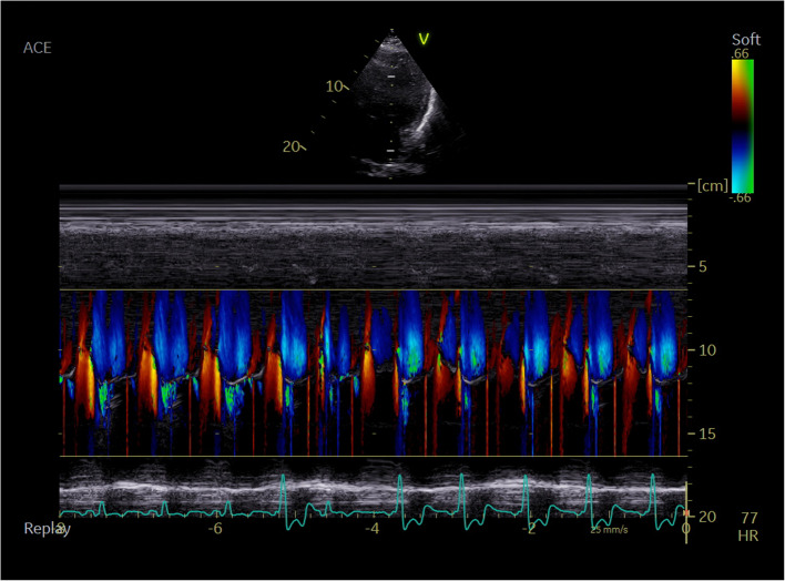
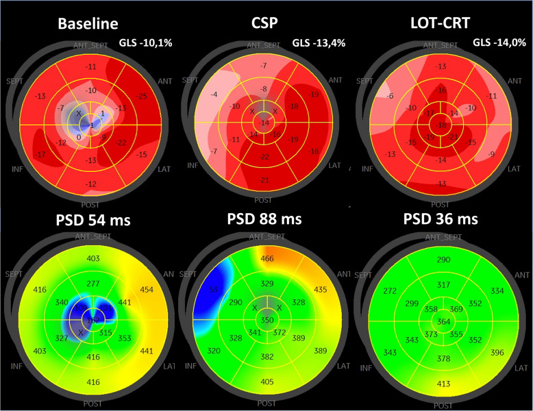
Conduction system pacing (CSP), encompassing His-bundle pacing (HBP) and left bundle branch area pacing (LBBAP), revolutionizes cardiac pacing, allowing a more physiological left ventricular activation than conventional right ventricular (RV) pacing through electrode placed in RV apex, interventricular septum or right ventricular outflow tract. Echocardiography plays a pivotal role in patient assessment, primarily by measuring left ventricular ejection fraction (LVEF) to determine the pacing strategy in alignment with current guidelines. Clinical data, simulations and ongoing trials on CSP explore CSP viability across various LVEF conditions. CSP is supposed to defer pacing-induced cardiomyopathy (PiCM) associated with conventional right ventricular pacing (RVP). This paper aims to review the current literature regarding the use of echocardiography in CSP. Images from our experience in the echocardiographic lab were used throughout this document to show our proposals of imaging in CSP. Echocardiography may help to determine lead localization within the interventricular septum (IVS), customizing pacing to individual anatomy and electromechanical indices (like atro-ventricular delay) and evaluates often-overlooked valvular function, a potential PiCM contributor. Three-dimensional (3-D) echocardiography widens the knowledge of lead localization and valvular dysfunction, as well as dyssynchrony assessment. Dyssynchrony, crucial both to resynchronization per se and physiological stimulation is quantified via echocardiography, especially using speckle-tracking imaging. Baseline LVEF and follow-up observation of CSP effects: early in Global Longitudinal Strain (GLS), afterwards in LV volumes and LVEF may improve the future proper qualification of patients. Limited left atrial (LA) and right atrial (RA) strain assessments hold potential in the CSP qualification and response assessment context. Echocardiography complements other imaging modalities for comprehensive patient evaluation. Echocardiography is integral in the CSP clinical use, from patient selection (by showing subtle changes in myocardial function) to post-procedure follow-up (tricuspid regurgitation, LV and RV function, leads and synchrony assessment). GLS, assessed by speckle tracking imaging and profound 2D and 3D (lead placement, septum morphology and global heart function under CSP) analyses show promise in CSP outcome assessment, though standardization is needed.
Keywords: Conduction system; Pacing; Echocardiography; Speckle tracking.
© 2025. The Author(s).
Conflict of interest statement
Ethics approval and consent to participate: Not applicable. Competing interests: The authors declare no competing interests.
Figures
References
-
- Burri H, Jastrzebski M, Cano Ó, et al. EHRA clinical consensus statement on conduction system pacing implantation: endorsed by the Asia Pacific Heart Rhythm Society (APHRS), Canadian Heart Rhythm Society (CHRS), and Latin American Heart Rhythm Society (LAHRS). EP Eur. 2023;25(4):1208–36. 10.1093/EUROPACE/EUAD043. - PMC - PubMed
-
- Glikson M, Nielsen JC, Kronborg MB, et al. 2021 ESC Guidelines on cardiac pacing and cardiac resynchronization therapy. Eur Heart J. 2021;42(35):3427–520. 10.1093/eurheartj/ehab364. - PubMed
-
- Vijayaraman P, Chelu MG, Curila K, et al. Cardiac Conduction System Pacing: A Comprehensive Update. JACC Clin Electrophysiol. Published online August 2023. 10.1016/J.JACEP.2023.06.005 - PubMed
-
- Naqvi TZ, Chao CJ. Adverse effects of right ventricular pacing on cardiac function: prevalence, prevention and treatment with physiologic pacing. Trends Cardiovasc Med. 2023;33(2):109–22. 10.1016/J.TCM.2021.10.013. - PubMed
Publication types
MeSH terms
LinkOut - more resources
Full Text Sources
Miscellaneous
