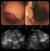
Multiple Succinate Dehydrogenase-Deficient Gastrointestinal Stromal Tumors of the Stomach: A Case Report
- PMID: 40382676
- PMCID: PMC12101094
- DOI: 10.12659/AJCR.947545
Multiple Succinate Dehydrogenase-Deficient Gastrointestinal Stromal Tumors of the Stomach: A Case Report
Abstract
BACKGROUND Gastrointestinal stromal tumors (GISTs) are a rare subset of gastrointestinal neoplasms, with approximately 85% of cases being characterized by genetic alterations in either the KIT gene or the platelet-derived growth factor receptor alpha (PDGFRA) gene. In contrast, succinate dehydrogenase-deficient gastrointestinal stromal tumors (SDH-deficient GIST) account for only 5-10% of cases. SDH-deficient GISTs is a rare form of gastrointestinal tumor, predominantly affecting young people and women. It typically presents with multifocal lesions, has a tendency to invade lymph nodes, follows an indolent course, and is poorly responsive to imatinib; sunitinib and regorafenib may be effective against it. For patients with resectable lesions, surgical intervention remains the cornerstone of treatment. CASE REPORT A 26-year-old male patient was admitted with the presenting symptom of melena. Subsequent diagnostic evaluations revealed the presence of multiple gastric neoplasms. He underwent laparoscopic distal gastrectomy for multiple gastric tumors and postoperative pathology was consistent with GIST, SDH-deficient type. Genetic testing for KIT, PDGFRA, KRAS, NRAS, BRAF, SDHA, SDHB, SDHC, SDHD, and NF1 showed no mutations. The patient is still being followed and no evidence of relapse has been found 6 months postoperatively. CONCLUSIONS Although SDH-deficient GISTs generally exhibit indolent biological behavior, clinically significant manifestations such as gastrointestinal bleeding, as observed in this case, occasionally occur. The postoperative resolution of hemorrhagic symptoms in this patient demonstrated the therapeutic efficacy of surgical intervention. This case underscores the importance of timely surgical management while highlighting the need for improved diagnostic precision and optimized treatment algorithms. The present report provides valuable clinical insights for prognostic evaluation and clinical decision-making in SDH-deficient GISTs, while also offering a reference for future investigations into novel targeted therapies.
Conflict of interest statement
Figures


Similar articles
-
Immunohistochemical loss of succinate dehydrogenase subunit A (SDHA) in gastrointestinal stromal tumors (GISTs) signals SDHA germline mutation.Am J Surg Pathol. 2013 Feb;37(2):234-40. doi: 10.1097/PAS.0b013e3182671178. Am J Surg Pathol. 2013. PMID: 23282968 Free PMC article.
-
Succinate dehydrogenase-deficient GISTs: a clinicopathologic, immunohistochemical, and molecular genetic study of 66 gastric GISTs with predilection to young age.Am J Surg Pathol. 2011 Nov;35(11):1712-21. doi: 10.1097/PAS.0b013e3182260752. Am J Surg Pathol. 2011. PMID: 21997692 Free PMC article.
-
Succinate dehydrogenase deficiency in a PDGFRA mutated GIST.BMC Cancer. 2017 Aug 2;17(1):512. doi: 10.1186/s12885-017-3499-7. BMC Cancer. 2017. PMID: 28768491 Free PMC article. Review.
-
Molecular Subtypes of KIT/PDGFRA Wild-Type Gastrointestinal Stromal Tumors: A Report From the National Institutes of Health Gastrointestinal Stromal Tumor Clinic.JAMA Oncol. 2016 Jul 1;2(7):922-8. doi: 10.1001/jamaoncol.2016.0256. JAMA Oncol. 2016. PMID: 27011036 Free PMC article.
-
Succinate dehydrogenase deficient gastrointestinal stromal tumors (GISTs) - a review.Int J Biochem Cell Biol. 2014 Aug;53:514-9. doi: 10.1016/j.biocel.2014.05.033. Epub 2014 Jun 2. Int J Biochem Cell Biol. 2014. PMID: 24886695 Free PMC article. Review.
References
-
- Blay JY, Kang YK, Nishida T, von Mehren M. Gastrointestinal stromal tumours. Nat Rev Dis Primers. 2021;7(1):22. - PubMed
Publication types
MeSH terms
Substances
LinkOut - more resources
Full Text Sources
Medical
Research Materials
Miscellaneous

