. 2025 Jul:57:102421.
doi: 10.1016/j.tranon.2025.102421. Epub 2025 May 17.

KRAS inhibition reverses chemotherapy resistance promoted by therapy-induced senescence-like in pancreatic ductal adenocarcinoma

Affiliations

KRAS inhibition reverses chemotherapy resistance promoted by therapy-induced senescence-like in pancreatic ductal adenocarcinoma

Analia Meilerman Abuelafia et al. Transl Oncol. 2025 Jul.

Abstract

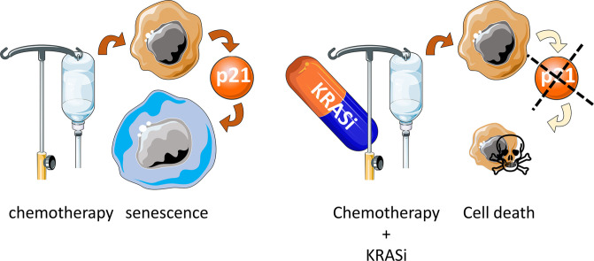
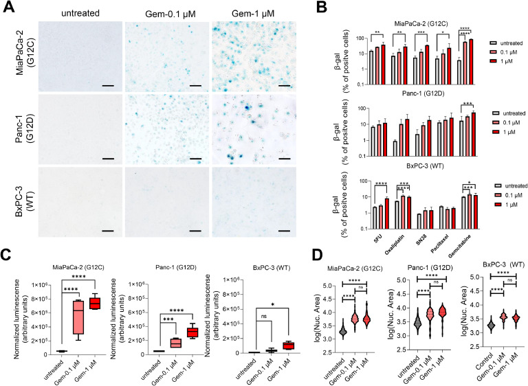
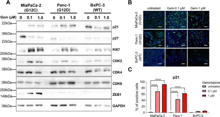
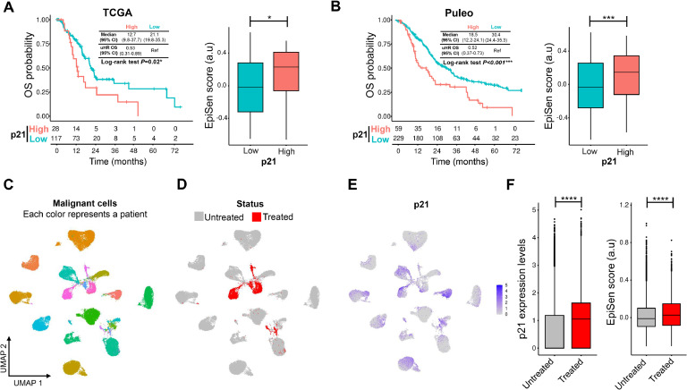
Background: Emerging evidence suggests that chemotherapy can accumulate senescent-like cells within tumor tissues, a phenomenon linked to therapy resistance. The aim of this study is to analyze the senescence-like state of after-treatment persistent cells associated with KRAS mutational status to offer a therapeutic strategy to target these cells in pancreatic ductal adenocarcinoma (PDAC).

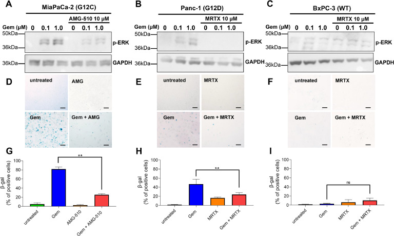
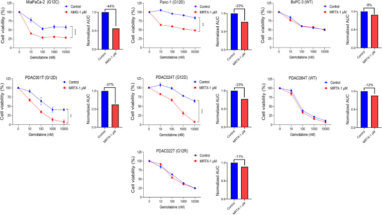
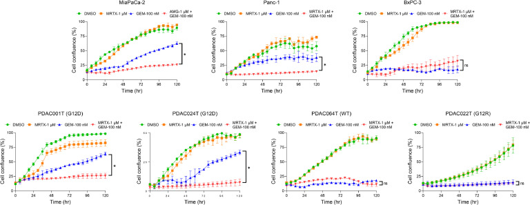
Experimental design: Three commercial cell lines and five patient-derived primary cell cultures with different KRAS statuses were studied following gemcitabine treatment. Senescence-like status was assessed using SA-β-gal, together with cell cycle regulators such as p21. Additionally, KRAS mutations were modulated using MRTX1133 and AMG-510, and the signaling pathways ERK and AKT were analyzed and modulated in vitro. Finally, p21 expression, associated with the senescence-like state, on patient outcomes and treatment response was analyzed in publicly available bulk RNA-seq and single-nucleus datasets.

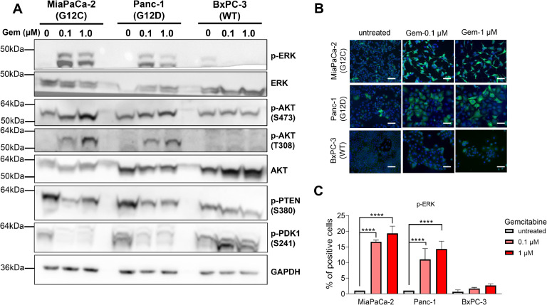
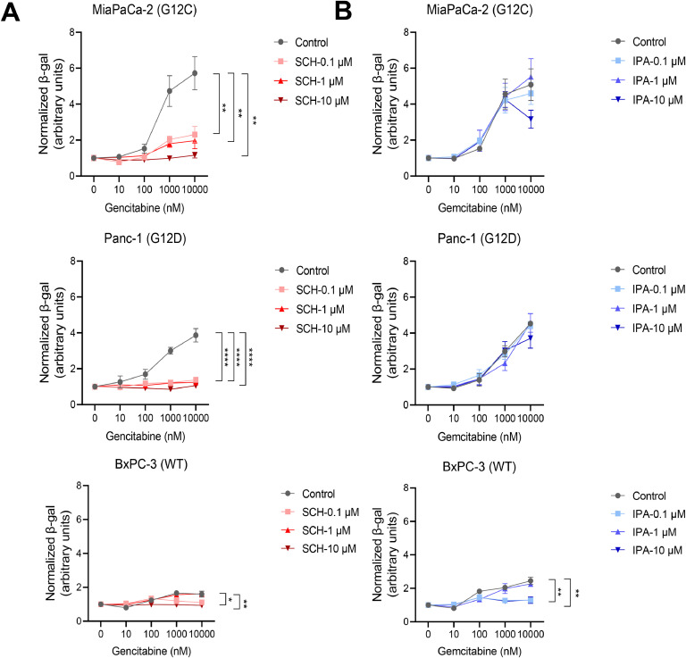
Results: We observed an overexpression of p21 alongside an increase in SA-β-gal signal in response to gemcitabine treatment, indicating the induction of a senescence-like state. Specific inhibition of KRAS G12D or G12C mutations reduced SA-β-gal signal and sensitized PDAC cells to gemcitabine. Moreover, ERK inhibition but not AKT inhibition decreased SA-β-gal signal. Additionally, we characterized p21 expression levels in relation to patient outcomes and found that they are modulated by treatment.

Conclusions: This dual-targeted therapeutic strategy holds promises for overcoming the challenges posed by KRAS-driven cancers, particularly in addressing the formidable obstacle of pancreatic cancer.

Keywords: Gemcitabine; MRTX1133; Mutated KRAS; PDAC; Resistance; Senescence-like.


Conflict of interest statement

Declaration of competing interest No potential conflicts of interest were disclosed. The funders had no role in the design of the study; in the collection, analyses, or interpretation of data, in the writing of the manuscript, or in the decision to publish the results.

Figures

Image, graphical abstract
Graphical abstract
Fig 1
Fig. 1
Increase of SA-β-galactosidase (SA-β-gal) levels after chemotherapeutic treatment in PDAC cells. A. Induction of SA-β-gal after the treatment with gemcitabine (72 h) 0.1 µM and 1 μM. B. Percentage of SA-β-gal cell after the treatment with gemcitabine, 5-fluorouracil (5FU), oxaliplatin, SN38 (the active metabolite of irinotecan), and paclitaxel at 0.1 and 1 µM for 72 h in MiaPaCa-2, Panc-1, and BxPC-3 cell lines. C. SA-β-gal expression measured by luminescence after the treatment by 72 h with gemcitabine 0.1 µM and 1 μM. D. Measurement of nuclear area after the treatment by 72 h with gemcitabine. Scale bars, 100 μm. *P < 0.01, **P < 0.01, ***P < 0.001, ****P< 0.0001 (n = 3).
Fig 2
Fig. 2
Senescence-like PDAC cells overexpress p21 after gemcitabine treatment. A. Immunoblotting of the cell cycle and epithelial-mesenchymal transition proteins after the treatment with gemcitabine at 0.1 µM and 1 μM for 72 h in MiaPaCa-2, Panc-1, and BxPC-3 cell lines. B. Immunofluorescent staining of p21 in MiaPaCa-2, Panc-1, and BxPC-3 after treatment with gemcitabine at 0.1 µM and 1 μM. C. Percentage of the p21 stained cells. Scale bars, 200 μm. ****P < 0.0001 (n = 3).
Fig 3
Fig. 3
Reduction of SA-β-gal levels after mutated KRAS inhibition. KRAS inhibitors AMG-510 and MRTX1133 reduce the phosphorylation of ERK in MiaPaCa-2 and Panc-1 (A and B) but not in BxPC-3 (C). SA-β-gal staining levels after the treatment with gemcitabine in MiaPaCa-2 (D) and Panc-1 (E) without significant effect on BxPC-3 (F). G, H and I. Percentage of the SA-β-gal stained cells. Scale bars, 100 μm. **P < 0.01, (n = 3).
Fig 4
Fig. 4
Inhibition of mutated KRAS sensitize PDAC cells to gemcitabine treatment. Three commercial cell lines MiaPaCa-2, Panc-1, and BxPC-3 and four patient-derived primary cell cultures were treated with increased concentrations of gemcitabine (from 0.01 μM to 10 μM) in combination with 1 μM of KRAS inhibitors, MRTX1133 (directed against G12D) or AMG-510 (directed against G12C) for 72 h. Cell viability was measured with PrestoBlue. Each experiment was repeated at least three times. Values were normalized and expressed as the percentage of the control (vehicle). Additionally, the reduction in area under the curve (AUC) is shown. **P< 0.01, ****P< 0.0001.
Fig 5
Fig. 5
Time-lapse experiment analyzing the sensitize effect KRAS inhibition on gemcitabine treatment. Incucyte video microscopy measured the cell viability of 3 commercial cell lines MiaPaCa-2, Panc-1, and BxPC-3 and four patient-derived primary cell cultures, treated with gemcitabine 0.1 μM, 1 μM of KRAS inhibitors, MRTX1133 (directed against G12D) or AMG-510 (directed against G12C) or the combination for 120 h. A control with DMSO (vehicle) was maintained. *P< 0.05, ***P< 0.001, ns, not significant (n = 2).
Fig 6
Fig. 6
Characterization of KRAS pathway in PDAC cells after the treatment with gemcitabine. A. Immunoblotting of ERK and AKT pathway in MiaPaCa-2, Panc-1, and BxPC-3 after treatment for 72 h with gemcitabine at 0.1 µM and 1 μM. B and C. Immunofluorescent staining of p-ERK and quantification of positive cells for p-ERK labeling after the treatment with gemcitabine at 0.1 µM and 1 μM for 72 h in MiaPaCa-2, Panc-1, and BxPC-3. Scale bars, 200 μm. ****P < 0.0001, (n = 3).
Fig 7
Fig. 7
ERK inhibitor reduces the levels of SA-β-gal in presence of gemcitabine. SA-β-gal level was measured by luminescence assay after the treatment with gemcitabine in increasing concentrations (from 0.01 μM to 10 μM) in combination with (A) the ERK (SCH772984; SCH) and (B) AKT (Ipatasertib; IPA) inhibitors (0.1 μM, 1 µM and 10 µM). *P < 0.05, **P < 0.01, ****P < 0.0001. (n = 3).
Fig 8
Fig. 8
Expression levels of p21 as mediator of the senescence-like state is associated with patient’s outcome and persistent cells after treatment. A. Survival analysis of p21 levels associated with overall survival (OS) and senescence-like signature EpiSen in TCGA-PAAD (A) and Puleo (B) cohorts. UMAP plots showing the patients studied with different colors (C), distinguishing untreated and treated groups (D), and indicating p21 expression levels (E). F. Expression of p21 and score of EpiSen signature before and after treatment in single nucleus Hwang cohort. *P < 0.05, ***P < 0.001, ****P < 0.0001. HR, Hazard ratio, IC, interval of confidence.

References

    1. Rahib L., Wehner M.R., Matrisian L.M., Nead K.T. Estimated projection of US cancer incidence and death to 2040. JAMA Netw. Open. Am. Med. Assoc. 2021;4 –e214708. - PMC - PubMed
    1. Neoptolemos J.P., Kleeff J., Michl P., Costello E., Greenhalf W., Palmer D.H. Therapeutic developments in pancreatic cancer: current and future perspectives. Nat. Rev. Gastroenterol. Hepatol. 2018;15:333–348. - PubMed
    1. Conroy T., Hammel P., Hebbar M., Ben Abdelghani M., Wei A.C., Raoul J.L., et al. FOLFIRINOX or gemcitabine as adjuvant therapy for pancreatic cancer. N. Engl. J. Med. 2018;379:2395–2406. - PubMed
    1. Fassnacht M., Terzolo M., Allolio B., Baudin E., Haak H., Berruti A., et al. Combination chemotherapy in advanced adrenocortical carcinoma. N. Engl. J. Med. 2012;366:2189–2197. - PubMed
    1. Feig C., Gopinathan A., Neesse A., Chan D.S., Cook N., Tuveson D.A. The pancreas cancer microenvironment. Clin. Cancer Res. 2012;18:4266–4276. - PMC - PubMed

LinkOut - more resources