

Baicalein based nano-delivery system restores mitochondrial homeostasis through PPAR signaling pathway to promote wound healing in diabetes
- PMID: 40383752
- PMCID: PMC12087252
- DOI: 10.1186/s12951-025-03427-6
Baicalein based nano-delivery system restores mitochondrial homeostasis through PPAR signaling pathway to promote wound healing in diabetes
Abstract
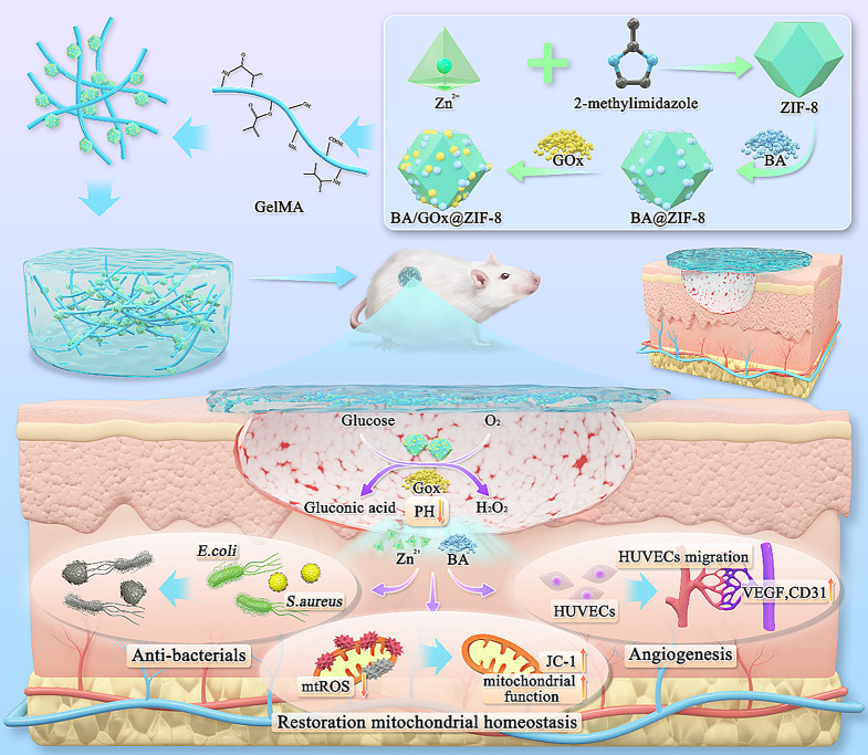
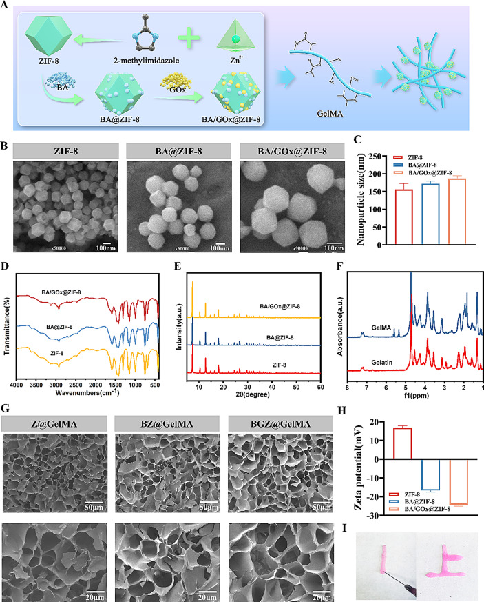
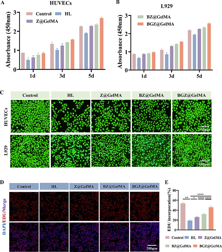
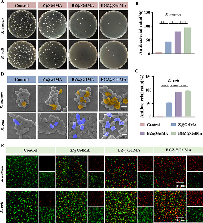
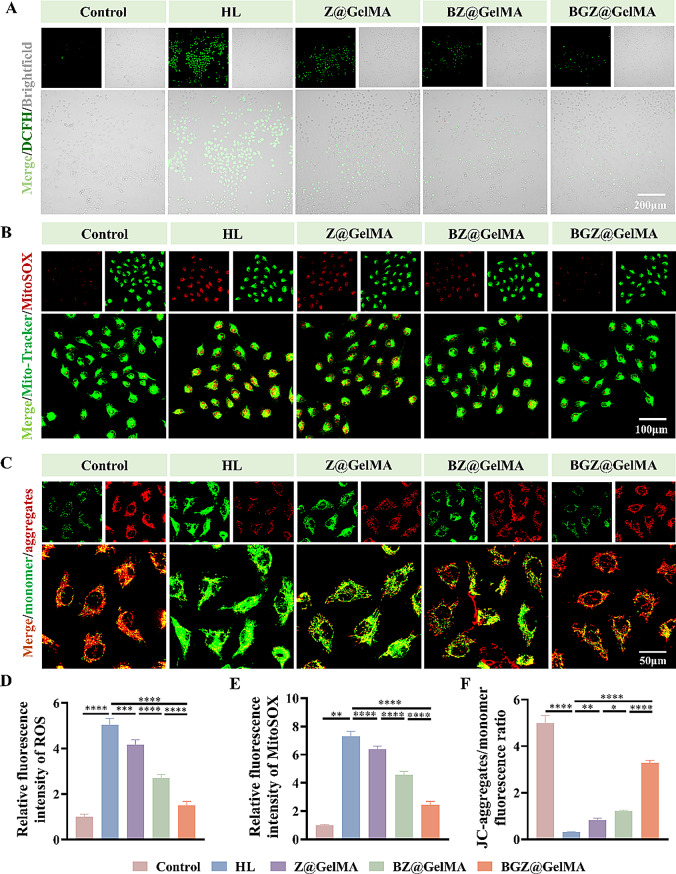
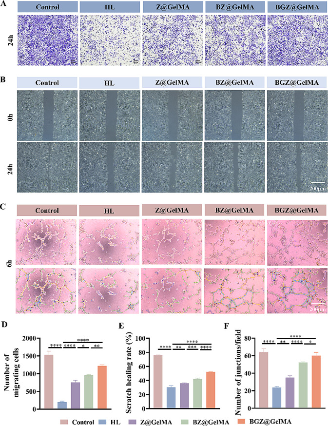
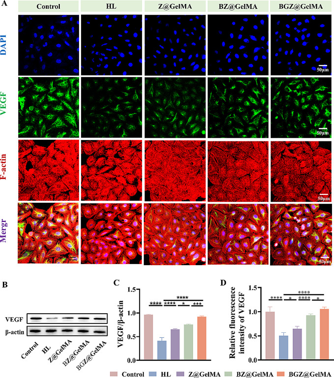
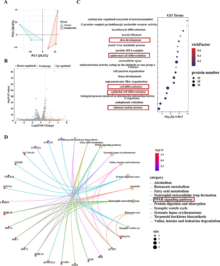
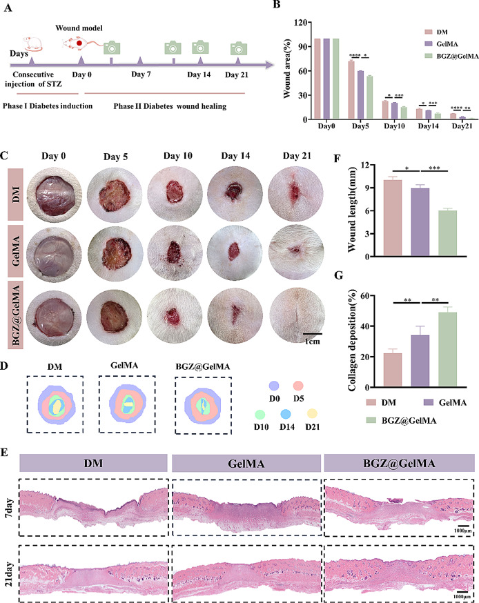
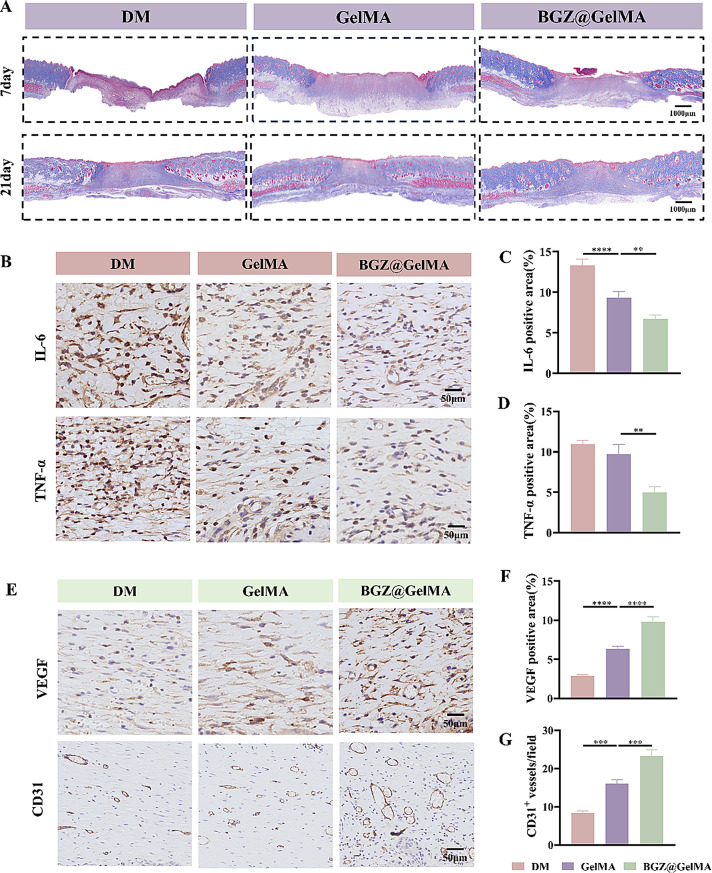
Wound healing in diabetes is a substantial clinical challenge due to the hyperglycemic microenvironment, high pH, bacterial infection, persistent inflammation, and impaired cellular functions, attributed to mitochondrial dysfunction. Here, we have developed an injectable photo-crosslinking nanocomposite hydrogel (BA/GOx@ZIF-8@GelMA, BGZ@GelMA) with baicalein (BA) and glucose oxidase (GOx) loaded Zinc metal-organic framework (ZIF-8) based on methacrylated gelatin (GelMA) to accelerate diabetic infected wound healing by regulating subcellular and cellular functions. The combination of ZIF-8 and BA gives the hydrogel excellent antibacterial properties. A high blood sugar environment triggers the release of GOx in BGZ@GelMA, reducing local glucose and pH, producing hydrogen peroxide (H2O2), and releasing BA and Zinc ions (Zn2+). This process provides a suitable microenvironment for wound healing. Zn2+ can significantly inhibit the proliferation of Staphylococcus aureus (S.aureus) and Escherichia coli (E.coli). The released BA can clear ROS in cells and mitochondria, restore mitochondrial function and stability, and make the hydrogel fundamentally improve the cell function damage induced by hyperglycemia, and ultimately promote cell proliferation, migration and angiogenesis. In general, our multifunctional nanocomposite hydrogel provides a new strategy for diabetes wound healing at the subcellular and cellular functional levels.
Keywords: Baicalein; Diabetic wound healing; Metal-organic framework; Mitochondrion.
© 2025. The Author(s).
Conflict of interest statement
Declarations. Ethics approval and consent to participate: All animal experiments were approved by the Animal Protection and Ethics Committee of Shanxi Medical University, School of Stomatology. Consent for publication: All authors consent for publication. Competing interests: The authors declare no competing interests.
Figures
Similar articles
-
A Zn-MOF-GOx-based cascade nanoreactor promotes diabetic infected wound healing by NO release and microenvironment regulation.Acta Biomater. 2024 Jul 1;182:245-259. doi: 10.1016/j.actbio.2024.05.015. Epub 2024 May 9. Acta Biomater. 2024. PMID: 38729545
-
Glucose-activated self-cascade antibacterial and pro-angiogenesis nanozyme-functionalized chitosan-arginine thermosensitive hydrogel for chronic diabetic wounds healing.Carbohydr Polym. 2025 Jan 15;348(Pt B):122894. doi: 10.1016/j.carbpol.2024.122894. Epub 2024 Oct 21. Carbohydr Polym. 2025. PMID: 39567166
-
Nanofibrous membrane/self-healing hydrogel double-layer nanocomposite dressings based on oxidized dextran and carboxymethyl chitosan loading ZIF-nanozyme for enhanced bacterial-infected diabetic wound healing.Int J Biol Macromol. 2025 Jun;313:144292. doi: 10.1016/j.ijbiomac.2025.144292. Epub 2025 May 16. Int J Biol Macromol. 2025. PMID: 40383329
-
Multifunctional self-healing and pH-responsive hydrogel dressing based on cationic guar gum and hyaluronic acid for on-demand drug release.Int J Biol Macromol. 2025 Apr;301:140326. doi: 10.1016/j.ijbiomac.2025.140326. Epub 2025 Jan 24. Int J Biol Macromol. 2025. PMID: 39864699 Review.
-
Hyaluronic acid and ZIF-8 nanocomposites for wound care.Int J Biol Macromol. 2025 Jun;315(Pt 2):144475. doi: 10.1016/j.ijbiomac.2025.144475. Epub 2025 May 23. Int J Biol Macromol. 2025. PMID: 40414391 Review.
References
MeSH terms
Substances
Grants and funding
- 2023L086/Scientific and Technological Innovation Programs of Higher Education Institutions in Shanxi (STIP)
- 2022L145/Scientific and Technological Innovation Programs of Higher Education Institutions in Shanxi (STIP)
- 202403021211121/Basic Research Program of Shanxi Province
- 202303021222335/Basic Research Program of Shanxi Province
LinkOut - more resources
Full Text Sources
