. 2025 May 18;16(1):4617.
doi: 10.1038/s41467-025-60035-6.

A broad antibody with enhanced HIV-1 neutralization via bispecific antibody-mediated prepositioning

Affiliations

A broad antibody with enhanced HIV-1 neutralization via bispecific antibody-mediated prepositioning

Soohyun Kim et al. Nat Commun. .

Abstract

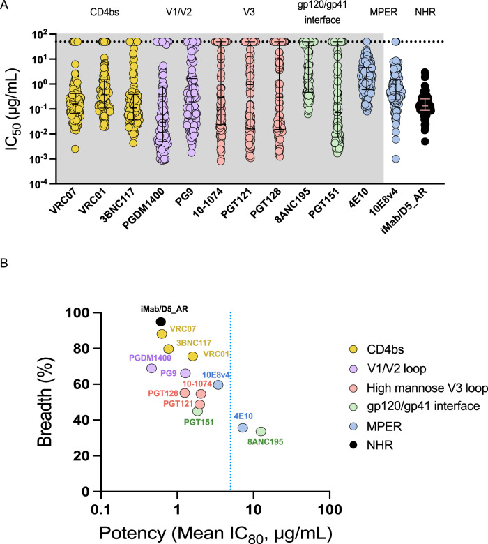
Antibodies targeting the highly conserved prehairpin intermediate (PHI) of class I viral membrane-fusion proteins are generally weakly neutralizing and are not considered viable therapeutic agents. We previously demonstrated that antibodies targeting the gp41 N-heptad repeat (NHR), which is transiently exposed in the HIV-1 PHI, exhibit enhanced broad neutralization in cells expressing the Fc receptor, FcγRI. To enhance neutralization in cells lacking FcγRI, we here develop a bispecific antibody (bsAb) by fusing an NHR-targeting antibody to an antibody against CD4, the HIV-1 receptor on T cells. The bsAb provides a 5000-fold neutralization enhancement and shows unprecedented neutralization breadth compared to existing broadly neutralizing antibodies. Importantly, the bsAb reduces viral load in HIV-1-infected humanized male mice, and viral envelope sequencing under bsAb pressure revealed an NHR mutation that potentially impairs viral fitness. These findings validate the NHR as a potential HIV-1 therapeutic target, setting the stage for a new class of broadly neutralizing antibodies.


Conflict of interest statement

Competing interests: The authors declare no competing interests.

Figures

Fig. 1
Fig. 1. The transiently exposed gp41 NHR is highly conserved.
A Overview of HIV-1 infection. Infection by HIV-1 is initiated by binding of envelope (Env) protein, a trimer composed of gp120 and gp41 subunits, to CD4 on the surface CD4 + T cells. Subsequently, the Env trimer binds to host cell receptors including CCR5 and CXCR4, and triggers membrane fusion. During the fusion process, Env undergoes a conformational change, forming a prehairpin intermediate (PHI) followed by formation of a trimer-of-hairpins. gp41 is composed of N-heptad repeat (NHR), C-heptad repeat (CHR), and membrane-proximal external region (MPER). The N-heptad repeat (NHR) becomes transiently exposed in the PHI, which is normally not readily accessible in the native pre-fusion state. B Domain structure of HIV-1 Env protein, which is cleaved into gp120 and gp41. Numbers on top of each domain show sequence identity (percentage of amino acids that are identical). NHR sequence conservation in the logo plot (inset) is based on Los Alamos 2021 HIV database (>8000 HIV-1 Env proteins). The epitope of the anti-NHR mAb D5 is highlighted in orange. C A table showing sequence identity of NHR and the epitope of D5 among HIV-1 sequences. D Sequence conservation of NHR from 65 clustered groups in the Los Alamos 2021 HIV database (Table S1) is overlaid on the structure of gp41 inner-core mimetic 5-helix (PDB: 2CMR). The epitopes of D5 are shown in spheres, colored based on the extent of conservation indicated by the heatmap scale.
Fig. 2
Fig. 2. Enhancing HIV-1 neutralization by NHR-targeting antibody through bispecific antibody-mediated prepositioning on host cells.
A Schematic of bispecific antibodies (bsAbs) generated by combining the antibody fragments of anti-NHR antibody D5_AR or anti-CD4 antibody iMab. mAb114 targeting unrelated Ebola glycoprotein is used as the control and is represented by the abbreviation ‘c’. A/B represents the antibody configuration, with antibody A contributing the knob arm (incorporating S354C and T366W substitutions) and antibody B contributing the hole arm (incorporating Y349C and T366S substitutions). B Flow cytometry to assess binding of bsAb to CD4-expressing TZM-bl cells and biotinylated NHR. Shown on the axes are the binding of the three antibodies to CD4 (probed with FITC-Fab fragment goat anti-human IgG, x-axis) and biotinylated NHR hydrophobic pocket mimetic IQN17 (probed with streptavidin-APC, y-axis). The configuration of the control Fab fragment is abbreviated as (c/) when on the knob arm and (/c) when on the hole arm. 10,000 cells were detected per measurement and results were plotted using FlowJo. The control shows binding to neither CCR5 nor NHR, while the bsAb binds both simultaneously. C Maximum % inhibition (left), IC50 (middle), and IC80 (right) for neutralization by iMab/D5_AR, its two control antibodies, and a mix of the two control antibodies, against a panel of 26 HIV-1 pseudotyped viruses representing diverse clades and tiers (see Table 1, source data are provided as a source data file). Each dot (n = 26) represents the average neutralization of biological duplicates of technical replicates for each pseudotyped virus. Maximum % inhibition values were calculated from the inhibition at the highest antibody concentration (50 μg/mL for iMab/D5_AR, and 50 μg/mL each for iMab/c and c/D5_AR in combination). IC50 and IC80 values were calculated using GraphPad Prism. Error bars indicate median +/- interquartile range. mAb114 Fab was used as a control Fab. The two-sided Wilcoxon-matched pairs signed test was used; **** indicates p < 0.0001. Black dotted lines indicate limit of quantification (50 μg/mL) of bsAb.
Fig. 3
Fig. 3. NHR-targeting antibody exhibits potent and broad neutralization against HIV-1 through bispecific antibody-mediated prepositioning.
A Neutralization potencies of iMab/D5_AR and 12 bnAbs against a panel of 119 HIV-1 pseudotyped viruses. Antibodies are grouped by color based on which of six epitopes they target (CD4-binding site, CD4bs; V1/V2 loop of gp120, V1/V2 loop; high-mannose V3 loop, High-mannose V3 loop; gp120/gp41 interface; the membrane-proximal external region, MPER; and gp41 N-heptad repeat, NHR). The black dotted line indicates the limit of quantification (50 μg/mL). Error bars indicate median +/- interquartile range. Neutralization potencies of antibodies, apart from iMab/D5_AR and 10E8v4, were obtained from published studies curated in the Los Alamos HIV database (shown in gray background), CATNAP. Source data is provided as a source data file. Each dot represents the average neutralization of technical replicates for each pseudotyped virus. B Neutralization potency vs. breadth curve against 119 HIV-1 pseudotyped viruses. Antibodies were grouped by the epitope targeted (colors in key). Breadth was defined as non-protective when IC80 > 5 μg/mL. The blue dotted line indicates IC80 of 5 μg/mL. Source data ARE provided as a source data file.
Fig. 4
Fig. 4. iMab/D5_AR potently neutralizes replication-competent HIV-1 viruses.
A A table showing neutralization potencies of iMab/D5_AR against six replication-competent HIV-1 viruses using the luciferase-based reporter assay. The IC80 values of bsAb at <5, 5–50, and > 50 μg/mL were assigned the colors of red, blue, and white, respectively. R5 and X4 tropisms indicate CCR5 and CXCR4, respectively. B Maximum % inhibition for neutralization by iMab/D5_AR, its control antibodies, and a mix of the two control antibodies against a panel of six replication-competent viruses on TZM-bl cells. Maximum % inhibition values were calculated from the inhibition at the highest antibody concentration (50 μg/mL for iMab/D5_AR, iMab/c, or c/D5_AR individually, and 50 μg/mL each for iMab/c and c/D5_AR in combination). Each dot represents an average of biological duplicates of technical replicates. Error bars indicate median +/− interquartile range. mAb114 Fab was used as a control Fab. Source data are provided as a source data file. C Neutralization of bsAb against NL4-3 replication-competent HIV-1 virus on human peripheral blood mononuclear cells (PBMCs). Each dot represents an average of technical replicates (n = 3 human donors). Error bars indicate mean +/- standard deviations (SD). Source data are provided as a source data file.
Fig. 5
Fig. 5. Identification of key residues for neutralization by iMab/D5_AR.
DMS maps of all sites in the BF520 Env ectodomain where mutations lead to resistance from (A) enfuvirtide- and (B) iMab/D5_AR-mediated viral neutralization. Positive values and negative values represent residues where mutations cause resistance or sensitivity, respectively. Individual points on the line plots represent the mean effect of mutations on escape at that site. Interactive versions of the resistance maps for enfuvirtide and iMab/D5_AR are available https://dms-vep.org/HIV_Envelope_BF520_DMS_iMab.D5_AR_Enfuvirtide/htmls/Enfuvirtide_mut_effect.html and https://dms-vep.org/HIV_Envelope_BF520_DMS_iMab.D5_AR_Enfuvirtide/htmls/iMab.D5_AR_mut_effect.html, respectively. C DMS-identified resistance sites are shown for enfuvirtide (blue) and iMab/D5_AR (purple) in NHR, mapped onto a structure of the gp41 NHR (PDB: 2CMR). D Neutralization of BF520 Env pseudotyped viruses with individual mutations by enfuvirtide, and iMab/D5_AR shown as maximum % inhibition calculated from the inhibition at the highest antibody concentration (50 μg/mL). Each dot represents an average of biological duplicates of technical replicates. E Table showing neutralization IC50 (nM) values of enfuvirtide and iMab/D5_AR against each of the four point mutations in the NHR hydrophobic pocket. F Neutralization by enfuvirtide, iMab/D5_AR, and control antibodies of HIV-1 isolate MVP-5180 pseudovirus with deviations from the known D5 epitope sequences at residues 574 and 577. Results are shown as mean from duplicate experiments. Error bars indicate mean +/− standard deviations (SD).
Fig. 6
Fig. 6. iMab/D5_AR suppresses HIV-1 viral load in vivo.
A Schematic representation of the experimental timeline for evaluating the efficacy of bsAbs in vivo. NOD/SCID/IL2rγnull (NSG) mice were implanted with human bone marrow-liver-thymus (BLT) tissues, followed by intravenous infection with 500 ng of NL4-3 replication-competent HIV-1 virus. Starting 1 week post-infection, mice were treated with weekly intraperitoneal injections of either iMab/D5_AR (2 mg), a combination of iMab/c (2 mg) and c/D5_AR (2 mg), or PBS as a control. Blood was drawn from the mice every two weeks for ten weeks for viral load (VL) titration. B Changes in plasma VL. Gray dashed lines indicate no change in VL from week 0. Black lines represent data from each mouse, and red lines represent the geometric mean of data from all mice (n = 8 for iMab/D5_AR, n = 4 for iMab/c + c/D5_AR, and n = 2 for PBS). C Geometric mean changes in plasma VL. Each line represents geometric changes in the VL on a log10 scale, and the error bars represent the standard deviation. iMab/D5_AR showed a statistical difference (p < 0.05 as determined by Mann-Whitney test) compared to the PBS group at weeks 2 and 4, whereas iMab/c + c/D5_AR did not show a significant difference from the PBS group (n = 8 for iMab/D5_AR, n = 4 for iMab/c + c/D5_AR, and n = 2 for PBS). D Plasma concentration of the bsAb in NSG mice. The gray dashed lines indicate the limit of quantification by ELISA at antibody concentration of 0.1 μg/mL. Black lines represent plasma concentration of the bsAbs from each mouse (n = 8 for iMab/D5_AR, n = 4 for iMab/c + c/D5_AR), with red lines representing the geometric mean. E The logo plots show the degree of conservation of the NHR from weeks 2–6 and week 10, with wild-type sequences shown in gray and mutation highlighted in blue.
Fig. 7
Fig. 7. Binding sites for broadly neutralizing antibodies.
Sites of vulnerability to broadly neutralizing antibodies on the HIV-1 Env trimer, shown from two perspectives (PDB: 7SKA). NHR is inaccessible in this state and is only transiently exposed in the prehairpin intermediate conformation.

References

    1. Orloff, G. M., Orloff, S. L., Kennedy, M. S., Maddon, P. J. & McDougal, J. S. Penetration of CD4 T cells by HIV-1. The CD4 receptor does not internalize with HIV, and CD4-related signal transduction events are not required for entry. J. Immunol.146, 2578–2587 (1991). - PubMed
    1. Moore, J. P., Trkola, A. & Dragic, T. Co-receptors for HIV-1 entry. Curr. Opin. Immunol.9, 551–562 (1997). - PubMed
    1. Chan, D. C. & Kim, P. S. HIV entry and its inhibition. Cell93, 681–684 (1998). - PubMed
    1. Eckert, D. M. & Kim, P. S. Mechanisms of viral membrane fusion and its inhibition. Annu. Rev. Biochem.70, 777–810 (2001). - PubMed
    1. Harrison, S. C. Viral membrane fusion. Nat. Struct. Mol. Biol.15, 690–698 (2008). - PMC - PubMed

MeSH terms