Case Reports
. 2025 May 18;19(1):231.
doi: 10.1186/s13256-025-05296-x.

Clinical application of vein visualization apparatus AccuVein®500 in breast cancer surgery: a case report

Affiliations
Case Reports

Clinical application of vein visualization apparatus AccuVein®500 in breast cancer surgery: a case report

Junta Sakakibara et al. J Med Case Rep. .

Abstract

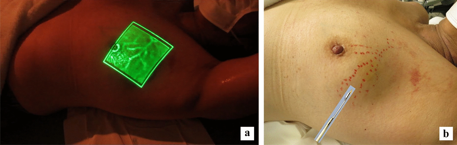
Background: AccuVein® can help visualize superficial veins and is generally used as an auxiliary device to identify patterns of veins that are difficult to locate for collecting blood and securing venous lines. Even when venous patterns are obscure via visual inspection and/or palpation, the clear projection/delineation of superficial veins using this apparatus facilitates safe venous puncture and helps secure venous lines. Therefore, this apparatus is widely used in clinical settings. AccuVein® can easily visualize not only superficial veins in the limbs but also the ones located throughout the body surface.

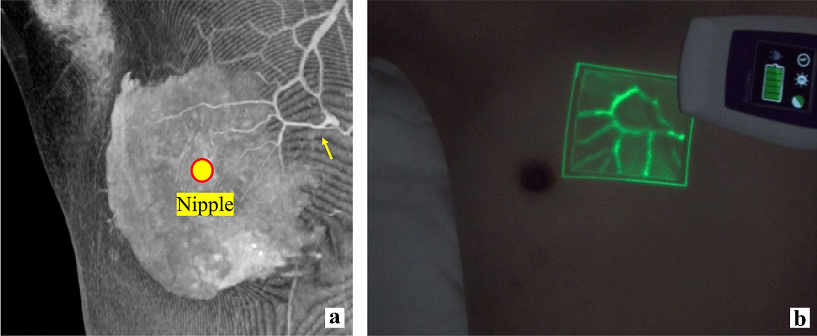
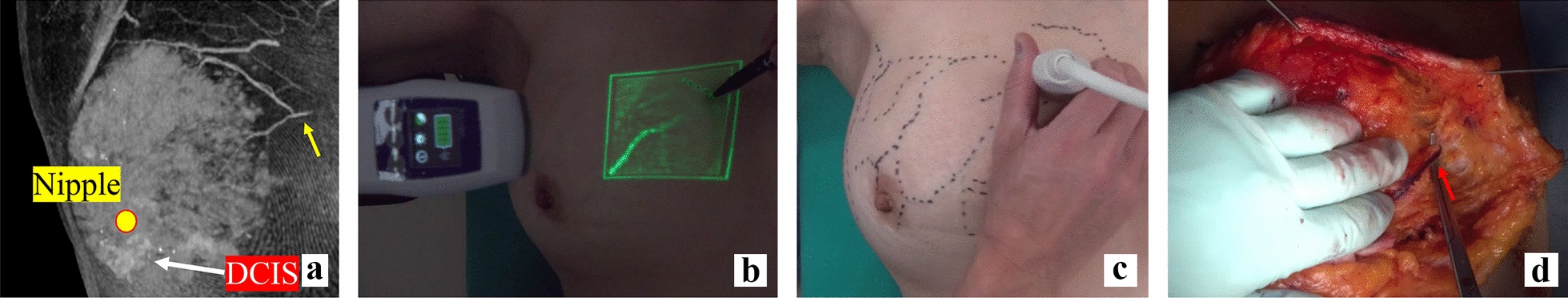
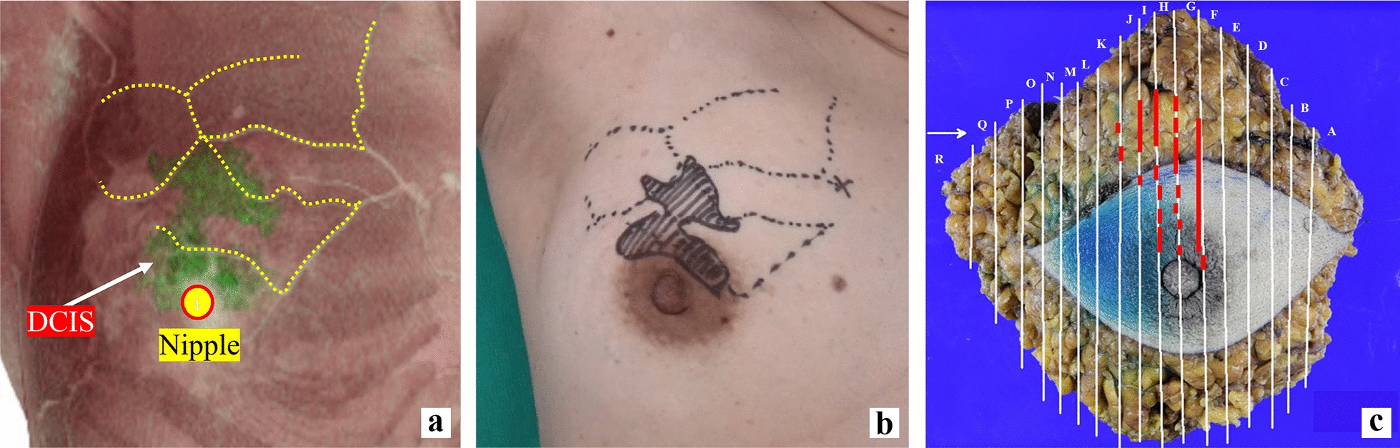
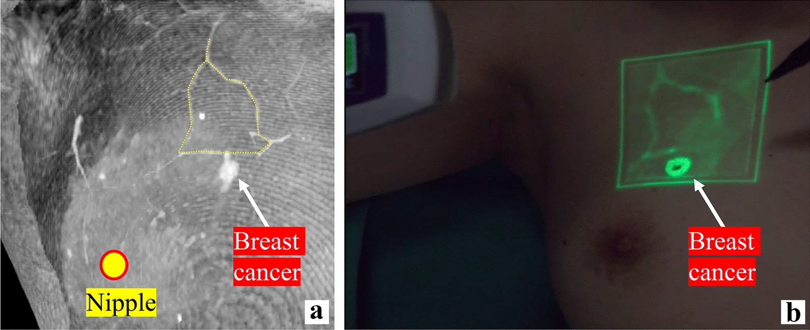
Case presentation: We report three cases of 68-year-old, 41-year-old, and 56-year-old Japanese women in whom superficial veins in the breasts were visualized using AccuVein®, and mastectomy and partial mastectomy were performed. All patients were of Japanese ethnicity. AccuVein® can enable the examiner to observe superficial veins in the breasts, irrespective of their skills. The examiner can, thus, secure detailed visualization of subcutaneous veins in the breasts. Furthermore, AccuVein® ensures reproducibility and subjectivity regardless of the examiners' experience. During a mastectomy, the perforating branches of the internal thoracic vein originating from the greater pectoral muscle are identified, ligated, and separated. The preoperative use of AccuVein® makes it possible to instantaneously identify their position. Visualizing the perforating branches to their root in patients with thin subcutaneous breast fat and their roots' proximity in patients with thick subcutaneous breast fat is possible. While the position and/or range of a breast cancer lesion may sometimes be unclear in ultrasonography, marking subcutaneous mammary veins around the lesion as the benchmark helps identify the lesion position. In this study, we inspected the patterns of subcutaneous mammary veins using AccuVein®. This manuscript reports the clinical application of this apparatus in breast cancer surgeries.

Conclusion: Understanding the vascular construction of subcutaneous mammary veins using the vein visualization apparatus AccuVein® may serve as an auxiliary technique for safely and securely identifying breast cancer lesions.

Keywords: AccuVein®; Breast cancer; Subcutaneous mammary veins; Surgery; Vein visualization apparatus.


Conflict of interest statement

Declarations. Ethics approval and consent to participate: All procedures adhered to the Helsinki Declaration of 1964 and later versions. Informed consent was obtained from all patients for being included in the study. Consent for publication: Written informed consent was obtained from the patient for publication of this case report and any accompanying images. A copy of the written consent is available for review by the Editor-in-Chief of this journal. Competing interests: The authors do not have any competing interests.

Figures

Fig. 1
Fig. 1
Consistency between contrast-enhanced computed tomography maximum intensity projection image and superficial mammary vein construction visualized via AccuVein®. a The contrast-enhanced computed tomography maximum intensity projection image shows the perforating branches of the internal thoracic vein (yellow arrow). b AccuVein® clearly visualizes the subcutaneous mammary veins. This is completely identical to the vascular construction shown in the contrast-enhanced computed tomography maximum intensity projection image
Fig. 2
Fig. 2
Consistency between superficial mammary veins visualized via AccuVein® and the corresponding blood vessels during surgery. a The contrast-enhanced computed tomography maximum intensity projection image shows the perforating branches of the internal thoracic vein (yellow arrow). b AccuVein® clearly visualized the subcutaneous mammary veins. c The marking of the vascular construction on the skin and the ultrasonography of the position of the perforating branches of the internal thoracic vein. d Safe ligation treatment of the perforating branches of the internal thoracic vein (red arrow); DCIS ductal carcinoma in situ
Fig. 3
Fig. 3
Marking of the breast cancer lesion using the subcutaneous mammary veins as the benchmark. a The positional relationship between the breast cancer lesion (green) in the contrast-enhance computed tomography maximum intensity projection image and the construction of the superficial mammary veins (yellow dotted line) is shown. b Using AccuVein®, the subcutaneous mammary veins were marked on the skin, and the positional relationship between the vascular construction and the breast cancer lesion in the contrast-enhanced computed tomography maximum intensity projection image was collated. This allowed us to visualize the breast cancer lesion in the contrast-enhanced computed tomography maximum intensity projection image on the skin. c In the removed specimen, the breast cancer lesion (red line) expanded as much as the lesion shown in a, b; DCIS ductal carcinoma in situ
Fig. 4
Fig. 4
Estimation of the breast cancer lesion using subcutaneous mammary veins as the benchmark (partial mastectomy). a The positional relationship between the breast cancer lesion in the contrast-enhanced computed tomography maximum intensity projection image (white arrow) and the bell-shaped construction of the superficial mammary veins (yellow dotted line) are shown. b The positional relationship between the subcutaneous veins visualized by AccuVein® and the ultrasonography-marked lesion after preoperative endocrine therapy was identical to that between the vascular construction and the lesion in the contrast-enhanced computed tomography maximum intensity projection image. This means that a breast cancer lesion can be estimated from vascular construction
Fig. 5
Fig. 5
Detailed visualization of subcutaneous mammary veins and avoidance of vascular damage during tissue biopsy. a Prior to tissue biopsy, the mammary subcutaneous veins are visualized using AccuVein®. b Visualization of the mammary subcutaneous veins makes it possible to avoid vascular damage caused by the biopsy needle

Similar articles

References

    1. Kaddoum RN, Anghelescu DL, Parish ME, Wright BB, Trujillo L, Wu J, et al. A randomized controlled trial comparing the AccuVein AV300 device to standard insertion technique for intravenous cannulation of anesthetized children. Paediatr Anaesth. 2012;22:884–9. - PubMed
    1. De Graaff JC, Cuper NJ, Mungra RA, Vlaardingerbroek K, Numan SC, Kalkman CJ. Near-infrared light to aid peripheral intravenous cannulation in children: a cluster randomized clinical trial of three devices. Anaesthesia. 2013;68:835–45. - PubMed
    1. Aulagnier J, Hoc C, Mathieu E, Dreyfus JF, Fischler M, Le Guen M. Efficacy of AccuVein to facilitate peripheral intravenous placement in adults presenting to an emergency department: a randomized clinical trial. Acad Emerg Med. 2014;21:858–63. - PubMed
    1. Lee H, Lee SH, Kim SJ, Choi WI, Lee JH, Choi IJ. Variations of the cubital superficial vein investigated by using the intravenous illuminator. Anat Cell Biol. 2015;48:62–5. - PMC - PubMed
    1. Rothbart A, Yu P, Müller-Lobeck L, Spies CD, Wernecke KD, Nachtigall I. Peripheral intravenous cannulation with support of infrared laser vein viewing system in a pre-operation setting in pediatric patients. BMC Res Notes. 2015;8:463. - PMC - PubMed

Publication types

LinkOut - more resources