Review
. 2025 May 19;13(1):103.
doi: 10.1186/s40478-025-01959-4.

Beyond the brain: exploring the impact of animal models of leptomeningeal disease from solid tumors

Affiliations
Review

Beyond the brain: exploring the impact of animal models of leptomeningeal disease from solid tumors

Jillyn R Turunen et al. Acta Neuropathol Commun. .

Abstract

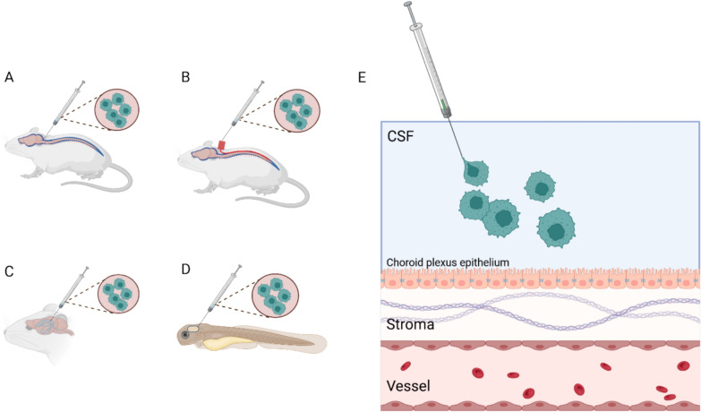
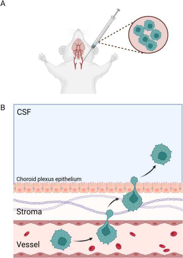
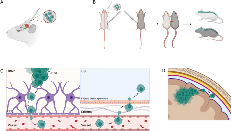
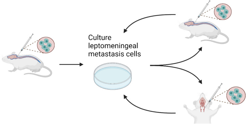
Leptomeningeal disease (LMD) is a devastating manifestation of late-stage cancer which currently suffers from a lack of effective therapeutics. Unfortunately, a significant obstacle preventing the widespread development and testing of therapeutics for LMD is the lack of biologically accurate animal models. We provide overviews of six types of animal models of leptomeningeal metastasis from solid tumors: injection of tumor cells into the cerebrospinal fluid (CSF), blood, or brain parenchyma; subcutaneous or mammary fat pad injection of tumor cells; the LeptoM/LM-phenotype model; and genetic manipulation. We identify the pros and cons of each model and suggest broad areas of future research that could improve each model in terms of its similarity to human LMD.

Keywords: Animal model; CSF; Cerebrospinal fluid; LMD; Leptomeningeal disease; Leptomeningeal metastasis.


Conflict of interest statement

Declarations. Ethics approval and consent to participate: Not applicable. Consent for publication: Not applicable. Competing interests: JRT and AUA declare no competing interests. PK declares: Consulting Fee (e.g., Advisory Board): Enclear Therapies, Affinia Therapeutics, Biocept, Janssen, Bioclinica, Novocure, Mirati, Servier, Roche, Telix Pharmaceuticals, PlusTherapeutics, Belay Diagnostics, Biodexa Data Safety Monitoring Community: BPGBio, In8bio Speaker's Bureau: Seagen Contracted Research: Genentech, Novocure, DNAtrix, Orbus Therapeutics, Servier, Plus Therapeutics Provisional Patent issued for the treatment of Leptomeningeal Carcinomatosis with ANG1005 Patent Number: WO2016/205367.

Figures

Fig. 1
Fig. 1
LMD imaging and pathology and hypotheses of spread. The leptomeningeal space consists of the pia mater, subarachnoid space, and arachnoid mater (A). Brain MRI with contrast showing leptomeningeal enhancement in supratentorial and infratentorial sulci, consistent with LMD (B). Spine MRI with contrast demonstrating linear enhancement along the cervical and thoracic regions of the spinal cord, consistent with LMD (C). CSF showing large multinucleated tumor cells, consistent with LMD (D). A main hypothesis of leptomeningeal spread is escape of tumor cells into the blood; cells cross the blood-CSF barrier, which is likely the choroid plexus, and enter the CSF, where they have access to seed the leptomeninges (E). Another hypothesis of leptomeningeal spread is contiguous spread from parenchymal metastasis (F). Another hypothesis of leptomeningeal spread is that tumor cells enter the CSF and blood during or after surgical resection of a parenchymal metastasis (G). Images A, E, F, G created in https://BioRender.com. Images B, C, D courtesy of PK
Fig. 2
Fig. 2
The intra-CSF injection model. Cancer cells can be injected into the CSF of rodents via injection into the subarachnoid space or cisterna magna (A), injection into a catheter extending from the cervical spine to the lumbar spine (B), or injection into the lateral ventricle (C). Cancer cells can also be injected into the CSF of zebrafish embryos via injection into the fourth ventricle (D). Cancer cells injected into the CSF have no obvious barriers to seeding the leptomeninges (E). Created in BioRender. Ahmed, A. (2025) https://BioRender.com/i85y916
Fig. 3
Fig. 3
The intracarotid injection model. Cancer cells can be injected into the bloodstream of rodents via injection into a carotid artery (A). Cancer cells injected into the bloodstream must cross the blood-CSF barrier, which may be the choroid plexus, to gain access to the CSF and leptomeninges (B). Created in BioRender. Ahmed. (2025) https://BioRender.com/h09i048
Fig. 4
Fig. 4
The intracranial injection model. Cancer cells can be injected into the brain parenchyma of rodents via injection into the frontal lobe, cerebellum (red), pons (green), or putamen (blue) (A). In the parabiosis method, one mouse is given an intraparenchymal injection of cancer cells; then the injected mouse and a non-injected mouse are incised and connected at the olecranon joint, allowing them to share a blood supply. Eventually, both mice may develop LMD (B). Cancer cells injected into the brain parenchyma may spread from the brain parenchyma to the blood, cross the blood-CSF barrier, and enter the CSF before seeding the leptomeninges (C). Alternatively, they may access the leptomeninges via contiguous spread (D). Created in BioRender. Ahmed. (2025) https://BioRender.com/k92d786
Fig. 5
Fig. 5
The subcutaneous/mammary fat pad injection model. Cancer cells can be injected subcutaneously or into the mammary fat pad of rodents; LMD development can occur after resection of the flank tumor or after resection of the flank tumor and metastases, culturing of the metastasis cells, and subcutaneous/mammary fat pad injection of the cultured metastasis cells into different mice (A). Cancer cells injected subcutaneously may travel from tissue to blood, cross the blood-CSF barrier, and enter the CSF before seeding the leptomeninges; alternatively, cancer cells may be released into the bloodstream during resection of the subcutaneous tumor (B). Created in BioRender. Ahmed. (2025) https://BioRender.com/t09i860
Fig. 6
Fig. 6
The LeptoM/LM Phenotype model. The LeptoM/LM phenotype model begins with injection of cancer cells into the cisterna magna of mice; leptomeningeal metastasis cells are cultured and injected into the cisterna magna or blood (via intracardiac or intracarotid injection) of mice without tumors; and the process is repeated until a cell line is established which is highly selective for metastasis to the leptomeninges. Created in BioRender. Ahmed (2025) https://BioRender.com/p94p748

Similar articles

References

    1. Adachi H, Ito H, Sawabata N (2022) Circulating tumor cells and the non-touch isolation technique in surgery for non-small-cell lung cancer. Cancers (Basel) 14:1448. 10.3390/cancers14061448 - PMC - PubMed
    1. Adile AA, Bakhshinyan D, Suk Y, Uehling D, Saini M, Aman A, Magolan J, Subapanditha MK, McKenna D, Chokshi C et al (2023) An effective kinase inhibition strategy for metastatic recurrent childhood medulloblastoma. J Neurooncol 163:635–645. 10.1007/s11060-023-04372-w - PubMed
    1. Ahn JH, Lee SH, Kim S, Joo J, Yoo H, Lee SH, Shin SH, Gwak HS (2012) Risk for leptomeningeal seeding after resection for brain metastases: implication of tumor location with mode of resection. J Neurosurg 116:984–993. 10.3171/2012.1.JNS111560 - PubMed
    1. Alder L, Trapani D, Bradbury C, Van Swearingen AED, Tolaney SM, Khasraw M, Anders CK, Lascola CD, Hsu L, Lin NU et al (2023) Durable responses in patients with HER2+ breast cancer and leptomeningeal metastases treated with trastuzumab deruxtecan. NPJ Breast Cancer 9:19. 10.1038/s41523-023-00519-0 - PMC - PubMed
    1. Alterman AL, Stackpole CW (1989) B16 melanoma spontaneous brain metastasis: occurrence and development within leptomeninges blood vessels. Clin Exp Metastasis 7:15–23. 10.1007/BF02057178 - PubMed

Publication types

LinkOut - more resources