Review
. 2025 May 19;10(1):170.
doi: 10.1038/s41392-025-02249-0.

Vascular endothelial growth factor signaling in health and disease: from molecular mechanisms to therapeutic perspectives

Affiliations
Review

Vascular endothelial growth factor signaling in health and disease: from molecular mechanisms to therapeutic perspectives

Chunsik Lee et al. Signal Transduct Target Ther. .

Abstract

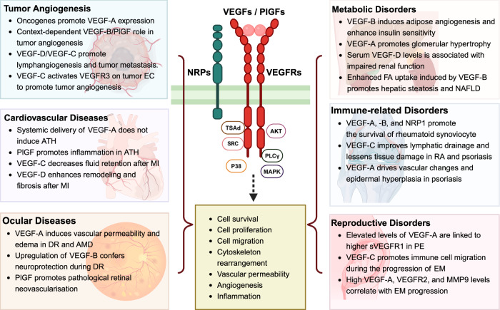
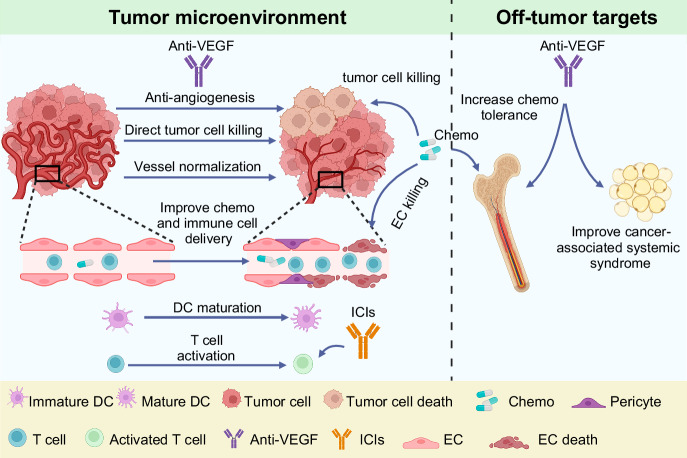
Vascular endothelial growth factor (VEGF) signaling is a critical regulator of vasculogenesis, angiogenesis, and lymphangiogenesis, processes that are vital for the development of vascular and lymphatic systems, tissue repair, and the maintenance of homeostasis. VEGF ligands and their receptors orchestrate endothelial cell proliferation, migration, and survival, playing a pivotal role in dynamic vascular remodeling. Dysregulated VEGF signaling drives diverse pathological conditions, including tumor angiogenesis, cardiovascular diseases, and ocular disorders. Excessive VEGF activity promotes tumor growth, invasion, and metastasis, while insufficient signaling contributes to impaired wound healing and ischemic diseases. VEGF-targeted therapies, such as monoclonal antibodies and tyrosine kinase inhibitors, have revolutionized the treatment of diseases involving pathological angiogenesis, offering significant clinical benefits in oncology and ophthalmology. These therapies inhibit angiogenesis and slow disease progression, but they often face challenges such as therapeutic resistance, suboptimal efficacy, and adverse effects. To further explore these issues, this review provides a comprehensive overview of VEGF ligands and receptors, elucidating their molecular mechanisms and regulatory networks. It evaluates the latest progress in VEGF-targeted therapies and examines strategies to address current challenges, such as resistance mechanisms. Moreover, the discussion includes emerging therapeutic strategies such as innovative drug delivery systems and combination therapies, highlighting the continuous efforts to improve the effectiveness and safety of VEGF-targeted treatments. This review highlights the translational potential of recent discoveries in VEGF biology for improving patient outcomes.


Conflict of interest statement

Competing interests: The authors declare no competing interests.

Figures

Fig. 1
Fig. 1
Timeline of key discoveries in angiogenesis research. The timeline begins with John Hunter’s observation of blood vessel growth and follows major advancements, including Folkman’s hypothesis, which links tumor growth to angiogenesis (1971). The significant milestones include the discovering the vascular permeability factor in 1983 and identifying vascular endothelial growth factor (VEGF), its receptors, and other VEGF family members between 1989 and 1997. It also highlights the approval of several anti-angiogenic drugs: pegaptanib for age-related macular degeneration (AMD) in 2004, bevacizumab for metastatic colorectal cancer in 2004, ranibizumab for AMD in 2006, aflibercept for AMD in 2011, ramucirumab for advanced gastric cancer in 2014, brolucizumab for AMD in 2019, the combination of atezolizumab and bevacizumab for hepatocellular carcinoma (HCC) in 2020, and faricimab for AMD in 2022. Created in BioRendender.com
Fig. 2
Fig. 2
Schematic representation of alternative splicing variants and proteolytic processing of vascular endothelial growth factors (VEGFs). a VEGF-A isoforms generated by alternative splicing including VEGF-A111, VEGF-A121, VEGF-A145, VEGF-A165, VEGF-A183, VEGF-A189, VEGF-A206, and VEGF165b. Each isoform is represented by its exon composition, highlighting the variations in exons 6A, 6B, 7, and 8 across the isoforms contributing to differences in receptor-binding affinities and functional properties. b Alternative splicing generates two VEGF-B isoforms, VEGF-B167 and VEGF-B186. These isoforms differ in their use of either exon 6A or 6B, which determines their specific molecular characteristics. c VEGF-C and VEGF-D exist in unprocessed dimerized forms, and their proteolytic processing occurs through cleavage by the protein convertases, kallikrein 3 (KLK3), cathepsin D (CTSD), or thrombin, resulting in mature, active forms with modified functional properties. Created in BioRendender.com
Fig. 3
Fig. 3
Structural domains of vascular endothelial growth factor receptors (VEGFRs) and isoforms of soluble VEGFR1. a Domain organization of VEGFR1, VEGFR2, and VEGFR3. Each receptor has key structural features, including Ig-like extracellular domains (D1-D7), transmembrane domain (TMD), juxtamembrane domain (JMD), kinase domain (KD), and C-terminal domain (CTD). b Alternative splicing generates five distinct splice variants of VEGFR1: the full-length VEGFR1, VEGFR1 with intron 13 retention (sVEGFR1-i13), VEGFR1 with intron 14 retention (sVEGFR1-i14), and the soluble isoforms sVEGFR1-e15a and sVEGFR1-e15b with alternative terminal exons. The schematic highlights the differences in domain configurations between full-length receptors and their soluble forms, illustrating the structural diversity resulting from alternative splicing. Created in BioRendender.com
Fig. 4
Fig. 4
Vascular endothelial growth factor (VEGF) family members and their receptors. Binding of VEGF-A, -B, -C, -D, and PlGF with their respective receptors, VEGFR1, VEGFR2, and VEGFR3, highlighting the primary receptor specificities. VEGF-A binds VEGFR1 and VEGFR2, VEGF-B and PlGF selectively interact with VEGFR1. VEGFR2 is the primary mediator of VEGF-A-driven angiogenesis, also binding VEGF-C. VEGFR3 predominantly binds VEGF-C and VEGF-D, regulating lymphangiogenesis. This binding schematic illustrates the selective affinities of VEGF family members for their receptors and their distinct roles in vascular and lymphatic regulation. Additionally, VEGF-B is depicted as a metabolic regulator that acts through VEGFR1, while PlGF is shown to modulate inflammation and vascular homeostasis. Created in BioRendender.com
Fig. 5
Fig. 5
Interactions of vascular endothelial growth factor receptors (VEGFR) with co-receptors and structural features of Neuropilin. a Interaction of VEGFR1 with co-receptors NRP1 and NRP2, α5β1 integrin, and FGFR1. The VEGFR1-NRP2 interaction facilitates VEGF-A121 binding, whereas VEGFR1 engagement with α5β1 promotes EC adhesion to the extracellular matrix. Additionally, the interaction of VEGFR1 with FGFR1 blocks FGF2-induced angiogenesis, highlighting the role of the VEGFR1 complex in modulating angiogenic signaling pathways and EC behavior. b Association of VEGFR2 with various co-receptors and adhesion molecules, including NRPs (NRP1 and NRP2), PDGFRβ, EGFR, CD44, EphA4, VE-Cadherin, and c-Met. These interactions contribute to the specificity and complexity of VEGF signaling, enabling crosstalk between VEGF receptors and other signaling pathways. Such an association with co-receptors and adhesion molecules enhances cellular responses, including EC migration, adhesion, and survival, supporting key physiological processes such as angiogenesis, vascular maturation, and cellular adhesion dynamics within the VEGF pathway. c Interaction between VEGFR3 and the co-receptor NRP2, which plays a critical role in promoting lymphangiogenesis and supporting EC survival. d NRP is shown with additional structural motifs, including the CUB, FV/VIII, MAM, and SEA domains. These domains contribute to receptor interactions and enhance signaling specificity within the VEGF pathway, potentially influencing binding affinities and receptor-ligand selectivity. NRP neuropilin, FGFR fibroblast growth factor receptor, PDGFR platelet-derived growth factor receptor, EGFR epidermal growth factor receptor. Created in BioRendender.com
Fig. 6
Fig. 6
Signal transduction of vascular endothelial growth factor receptors. a VEGFR1: Schematic representation of critical phosphorylation sites and their roles in signaling. Y794 in the juxtamembrane domain facilitates PLCγ1 binding and eNOS activation, promoting nitric oxide (NO) release and tubular structure formation. Y1169 recruits PLCγ1, while Y1213 enables binding of SH2 domain proteins such as PLCγ1, SHP2, and GRB2, supporting PI3K-mediated cell survival and proliferation. Y1242 and Y1333 support PLCγ1 phosphorylation but are insufficient for full mitogenic signaling. VEGF165b inhibits Y1333 phosphorylation, suppressing VEGFR1-STAT3 signaling, which can be restored to enhance angiogenesis in peripheral artery disease. b VEGFR2: Key phosphorylation sites and their roles in vascular functions. Y801 and Y1214 activate GAB1, promoting PI3K/AKT signaling, endothelial survival, migration, and NO production. Y951 (Y949 in mice) binds TSAd, regulating vascular permeability via the VEGFR2-TSAd-SRC complex. Y1054 and Y1059 are essential for full kinase activity, while Y1175 recruits adaptor proteins such as SHB and PLCγ1. Serine phosphorylation at S1183 and S1188 in mice (S1185 and S1190 in humans) promotes VEGFR2 degradation via β-TRCP1-mediated ubiquitination, regulating receptor stability. c VEGFR3: Schematic representation of essential phosphorylation sites and their roles in cell signaling. Phosphorylation at Y1063 recruits CRK I/II, activating the JNK pathway to support cell survival. Y1230 and Y1231 facilitate GRB2 recruitment, initiating ERK and AKT signaling pathways that promote cell proliferation and migration. Y1337 acts as a docking site for the GRB2-SHC complex, driving RAS-mediated signaling. PLCγ1 Phospholipase C gamma 1, GAB1 GRB2-associated binding protein 1, SHP2 Src homology-2 domain-containing protein tyrosine phosphatase-2, GRB2 growth factor receptor-bound protein 2, PI3K Phosphoinositide 3-kinase, TSad T cell-specific adapter protein, SHC SH2 domain protein C1, SHB SH2 domain-containing adapter protein B, SCK SHC-like protein, CRK I/II CT10 regulator of kinase I and II. Created in BioRendender.com
Fig. 7
Fig. 7
Negative regulation and fine-tuning of VEGFR2-mediated signaling pathways. a VE-PTP regulates TIE2 and VEGFR2 dephosphorylation to balance angiogenic signaling and support vessel integrity. Activin A enhances VE-PTP expression and reduces VEGF-induced permeability, which may help control excessive vessel leakage in conditions such as diabetic retinopathy. b PTP1B regulates VEGFR2 signaling by binding to its cytoplasmic domain and dephosphorylating it, thereby acting as a negative regulator. c SHP1 acts as a negative regulator of VEGFR2 by dephosphorylating key tyrosine residues (Y996, Y1059, and Y1175) essential for endothelial cell proliferation. d TSP-1 modulates VEGFR2 activity by binding to CD36 and recruiting SHP1, which dephosphorylates VEGFR2 at Y1175. This action suppresses VEGF signaling and inhibits angiogenesis. e Notch signaling coordinates angiogenesis by balancing the roles of tip and stalk cells. Dll4 on tip cells activates Notch in stalk cells, reducing VEGFR expression and VEGF sensitivity to stabilize newly formed vessels. Fringe, a glycosyltransferase in stalk cells, enhances Dll4-Notch signaling, further reducing VEGFR levels and reinforcing stalk cell quiescence. Meanwhile, Jagged1 counteracts Dll4 by modulating Notch in stalk cells, allowing some cells to remain responsive to VEGF, supporting sprouting. This interplay ensures selective sprouting while maintaining vessel stability. Notably, the Notch-VEGFR3 pathway can also promote angiogenesis independently of VEGF-A/VEGFR2. VE-PTP vascular endothelial protein tyrosine phosphatase, VEGFR vascular endothelial growth factor receptor, PTP1B protein tyrosine phosphatase 1B, SHP-1 Src homology 2 domain-containing phosphatase-1, TSP-1 Thrombospondin-1. Created in BioRendender.com
Fig. 8
Fig. 8
VEGF/VEGFR signaling controls angiogenesis and vascular permeability. a Role of VEGF/VEGFR signaling pathway in angiogenesis. (1) The vascular stability of quiescent microvessels covered by pericytes and basement membrane (BM) is maintained by non-VEGF/VEGFR signaling pathways such as Ang/TIE signaling. (2) Upon VEGF ligand stimulation, the BM degrades and releases bio-unavailable VEGF ligands. Endothelial cells (ECs) lose structural support and key adhesion molecules such as VE-cadherin and undergo tip cell selection. (3) VEGF signaling strongly induces the tip cell phenotype and metabolic characteristics, providing guidance cue for tip cells, whereas VEGFR2 and VEGFR3 are highly expressed in tip cells. Tip cells inhibit VEGFR2 and VEGFR3 and increase VEGFR1 and sVEGFR1 in stalk cells. VEGF supports perpendicular proliferation of stalk cells and lumen formation. (4) Tip cells recognize each other and induce sprout anastomosis. sVEGFR1, possibly produced by macrophages, regulates this process. VEGF negatively regulates vessel maturation. b Role of VEGF/VEGFR signaling in vascular permeability. The vascular permeability of quiescent microvessels is maintained by pores in ECs, glycocalyx, and EC junctions. VEGFR2 Y949 phosphosite in mice downregulates VE-cadherin via SRC. VEGFR2 also promotes nitric oxide (NO) production to reduce the expression of adhesion molecules required for paracellular permeability. VEGF has also been shown to stimulate vesicovacuolar organelles (VVO) and fenestrae formation for transcellular permeability. However, whether the glycocalyx can be cleaved by VEGF stimulation is not fully understood. Created in BioRendender.com
Fig. 9
Fig. 9
VEGF/VEGFR signaling under pathological conditions. The diverse roles of VEGF ligands and their receptors drive pathological processes, including tumor angiogenesis, cardiovascular diseases, ocular diseases, and metabolic/immune-related/reproductive disorders. Key molecular interactions and downstream signaling pathways are upregulated or dysregulated in disease states, thereby promoting cell survival, migration, and proliferation. These pathways highlight the therapeutic potential of targeting VEGF/VEGFR signaling across various disease contexts. ATH atherosclerosis, MI myocardial ischemia, DR diabetic retinopathy, AMD age-related macular degeneration, FA fatty acid, NAFLD non-alcoholic fatty liver disease, RA rheumatoid arthritis, PE pre-eclampsia, EM endometriosis. Created in BioRendender.com
Fig. 10
Fig. 10
Mechanisms of action of anti-VEGF/VEGFR therapies in combination therapy. In the tumor microenvironment (TME), the mechanisms of action of anti-VEGF/VEGFR therapies combined with chemotherapy or immune checkpoint inhibitors (ICIs) include reducing angiogenicity of vessels for tumor inhibition, inducing vessel normalization for better chemo-drug or immune cell delivery, directly inhibiting tumor cell proliferation, increasing anti-endothelial cell effect of chemotherapy, and modulating immune cells for better dendritic cell (DC) differentiation and T-cell activation. Outside the TME, the anti-VEGF/VEGFR therapies may reduce chemotoxicity and improve cancer-associated systemic syndromes, thereby improving therapeutic efficacy. Created in BioRendender.com

Similar articles

Cited by

References

    1. Carmeliet, P. Angiogenesis in life, disease and medicine. Nature438, 932–936 (2005). - PubMed
    1. Ferrara, N. & Kerbel, R. S. Angiogenesis as a therapeutic target. Nature438, 967–974 (2005). - PubMed
    1. Apte, R. S., Chen, D. S. & Ferrara, N. VEGF in signaling and disease: beyond discovery and development. Cell176, 1248–1264 (2019). - PMC - PubMed
    1. Stephenson, J. A., Goddard, J. C., Al-Taan, O., Dennison, A. R. & Morgan, B. Tumour angiogenesis: a growth area—from John Hunter to Judah Folkman and beyond. J. Cancer Res.2013, 895019 (2013).
    1. Lenzi, P., Bocci, G. & Natale, G. John Hunter and the origin of the term “angiogenesis”. Angiogenesis19, 255–256 (2016). - PubMed

MeSH terms

Substances

LinkOut - more resources