. 2025 Dec;17(1):2506192.
doi: 10.1080/19490976.2025.2506192. Epub 2025 May 18.

CD44 is a macrophage receptor for TcdB from Clostridioides difficile that via its lysine-158 succinylation contributes to inflammation

Affiliations

CD44 is a macrophage receptor for TcdB from Clostridioides difficile that via its lysine-158 succinylation contributes to inflammation

Zhuo Chen et al. Gut Microbes. 2025 Dec.

Erratum in

  • Correction.
    [No authors listed] [No authors listed] Gut Microbes. 2025 Dec;17(1):2532985. doi: 10.1080/19490976.2025.2532985. Epub 2025 Jul 11. Gut Microbes. 2025. PMID: 40650384 Free PMC article. No abstract available.

Abstract

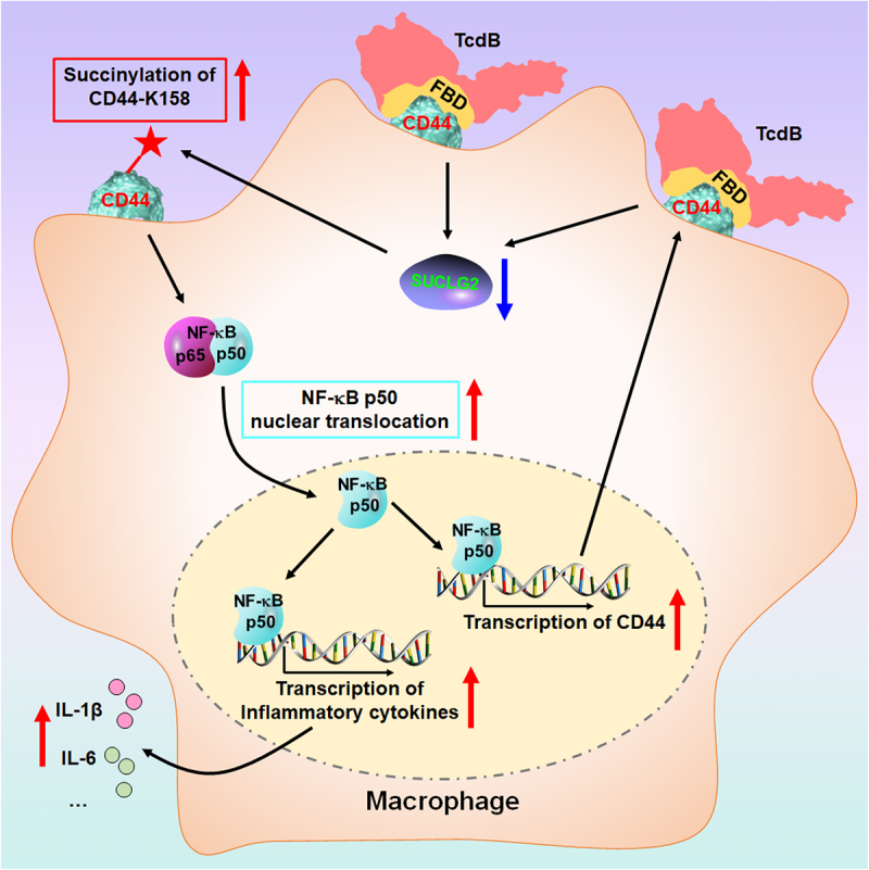
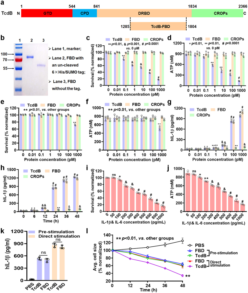
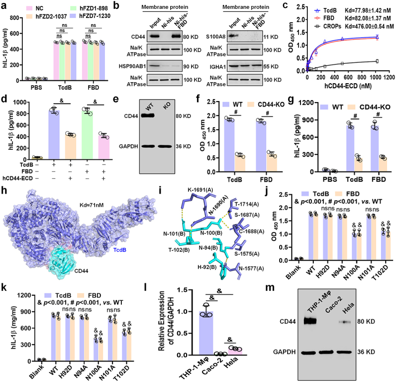
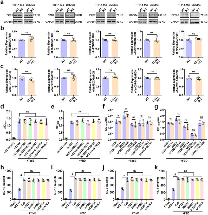
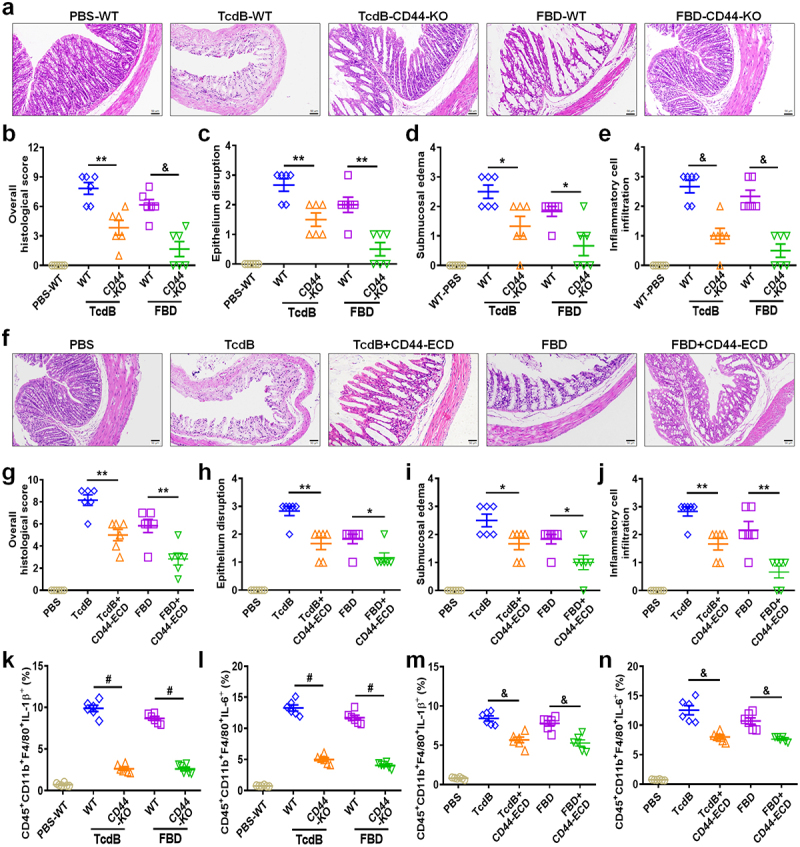
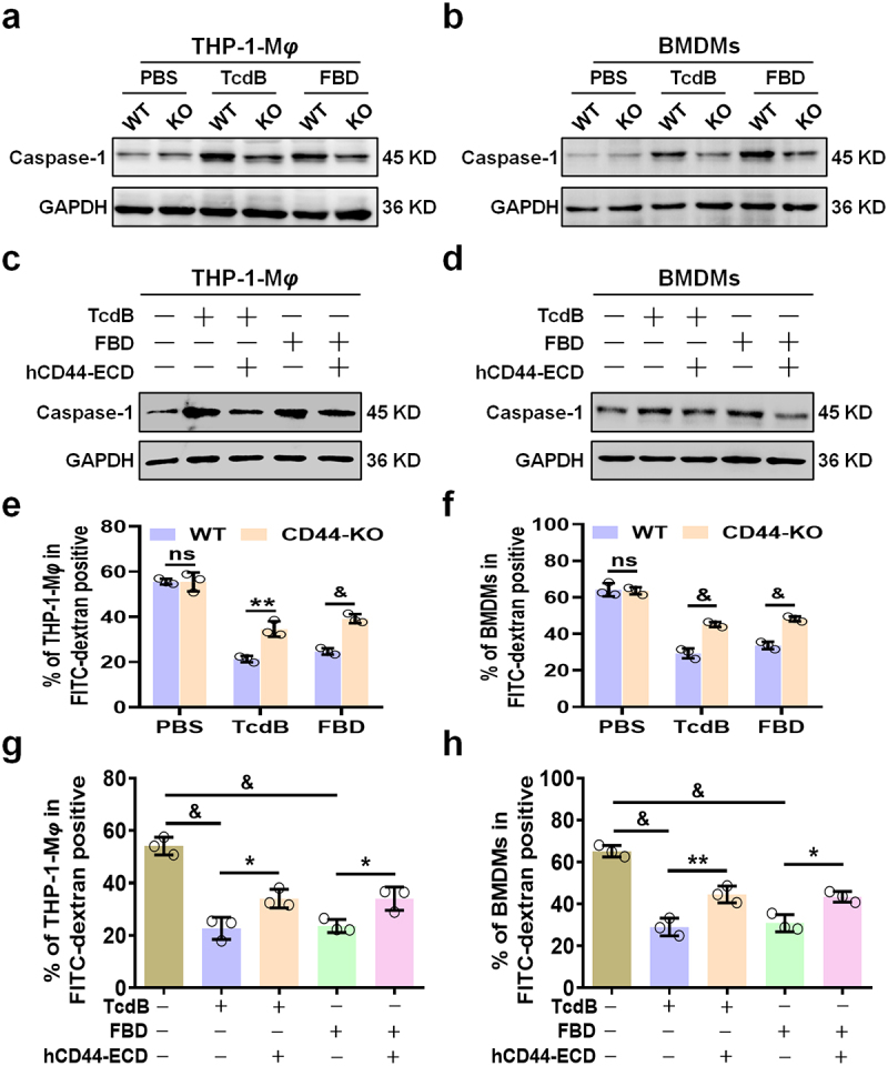
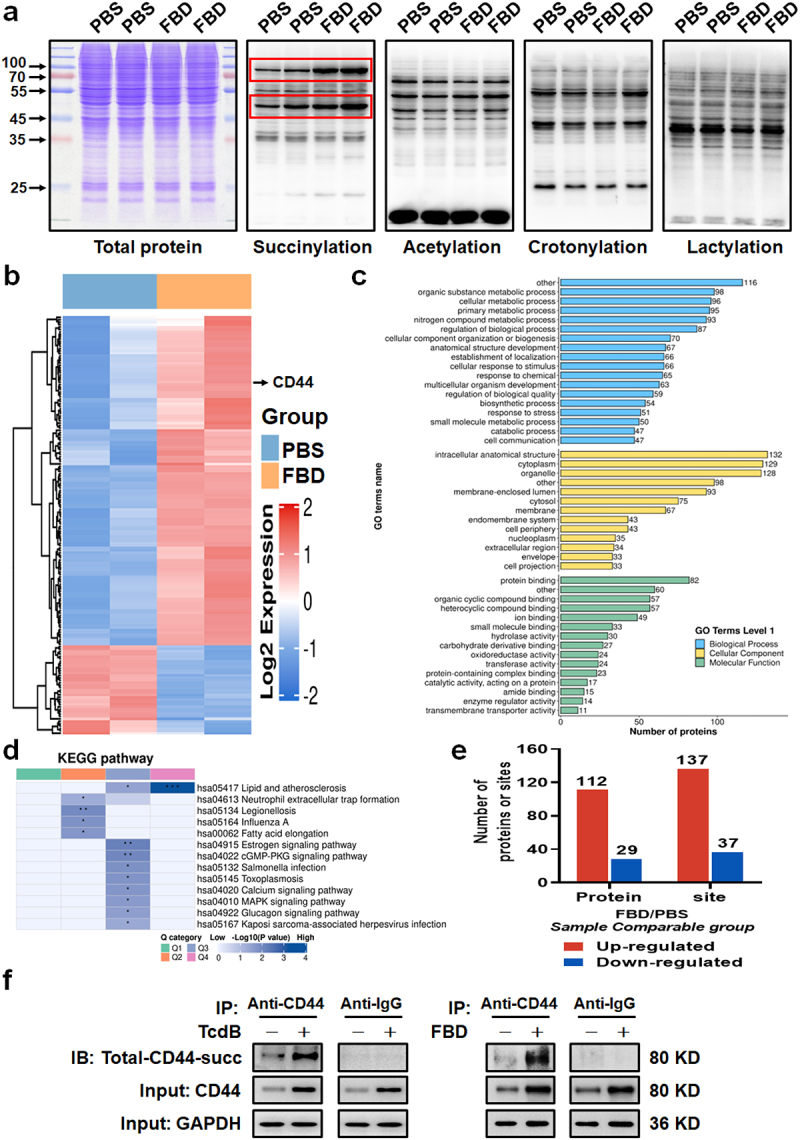
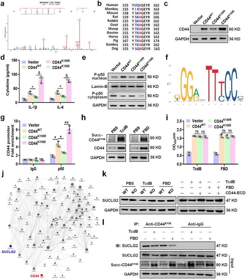
Toxin B (TcdB) is a critical virulence factor in Clostridioides difficile-associated disease (CDAD), which activates macrophages to promote inflammation and epithelial damage. However, the mechanism by which TcdB targets inflammation-related receptors on the macrophage surface and the underlying molecular mechanisms remain unknown. The frizzled-binding domain of TcdB (TcdB-FBD) is a promising target of TcdB. Here, FBD was found to trigger macrophage inflammation, similar to TcdB, but did not induce cytotoxicity. Thus, using FBD as a bait protein, macrophage CD44 was identified as an inflammation-related receptor for TcdB/FBD. The role of CD44 was confirmed by CRISPR/Cas9-mediated gene knockout in macrophages and CD44 knockout mice. Using 4-D label-free succinylation quantitative modification proteomics, we demonstrated that TcdB/FBD binds to CD44 in macrophages, promotes CD44 K158 succinylation via SUCLG2 suppression, and enhances NF-κB translocation/transcriptional activity, thereby driving inflammation. Finally, blocking the binding of TcdB to CD44 was demonstrated as a favorable strategy for inhibiting TcdB-mediated macrophage inflammation. This study not only provides a new therapeutic target for the prevention and treatment of CDAD but also elucidates a new molecular mechanism underlying the inflammatory effect of TcdB via the TcdB/FBD-CD44 axis.

Keywords: CD44; TcdB; inflammation; receptor; succinylation.


Conflict of interest statement

No potential conflict of interest was reported by the author(s).

Figures

None
Graphical abstract
Figure 1.
Figure 1.
Effect of FBD-mediated macrophage secretion of inflammatory cytokines is comparable to that of TcdB. (a) Schematic diagram showing the domain organization of TcdB. GTD, glucosyltransferase domain (red); CPD, cysteine protease domain (light blue); DRBD, delivery and receptor-binding domain (orange); CROPs, combined repetitive oligopeptides domain (green). (b) The FBD extracted was analyzed by SDS−PAGE. (c–f) cells were treated with a range of TcdB/FBD/CROPs concentrations, and then the survival (c) or ATP levels (d) in caco-2 cells, the survival (e) or ATP levels (f) in THP-1-Mφwere assessed. (g) THP-1-Mφ were treated with different proteins for 24 h and then production of IL-1β were measured by an ELISA. (h) THP-1-Mφ were treated with 100 pM of TcdB, FBD, or CROPs, and the levels of IL-1β was assessed at different time points. (i–j) caco-2 cells were stimulated with different concentrations of inflammatory cytokines (IL-1β and IL-6) for 48 h. The cell (i) viability, and (j) production of ATP by the cells was determined. (k) THP-1-Mφ were treated with TcdB/FBD (100 pM), after being pre-stimulated or stimulated directly as indicated for 24 h, and the levels of IL-1β were measured. (l) Normalized average cell size over time of caco-2 cells co-cultured with THP-1-Mφ, which were pre-stimulated or stimulated directly with TcdB/FBD, were analyzed by an HCI system. All data in parts (c–l) are shown as the mean ± SEM (n = 3). (ns: p > 0.05, *p < 0.05, **p < 0.01, &;p < 0.001, #p < 0.0001; g: vs 0 pM; h: vs 0 h; i – j: vs 0 pg/mL).
Figure 2.
Figure 2.
FBD/TcdB binds to CD44 on macrophages to induce inflammatory cytokine production. (a) THP-1-Mφ were treated with TcdB or FBD (100 pM) for 24 h after knockdown of FZD1/2/7. Levels of IL-1β were measured by an ELISA. (b) An FBD-bead pull-down experiment using membrane extracts of THP-1-Mφ was assessed by MALDT-TOF-MS, and the identified proteins related to inflammation were analyzed with a WB. (c) ELISA of the binding of human CD44-ECD protein to TcdB/FBD/CROPs. Various concentrations (1–1024 nM) of hCD44-ECD (labeled with fc) were incubated in wells coated with TcdB/FBD/CROPs. (d) THP-1-Mφ were treated with TcdB/FBD (100 pM) and CD44-ECD (1 μM) for 24 h, and then the levels of IL-1β were measured. (e) Wild-type (WT) and CD44-knockout (CD44-KO) THP-1-Mφ were identified by WB. (f) Binding of TcdB or FBD to membrane extracts of WT or CD44-KO THP-1-Mφ as shown by ELISA. (g) WT or CD44-KO THP-1-Mφ were treated with TcdB or FBD (100 pM) for 24 h, and the levels of IL-1β were measured by an ELISA. Molecular docking predicts the structure and dissociation constant kd value (h) and the interaction sites of TcdB (blue purple) and hCD44 (cyan) complexes (i). (j) Mutations in CD44 that disrupt the interactions of THP-1-Mφ membrane extracts with TcdB/FBD were demonstrated by ELISA. (k) Wild-type or CD44-mutated THP-1-Mφ were treated with TcdB or FBD (100 pM) for 24 h, and the levels of IL-1β were measured by an ELISA. (l – m) the expression of CD44 in different cells by (l) qRT-PCR and (m) WB. All data in (a), (c – d), (f – g) and (j – l) are shown as the mean ± SEM (n = 3). (ns: p > 0.05, &p < 0.001, #p < 0.0001).
Figure 3.
Figure 3.
CD44 functions as an independent receptor for TcdB/FBD in macrophages. (a – c) expression of known receptors (FZD1/2/7, CSPG4, PVRL3) in WT versus CD44-KO macrophages analyzed by (a) immunoblotting and (b, c) q-PCR. (d – g) competitive binding assays: (d, e) FZD1/2/7, CSPG4, and PVRL3 did not inhibit CD44 binding of (d) TcdB or (e) FBD, while (f, g) CD44 similarly did not interfere with TcdB/FBD binding to other receptors. (ELISA protocol: wells coated with 1 μM target protein [CD44-ECD or other receptors] were incubated with 1 μM TcdB/FBD, followed by competitor proteins. Binding was quantified by absorbance measurement). (h – k) IL-1β (h, i) and IL-6 (j, k) levels in THP-1-Mφ treated with TcdB/FBD (100 pM) and receptor proteins (1 μM) for 24 h. All data in parts (b – k) are shown as the mean ± SEM (n = 3). ((ns: p > 0.05, **p < 0.01, &p < 0.001, #p < 0.0001).
Figure 4.
Figure 4.
CD44 is an inflammatory-related receptor for TcdB/FBD in vivo. (a – j) mouse colonic tissues harvested after intrarectal instillation assays were assessed for pathology through H&E staining. (a) Mice colons were exposed to high concentrations of TcdB/FBD (100 pM) after being stimulated as indicated for 48 h, and representative H&E images are shown. Scale bar represents 50 μm. (b) Overall histology scores are graphed. (c – e) histopathological scores (n = 6 mice) for (a) were assessed based on indicated pathological features for epithelium disruption (c), submucosal oedema (d), and inflammatory cell infiltration (e). (f) Mice were exposed to high concentration of TcdB/FBD (100 pM) and supersaturated CD44-ECD (1 μM), after being stimulated as indicated 48 h, and representative H&E images are shown. The scale bar represents 50 μm. (g) Overall histology scores are graphed. (h – j) histopathological scores for (f) were assessed based on indicated pathological features for epithelium disruption (h), submucosal oedema (i), and inflammatory cell infiltration (j). (k – n) FCM statistical analysis of the TcdB- or FBD-induced IL-1β (k, m) or IL-6 (l, n) production by intestinal macrophages in mice. All data in parts (b – e) and (g – n) are shown as the mean ± SEM (n = 6). (*p < 0.05, **p < 0.01, &p < 0.001, #p < 0.0001).
Figure 5.
Figure 5.
TcdB/FBD induces CD44-dependent inflammasome activation and modulates macrophage phagocytosis. (a – b) Western blot analysis of caspase-1 in (a) THP-1-Mφ and (b) BMDMs from WT and CD44-knockout macrophages after 24-hour stimulation with 100 pM TcdB or FBD. (c – d) immunoblot showing caspase-1 expression in (c) THP-1-Mφ and (d) BMDMs treated with 100 pM TcdB/FBD in combination with 1 μM CD44-ECD for 24 hours. (e – f) FITC-dextran uptake assay in (e) THP-1-Mφ and (f) BMDMs from WT and CD44-knockout macrophages. Cells were treated with 100 pM TcdB or FBD for 24 hours, followed by exposure to FITC-dextran (250 μg/ml) for 1 hour. (g – h) FITC-dextran uptake assay in (g) THP-1-Mφ and (h) BMDMs co-treated with 100 pM TcdB/FBD and 1 μM CD44-ECD for 24 hours, then incubated with FITC-dextran (250 μg/ml) for 1 hour. Fluorescence intensity was quantified by flow cytometry. Statistical analysis of fluorescence signals. All data are shown as the mean ± SEM (n = 3). (ns: p > 0.05, *p < 0.05, **p < 0.01, &p < 0.001).
Figure 6.
Figure 6.
FBD promotes succinylation of CD44 in macrophages. (a) Changes in common PTM levels in THP-1-Mφ cells caused by FBD were identified. THP-1-Mφ were treated with FBD (100 pM) for 24 h, and then were detected by WB using specific antibodies against multiple PTMs, including succinylation, acetylation, crotonylation, and lactylation. Red box represents the bands with significant differences, and Coomassie brilliant blue staining was used as a loading control. (b) Heat map for protein succinylation omics screening. THP-1-Mφ were treated with FBD (100 pM) for 24 h. (c – d) global landscape and functional annotation of FBD-regulated succinylation in THP-1-Mφ. (c) GO enrichment of quantified differentially modified proteins. Vertical axis represents the secondary functional classification in the primary classification of GO, the horizontal axis represents the number of differentially succinylated modified proteins in the classification, and the different colors represent the primary classification of GO. (d) KEGG pathway enrichment analysis of four groups of samples, Q1 < 0.5, Q2 (0.5–0.667), Q3 (1.5–2.0), and Q4 > 2.0. (e) Summary of differentially quantified sites and proteins. (f) The immunoprecipitation analysis of the total succinylation level of CD44 in THP-1-Mφ induced by TcdB (left) or FBD (right). Representative western blot lines of total succinyl-CD44 and CD44 proteins in THP-1-Mφ. The result of the anti-IgG was indicated as a negative control. GAPDH was used as the loading control. Succinylation pan antibodies were used.
Figure 7.
Figure 7.
Desuccinylase SUCLG2 mediated CD44 succinylation at Lys158 enhances macrophage inflammatory cytokine production via NF-κB activation. (a) Succinylation of CD44 in THP-1-Mφ, and mass spectrometric verification of CD44 succinylation at K158 (KsuccYVQKGEYR). (b) K158 (marked in red), a CD44 ortholog, is highly conserved in mammals. (c – e) expression of CD44 was measured by WB (c), production of IL-1β and IL-6 was determined by an ELISA (d), and the expression of phosphorylated p50 in the nucleus and cytoplasm was detected by WB (e) in wild-type or CD44-mutated THP-1-Mφ. (f) Results show the NF-κB-JASPAR binding motif. (g) ChIP assay coupled with qRT-PCR analysis revealed the relative enrichment of NF-κB p50 on the CD44 promoters in THP-1-Mφ. The Fold enrichment of the ChIP assay was calculated with reference to the control IgG after normalization to the input DNA. (h) Representative western blot lines of succ-CD44Lys158 and CD44 proteins in THP-1-Mφ after treatment with TcdB/FBD. (i) Mutations in CD44 that did not disrupt the binding of THP-1-Mφ membrane extracts with TcdB or FBD were demonstrated by ELISA. (j) The protein-protein interaction (PPI) network was constructed using the STRING database based on post-translational modification data. CD44 and SUCLG2 are highlighted in red and blue, respectively. Connecting lines indicate predicted binding, interaction, or complex formation between proteins. (k) Representative western blot bands showing SUCLG2 expression in THP-1-Mφ. GAPDH served as the loading control. (l) Immunoprecipitation analysis of SUCLG2 binding to succ-CD44Lys158 in TcdB/FBD-treated macrophages. Anti-IgG served as a negative control, with GAPDH as the loading control. All data in (d) and (g – h) are shown as the mean ± SEM (n = 3). (ns: p > 0.05, *p < 0.05, **p < 0.01, &p < 0.001).

References

    1. Di Bella S, Sanson G, Monticelli J, Zerbato V, Principe L, Giuffre M, Pipitone G, Luzzati R, Staley C.. Clostridioides difficile infection: history, epidemiology, risk factors, prevention, clinical manifestations, treatment, and future options. Clin Microbiol Rev. 2024;37(2):e0013523. doi: 10.1128/cmr.00135-23. - DOI - PMC - PubMed
    1. Kordus SL, Thomas AK, Lacy DB. Clostridioides difficile toxins: mechanisms of action and antitoxin therapeutics. Nat Rev Microbiol. 2022;20(5):285–23. doi: 10.1038/s41579-021-00660-2. - DOI - PMC - PubMed
    1. Walter J, Shanahan F. Fecal microbiota-based treatment for recurrent Clostridioides difficile infection. Cell. 2023;186(6):1087. doi: 10.1016/j.cell.2023.02.034. - DOI - PubMed
    1. Kuehne SA, Cartman ST, Heap JT, Kelly ML, Cockayne A, Minton NP. The role of toxin a and toxin B in clostridium difficile infection. Nature. 2010;467(7316):711–713. doi: 10.1038/nature09397. - DOI - PubMed
    1. Lyras D, O’Connor JR, Howarth PM, Sambol SP, Carter GP, Phumoonna T, Poon R, Adams V, Vedantam G, Johnson S, et al. Toxin B is essential for virulence of Clostridium difficile. Nature. 2009;458(7242):1176–1179. doi: 10.1038/nature07822. - DOI - PMC - PubMed

LinkOut - more resources